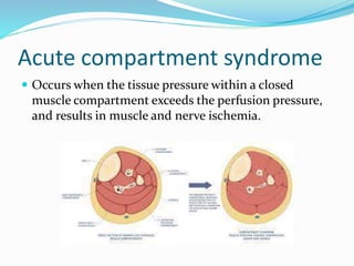
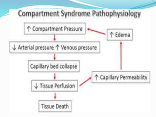
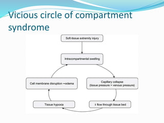
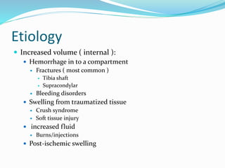
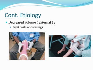
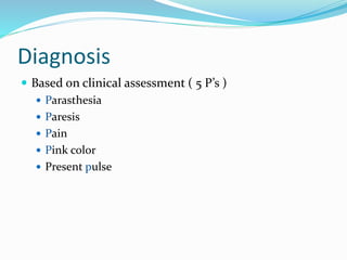
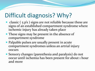
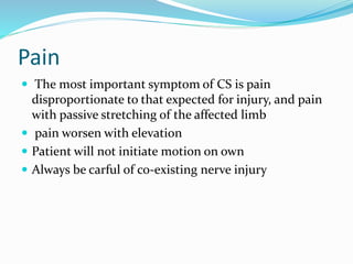
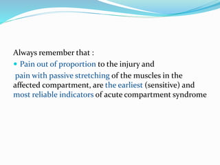
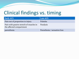
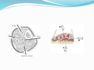
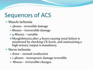
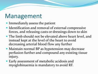
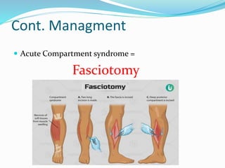
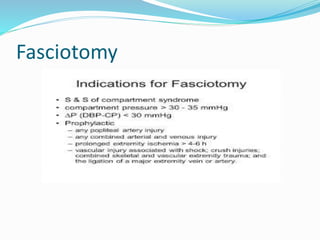
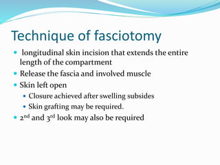
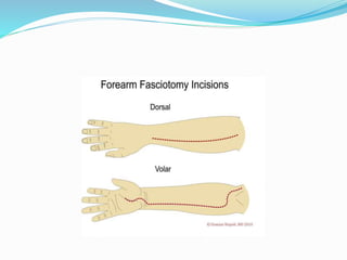
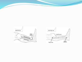
Presented by : Dr. Mohamed Adel Al-Weshali
Resident of G.surgury, orthopedic arabic board
48 modern hospital
Under supervision of :
Dr. Ali Mohamed Saled
Dr. Abdo shamsan
Dr. Mohamed Essa
Dr. Raidan Al-Eryani
Types of compartment syndrome
 Acute
or
 Chronic ( Exertional )
Acute compartment syndrome
 Occurs when the tissue pressure within a closed
muscle compartment exceeds the perfusion pressure,
and results in muscle and nerve ischemia.
Vicious circle of compartment
syndrome
Etiology
 Increased volume ( internal ):
 Hemorrhage in to a compartment
 Fractures ( most common )
 Tibia shaft
 Supracondylar
 Bleeding disorders
 Swelling from traumatized tissue
 Crush syndrome
 Soft tissue injury
 increased fluid
 Burns/injections
 Post-ischemic swelling
Cont. Etiology
 Decreased volume ( external ) :
 tight casts or dressings.
Arterial injury
 Post ischemic swelling
 Reperfusion injury :
 Ischemia causes damage to cellular basement membrane
that results in edema.
 With re-establishment of flow, fluid leaks into the
compartment, increasing the pressure.
Fracture treatment increases IMP
 Splinting / casting.
 Manipulation.
 Traction.
 Spanning Ex. Fix.
 Nailing.
Incidence of ACS
 2-10% tibial fractures
 10% calcaneal fractures
 18% Schatzker VI plateau fractures
 41% foot crush injuries
 48% segmental tibia fractures
 53% medial knee fractures/dislocations
Diagnosis
 Based on clinical assessment ( 5 P’s )
 Parasthesia
 Paresis
 Pain
 Pink color
 Present pulse
Difficult diagnosis? Why?
 classic ( 5 p’s ) signs are not reliable because these are
signs of an established compartment syndrome where
ischemic injury has already taken place
 These signs may be present in the absence of
compartment syndrome
 Palpable pulses are usually present in acute
compartment syndromes unless an artrial injury
oocurs
 Sensory changes (paresthesia and paralysis) do not
oocur until ischemia has been present for about 1 hour
and more
Pain
 The most important symptom of CS is pain
disproportionate to that expected for injury, and pain
with passive stretching of the affected limb
 pain worsen with elevation
 Patient will not initiate motion on own
 Always be carful of co-existing nerve injury
Always remember that :
 Pain out of proportion to the injury and
pain with passive stretching of the muscles in the
affected compartment, are the earliest (sensitive) and
most reliable indicators of acute compartment syndrome
Clinical findings vs. timing
Early ACS Late ACS
Pain out of proportion to injury Pulselss
Pain with passive stretch of muscles in
the affected compartment
Paralysis
paresthesia Paresthesia / sensation loss
Pediatric ACS
 5 P’s are not reliable
 3 A’s instead
 Increasing Analgesic reqirement
 Anxiety
 Agitation
Physical signs of ACS
 Pain with passive stretch of limb.
 A firm and tense compartment with a firm “wood-like”
feeling.
 Diminished sensation.
 Muscle weakness (onset within two to four hours of
ACS)
 Paralysis (late finding)
Who is at high risk?
 inability to obtain history and physical exam such as:
 Head trauma
 Drug/Alcohol intake
 pediatric
 Multiple trauma patients with hypoxia and hypotension
 Compartment syndrome can occur at lower absolute pressure
 high energy fractures
 Severe comminution
 Segmental injuries
 widely displaced
 Bilateral
 Floating knee
 Open fractures
 Presence of an open fracture does not role out the presence of a C.S.
 Impaired sensorium
 Alcohol
 Drug
 Decreased GCS
 Unconscious
 Neurologic deficit
 Post operative patients on analgesia that mask the development of pain
Intramuscular pressure (IMP)
measurment
 Adjunct to clinical examination
 By using Stryker device
 Needed for comatose or otherwise non-evaluable
patient:
 Anasthesia
 Head injury
 Sedated
 Intoxicated
 Pediatric Pts.
Sequences of ACS
 Muscle ischemia
 4hours - reversible damage
 8hours – irreversible damage
 4-8hours – variable
Myoglobinuria after 4 hours causing renal failure is
monitored by checking CK levels, and maintaining a
high urinary output is mandatory.
 Nerve ischemia
 1hour – normal conduction
 1-4hours - neuropraxic damage reversible
 8hours – irriversible changes.
Management
 Immediately assess the patient
 Identification and removal of external compressive
forces, and releasing casts or dressings down to skin
 The limb should not be elevated above heart level, and
instead kept at the level of the heart to avoid
decreasing arterial blood flow any further
 Maintain normal BP as hypotension may decrease
perfusion further and compound any existing tissue
injury.
 Early assessment of metabolic acidosis and
myoglobinaemia is mandatory to avoid RF.
Cont. Managment
 Acute Compartment syndrome =
Fasciotomy
Fasciotomy
Technique of fasciotomy
 longitudinal skin incision that extends the entire
length of the compartment
 Release the fascia and involved muscle
 Skin left open
 Closure achieved after swelling subsides
 Skin grafting may be required.
 2nd and 3rd look may also be required
Thank you…
References
Orthopaedic trauma association.
Orthobullets.
American Academy of orthopaedic surgeons.
AAOS (American association for orthpaedic surgeons).

acute Compartment syndrome.pptx

  • 1.
    Presented by :Dr. Mohamed Adel Al-Weshali Resident of G.surgury, orthopedic arabic board 48 modern hospital Under supervision of : Dr. Ali Mohamed Saled Dr. Abdo shamsan Dr. Mohamed Essa Dr. Raidan Al-Eryani
  • 2.
    Types of compartmentsyndrome  Acute or  Chronic ( Exertional )
  • 3.
    Acute compartment syndrome Occurs when the tissue pressure within a closed muscle compartment exceeds the perfusion pressure, and results in muscle and nerve ischemia.
  • 5.
    Vicious circle ofcompartment syndrome
  • 6.
    Etiology  Increased volume( internal ):  Hemorrhage in to a compartment  Fractures ( most common )  Tibia shaft  Supracondylar  Bleeding disorders  Swelling from traumatized tissue  Crush syndrome  Soft tissue injury  increased fluid  Burns/injections  Post-ischemic swelling
  • 7.
    Cont. Etiology  Decreasedvolume ( external ) :  tight casts or dressings.
  • 8.
    Arterial injury  Postischemic swelling  Reperfusion injury :  Ischemia causes damage to cellular basement membrane that results in edema.  With re-establishment of flow, fluid leaks into the compartment, increasing the pressure.
  • 9.
    Fracture treatment increasesIMP  Splinting / casting.  Manipulation.  Traction.  Spanning Ex. Fix.  Nailing.
  • 10.
    Incidence of ACS 2-10% tibial fractures  10% calcaneal fractures  18% Schatzker VI plateau fractures  41% foot crush injuries  48% segmental tibia fractures  53% medial knee fractures/dislocations
  • 11.
    Diagnosis  Based onclinical assessment ( 5 P’s )  Parasthesia  Paresis  Pain  Pink color  Present pulse
  • 12.
    Difficult diagnosis? Why? classic ( 5 p’s ) signs are not reliable because these are signs of an established compartment syndrome where ischemic injury has already taken place  These signs may be present in the absence of compartment syndrome  Palpable pulses are usually present in acute compartment syndromes unless an artrial injury oocurs  Sensory changes (paresthesia and paralysis) do not oocur until ischemia has been present for about 1 hour and more
  • 13.
    Pain  The mostimportant symptom of CS is pain disproportionate to that expected for injury, and pain with passive stretching of the affected limb  pain worsen with elevation  Patient will not initiate motion on own  Always be carful of co-existing nerve injury
  • 14.
    Always remember that:  Pain out of proportion to the injury and pain with passive stretching of the muscles in the affected compartment, are the earliest (sensitive) and most reliable indicators of acute compartment syndrome
  • 15.
    Clinical findings vs.timing Early ACS Late ACS Pain out of proportion to injury Pulselss Pain with passive stretch of muscles in the affected compartment Paralysis paresthesia Paresthesia / sensation loss
  • 16.
    Pediatric ACS  5P’s are not reliable  3 A’s instead  Increasing Analgesic reqirement  Anxiety  Agitation
  • 17.
    Physical signs ofACS  Pain with passive stretch of limb.  A firm and tense compartment with a firm “wood-like” feeling.  Diminished sensation.  Muscle weakness (onset within two to four hours of ACS)  Paralysis (late finding)
  • 18.
    Who is athigh risk?  inability to obtain history and physical exam such as:  Head trauma  Drug/Alcohol intake  pediatric  Multiple trauma patients with hypoxia and hypotension  Compartment syndrome can occur at lower absolute pressure  high energy fractures  Severe comminution  Segmental injuries  widely displaced  Bilateral  Floating knee  Open fractures  Presence of an open fracture does not role out the presence of a C.S.  Impaired sensorium  Alcohol  Drug  Decreased GCS  Unconscious  Neurologic deficit  Post operative patients on analgesia that mask the development of pain
  • 19.
    Intramuscular pressure (IMP) measurment Adjunct to clinical examination  By using Stryker device  Needed for comatose or otherwise non-evaluable patient:  Anasthesia  Head injury  Sedated  Intoxicated  Pediatric Pts.
  • 21.
    Sequences of ACS Muscle ischemia  4hours - reversible damage  8hours – irreversible damage  4-8hours – variable Myoglobinuria after 4 hours causing renal failure is monitored by checking CK levels, and maintaining a high urinary output is mandatory.  Nerve ischemia  1hour – normal conduction  1-4hours - neuropraxic damage reversible  8hours – irriversible changes.
  • 22.
    Management  Immediately assessthe patient  Identification and removal of external compressive forces, and releasing casts or dressings down to skin  The limb should not be elevated above heart level, and instead kept at the level of the heart to avoid decreasing arterial blood flow any further  Maintain normal BP as hypotension may decrease perfusion further and compound any existing tissue injury.  Early assessment of metabolic acidosis and myoglobinaemia is mandatory to avoid RF.
  • 23.
    Cont. Managment  AcuteCompartment syndrome = Fasciotomy
  • 24.
  • 25.
    Technique of fasciotomy longitudinal skin incision that extends the entire length of the compartment  Release the fascia and involved muscle  Skin left open  Closure achieved after swelling subsides  Skin grafting may be required.  2nd and 3rd look may also be required
  • 28.
  • 29.
    References Orthopaedic trauma association. Orthobullets. AmericanAcademy of orthopaedic surgeons. AAOS (American association for orthpaedic surgeons).