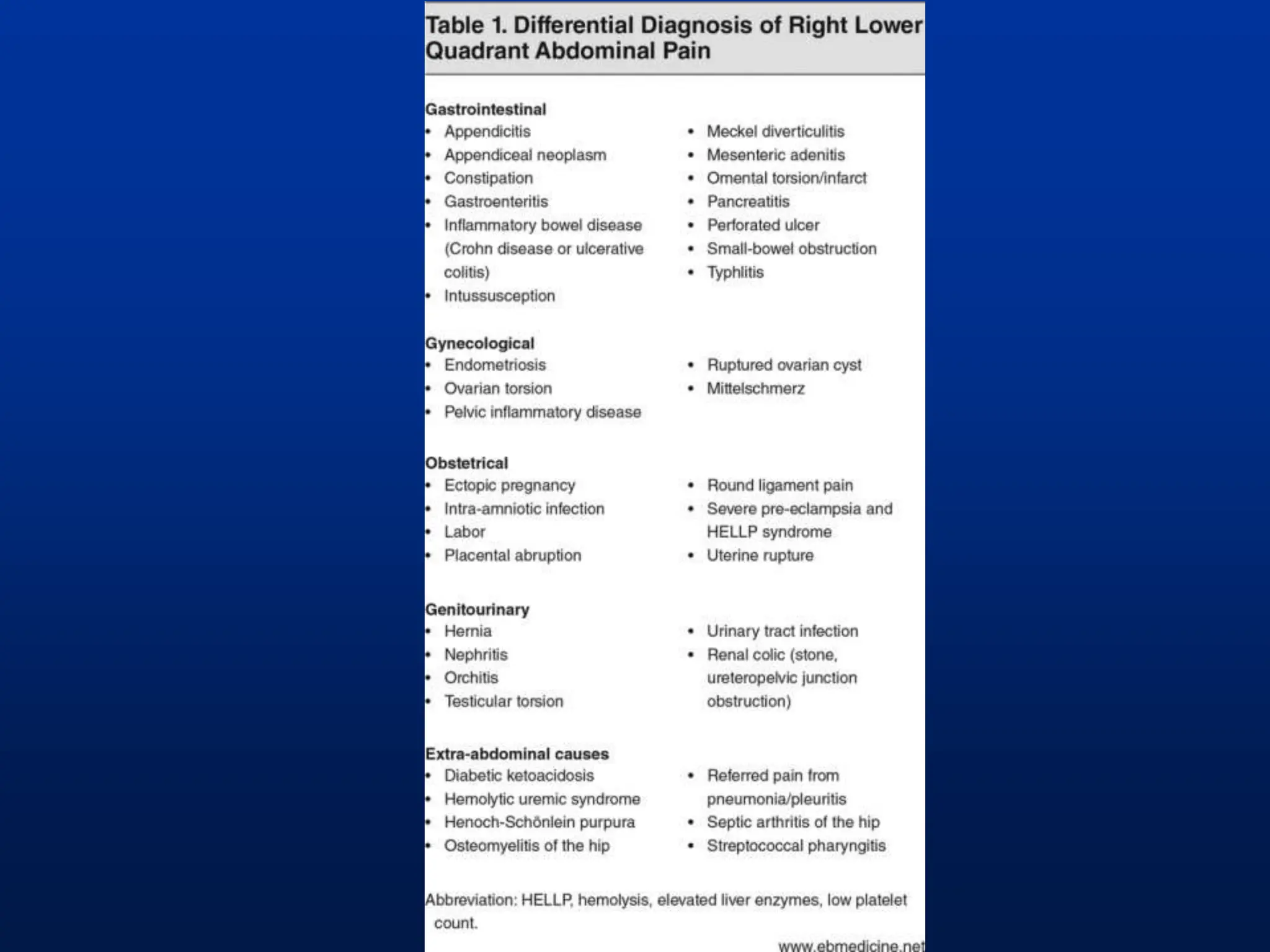
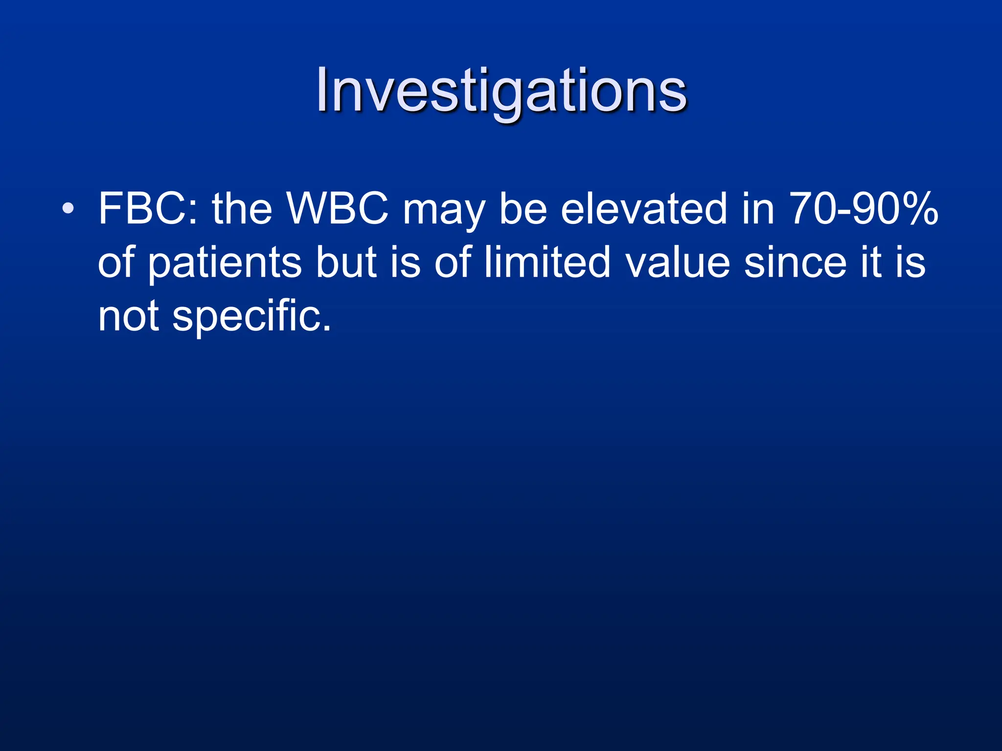
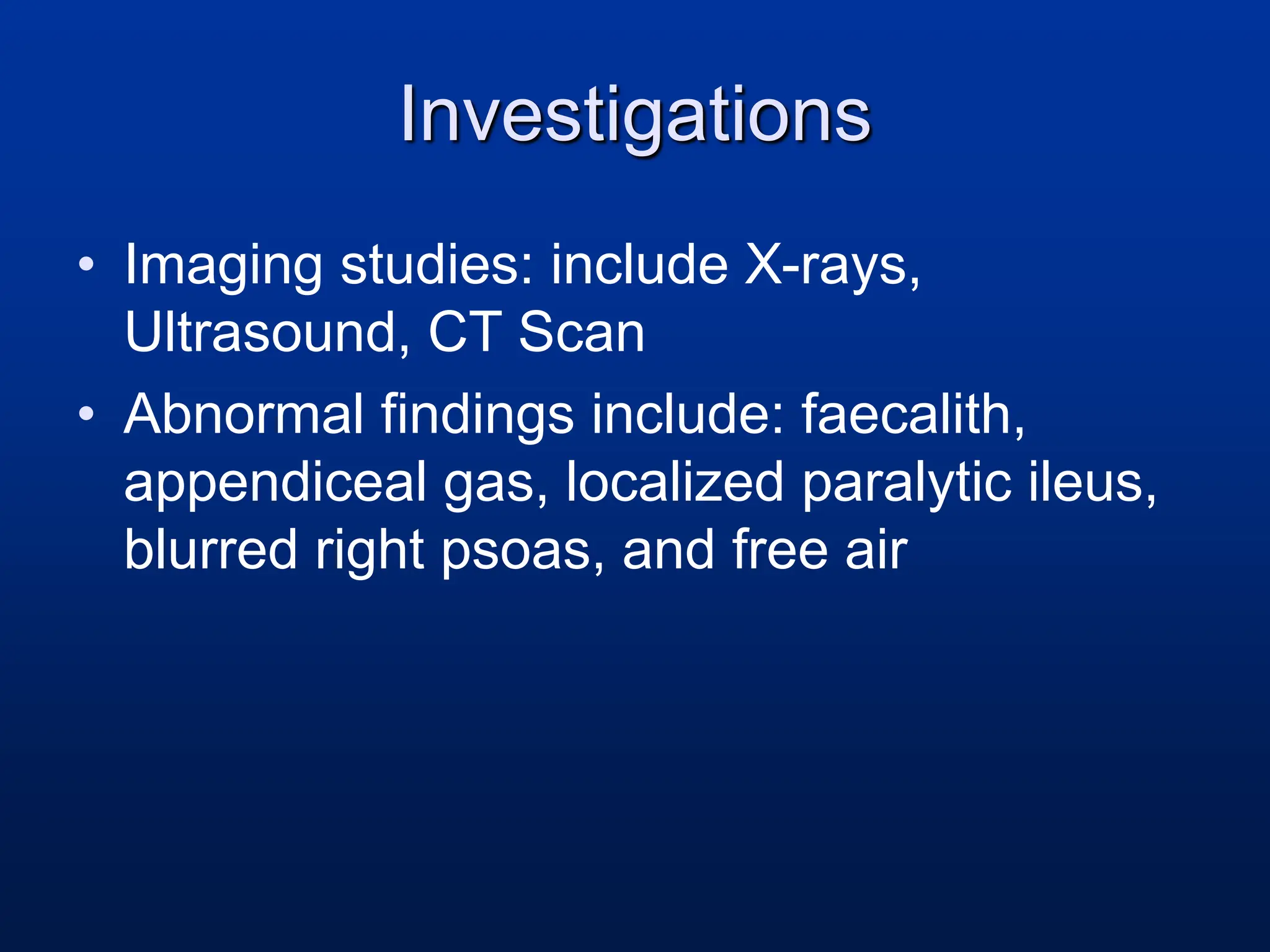
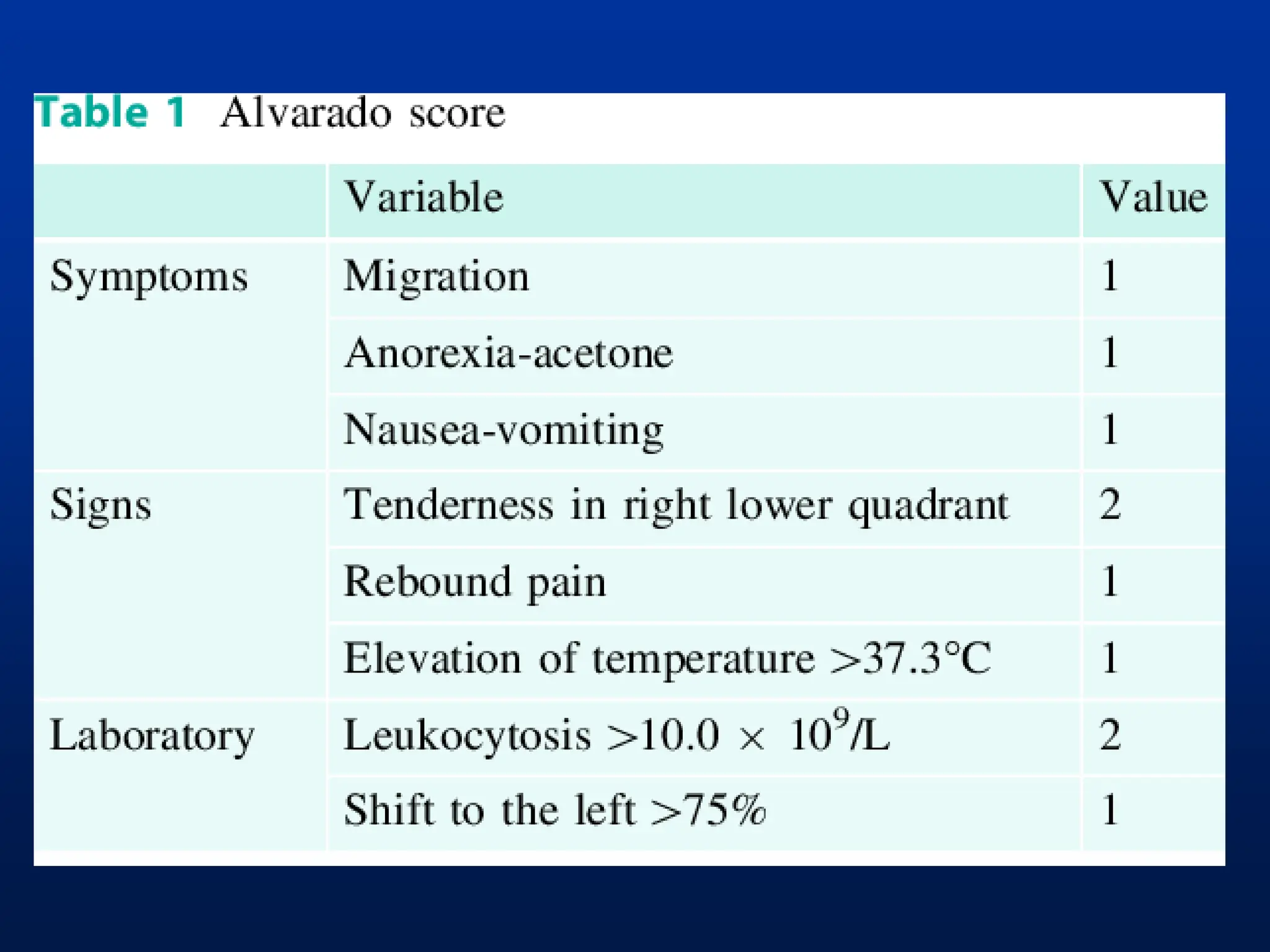
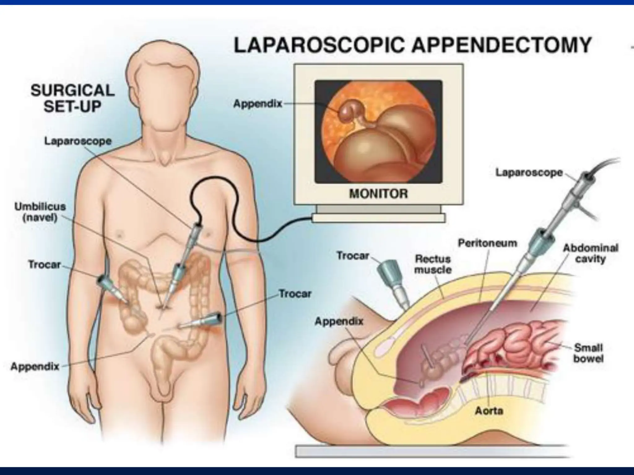
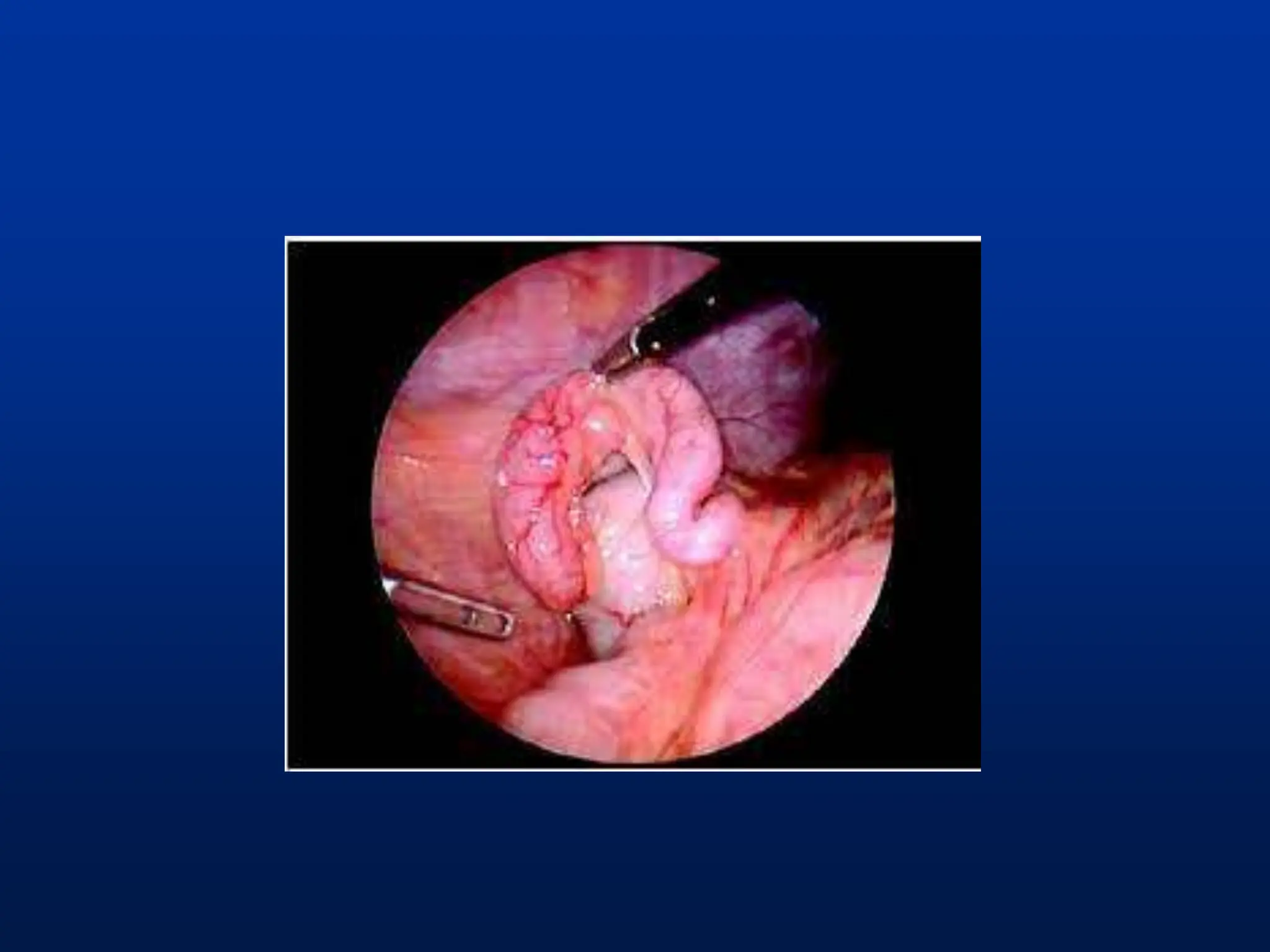
Appendicitis and
Appendicectomy
Mr S. Chinyowa
MBChB III 2021
Anatomy
• Average adult length is 9cm.
• Base of appendix is constant – at the base of the
caecum.
• Position of tip varies
widely.
Function of Appendix
• It is an immunological organ
• Secretes IgA
• Function not essential to life -> removal
of the appendix does not lead to
compromised immunity or sepsis
Acute Appendicitis
• Definition: Acute Inflammation of
the appendix.
Epidemiology
• Appendicitis M:F is 1.2:1
• Typically 2nd – 4th decade of life.
• Lifetime incidence of appendicectomy is
15% Men and 25% Women
Pathophysiology
• Obstruction of the lumen from faecal
matter, adhesions, or lymphoid
hyperplasia
• Mucosal secretions continue to increase
intraluminal pressure
• Increased pressure → Vascular
compromise → Bacterial invasion →
Gangrene → Perforation → Peritonitis
History
• Abdominal Pain
• Initial luminal distention causes pain which
is generally vague and felt in the
periumbilical or epigastric area.
• As inflammation continues, the serosa and
adjacent structures become inflamed and
this triggers somatic pain causing pain in
the Right Lower Quadrant (RLQ)
History
Associated symptoms:
• Anorexia
• Nausea and vomiting
• Indigestion
• Flatulence
• Urge to defecate
• Constipation
• Diarrhoea
• Dysuria
Physical Exam
• Findings depend on duration of illness
prior to exam.
• Early on patients may not have localized
tenderness
• With progression there is tenderness to
deep palpation in RLQ
McBurney’s point
Physical Exam
• Fever: a late finding.
• At the onset of pain, fever is usually not
found.
• Temperatures >38 C are uncommon in first
24 h, but not uncommon after rupture
Physical Exam
• Additional components: rebound
tenderness, guarding, muscular rigidity,
tenderness on rectal exam
• Rovsing sign
• Psoas sign
• Obturator sign
Investigations
• FBC: the WBC may be elevated in 70-90%
of patients but is of limited value since it is
not specific.
Investigations
• Imaging studies: include X-rays,
Ultrasound, CT Scan
• Abnormal findings include: faecalith,
appendiceal gas, localized paralytic ileus,
blurred right psoas, and free air
Special Populations
• Very young, very old, pregnant, and HIV
patients present atypically and often have
delayed diagnosis
• High index of suspicion is needed in the
these groups to get an accurate diagnosis
Treatment
• Appendicectomy is the standard of care
• Patients should be NPO, given IV fluids,
and preoperative antibiotics
Nursing responsibilities
• Relieving pain
• Reducing anxiety
• Preventing fluid volume deficit
• Eliminating infection
• Maintaining skin integrity
• Attaining optimal nutrition
Complications of Appendicitis
• Perforation
• Abscess formation
• Peritonitis
Appendicectomy
ā— Definition: Removal of the appendix
ā— It is performed as soon as possible to decrease the
risk of perforation.
ā— Can be performed at laparotomy or laparoscopy
How is it done? Laparotomy
• An incision through the skin and the layers
of the abdominal wall is made in the area
overlying the appendix.
• The surgeon enters the abdomen and
looks for the appendix. (usually RLQ)
• After examining the area around the
appendix to make sure there are no
additional problems, the appendix is
removed and its stump is sutured.
How is it done? Laparotomy
• If an abscess is present, the pus is drained
via rubber tubes that go from the abscess
out through the skin.
• The abdominal incision is then closed.
How is it done? Laparoscopy
• Newer techniques involve using a
Laparoscope.
• This is a thin telescope that is introduced
into the abdomen via a small puncture
wound in the abdominal wall.
• The appendix is then removed using
special instruments that are also passed
into the abdomen via small puncture
wounds.
Laparoscopy: Advantages
• Less post-operative pain.
• Quicker recovery.
• Allows the surgeon to look inside the
abdomen and make a clear diagnosis in
cases where the diagnosis of appendicitis
is in doubt.
Post-op care and Nursing
Management
• Monitor vital signs for evidence of infection
or shock such as fever, tachycardia and
hypotension.
• Monitor input and output for signs of
imbalance, dehydration and shock.
• Evaluate dressing and incision
• Evaluate the passing of flatus or stool
Post-op care and Nursing
Management
• Monitor for nausea and vomiting.
• Wound drains, iv lines and all other
catheters as evaluated for signs of
infection.
• Administration of drugs as ordered
• Diet is advanced as ordered
Thank you

Acute Appendicitis A comprehensive guide.ppt

  • 1.
  • 2.
    Anatomy • Average adultlength is 9cm. • Base of appendix is constant – at the base of the caecum. • Position of tip varies widely.
  • 3.
    Function of Appendix •It is an immunological organ • Secretes IgA • Function not essential to life -> removal of the appendix does not lead to compromised immunity or sepsis
  • 4.
    Acute Appendicitis • Definition:Acute Inflammation of the appendix.
  • 5.
    Epidemiology • Appendicitis M:Fis 1.2:1 • Typically 2nd – 4th decade of life. • Lifetime incidence of appendicectomy is 15% Men and 25% Women
  • 6.
    Pathophysiology • Obstruction ofthe lumen from faecal matter, adhesions, or lymphoid hyperplasia • Mucosal secretions continue to increase intraluminal pressure • Increased pressure → Vascular compromise → Bacterial invasion → Gangrene → Perforation → Peritonitis
  • 7.
    History • Abdominal Pain •Initial luminal distention causes pain which is generally vague and felt in the periumbilical or epigastric area. • As inflammation continues, the serosa and adjacent structures become inflamed and this triggers somatic pain causing pain in the Right Lower Quadrant (RLQ)
  • 8.
    History Associated symptoms: • Anorexia •Nausea and vomiting • Indigestion • Flatulence • Urge to defecate • Constipation • Diarrhoea • Dysuria
  • 9.
    Physical Exam • Findingsdepend on duration of illness prior to exam. • Early on patients may not have localized tenderness • With progression there is tenderness to deep palpation in RLQ
  • 10.
  • 11.
    Physical Exam • Fever:a late finding. • At the onset of pain, fever is usually not found. • Temperatures >38 C are uncommon in first 24 h, but not uncommon after rupture
  • 12.
    Physical Exam • Additionalcomponents: rebound tenderness, guarding, muscular rigidity, tenderness on rectal exam • Rovsing sign • Psoas sign • Obturator sign
  • 14.
    Investigations • FBC: theWBC may be elevated in 70-90% of patients but is of limited value since it is not specific.
  • 15.
    Investigations • Imaging studies:include X-rays, Ultrasound, CT Scan • Abnormal findings include: faecalith, appendiceal gas, localized paralytic ileus, blurred right psoas, and free air
  • 17.
    Special Populations • Veryyoung, very old, pregnant, and HIV patients present atypically and often have delayed diagnosis • High index of suspicion is needed in the these groups to get an accurate diagnosis
  • 18.
    Treatment • Appendicectomy isthe standard of care • Patients should be NPO, given IV fluids, and preoperative antibiotics
  • 19.
    Nursing responsibilities • Relievingpain • Reducing anxiety • Preventing fluid volume deficit • Eliminating infection • Maintaining skin integrity • Attaining optimal nutrition
  • 20.
    Complications of Appendicitis •Perforation • Abscess formation • Peritonitis
  • 21.
    Appendicectomy ā— Definition: Removalof the appendix ā— It is performed as soon as possible to decrease the risk of perforation. ā— Can be performed at laparotomy or laparoscopy
  • 22.
    How is itdone? Laparotomy • An incision through the skin and the layers of the abdominal wall is made in the area overlying the appendix. • The surgeon enters the abdomen and looks for the appendix. (usually RLQ) • After examining the area around the appendix to make sure there are no additional problems, the appendix is removed and its stump is sutured.
  • 23.
    How is itdone? Laparotomy • If an abscess is present, the pus is drained via rubber tubes that go from the abscess out through the skin. • The abdominal incision is then closed.
  • 25.
    How is itdone? Laparoscopy • Newer techniques involve using a Laparoscope. • This is a thin telescope that is introduced into the abdomen via a small puncture wound in the abdominal wall. • The appendix is then removed using special instruments that are also passed into the abdomen via small puncture wounds.
  • 28.
    Laparoscopy: Advantages • Lesspost-operative pain. • Quicker recovery. • Allows the surgeon to look inside the abdomen and make a clear diagnosis in cases where the diagnosis of appendicitis is in doubt.
  • 29.
    Post-op care andNursing Management • Monitor vital signs for evidence of infection or shock such as fever, tachycardia and hypotension. • Monitor input and output for signs of imbalance, dehydration and shock. • Evaluate dressing and incision • Evaluate the passing of flatus or stool
  • 30.
    Post-op care andNursing Management • Monitor for nausea and vomiting. • Wound drains, iv lines and all other catheters as evaluated for signs of infection. • Administration of drugs as ordered • Diet is advanced as ordered
  • 31.