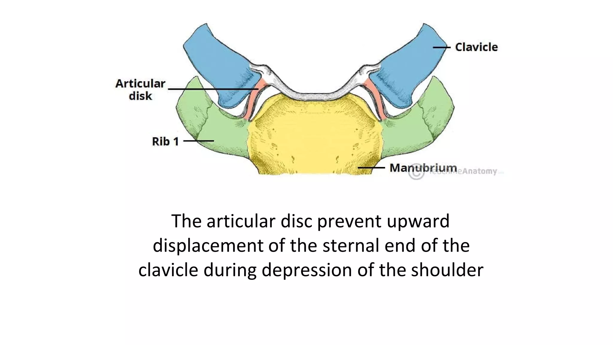
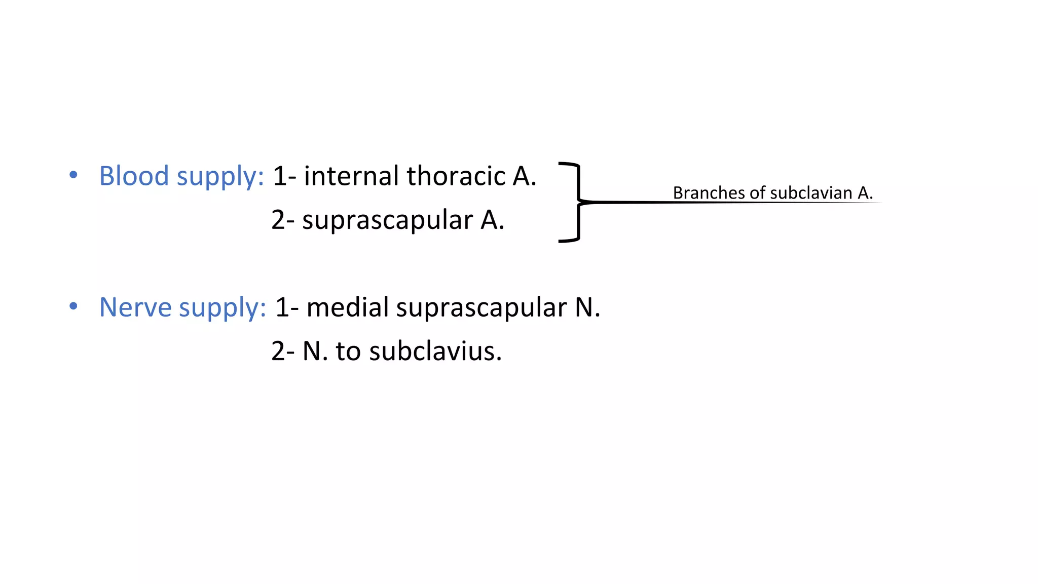
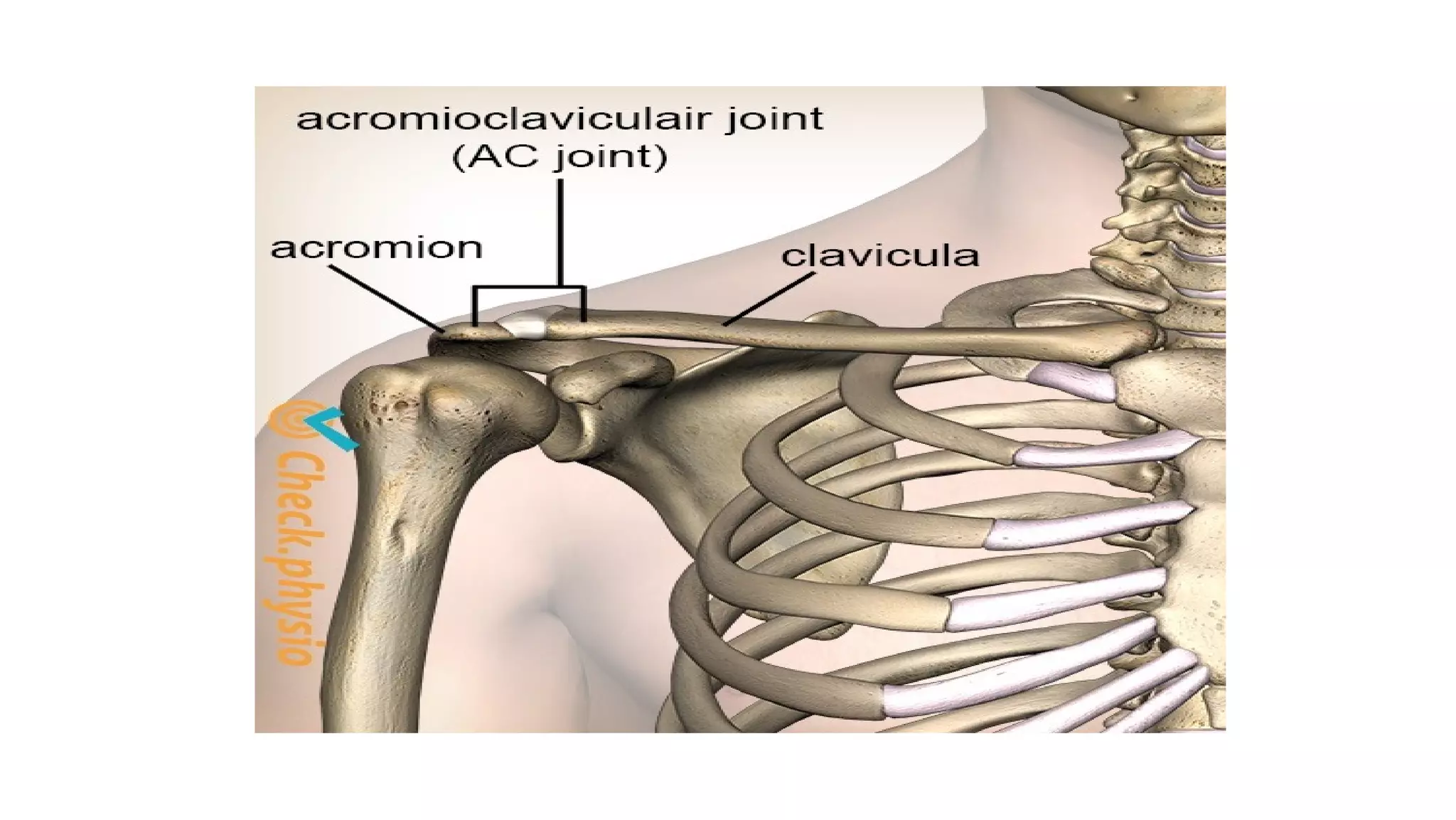
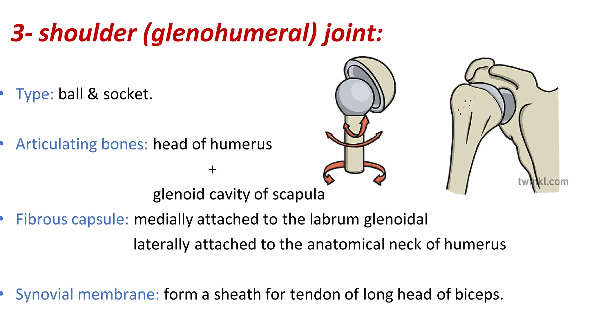
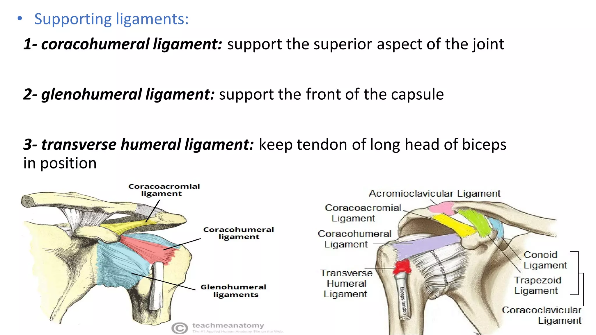
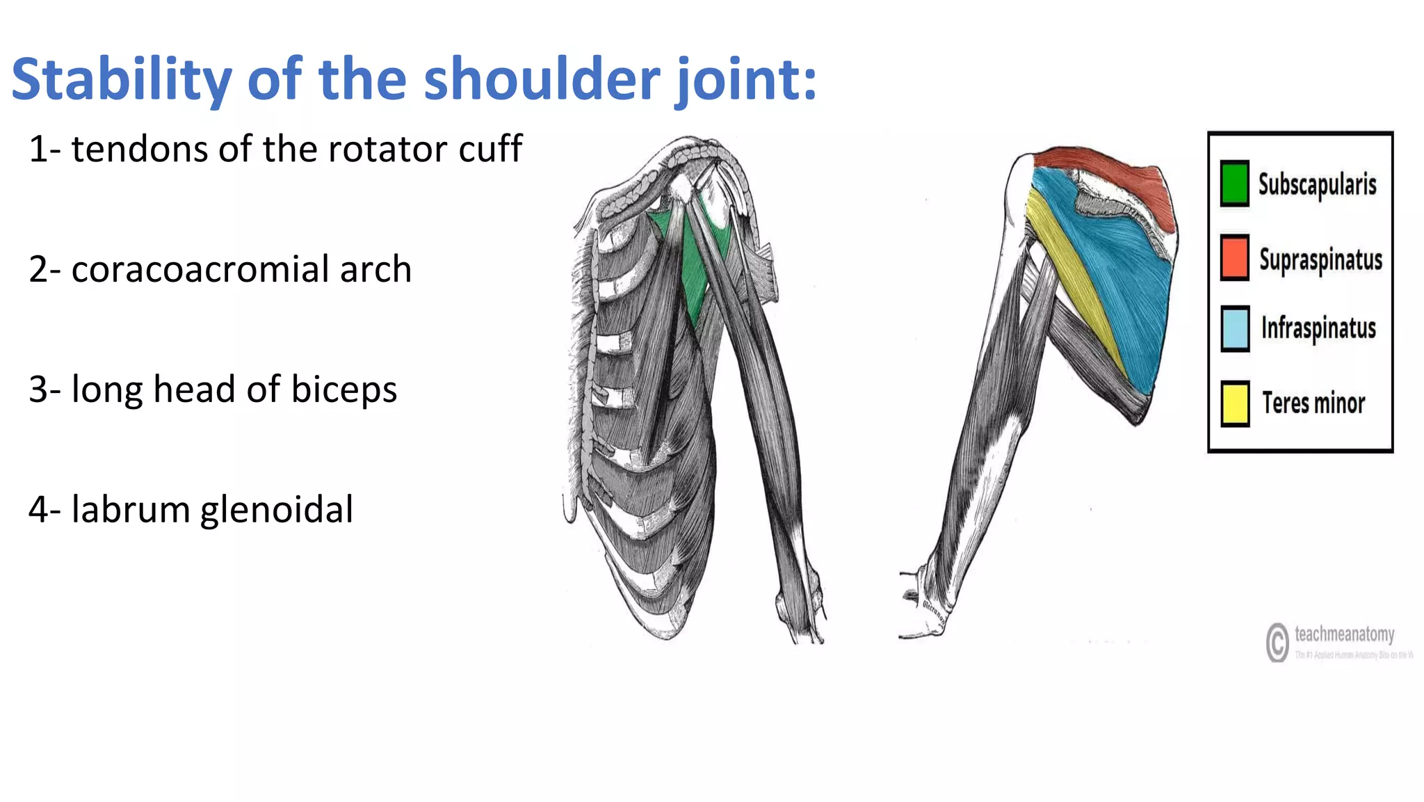
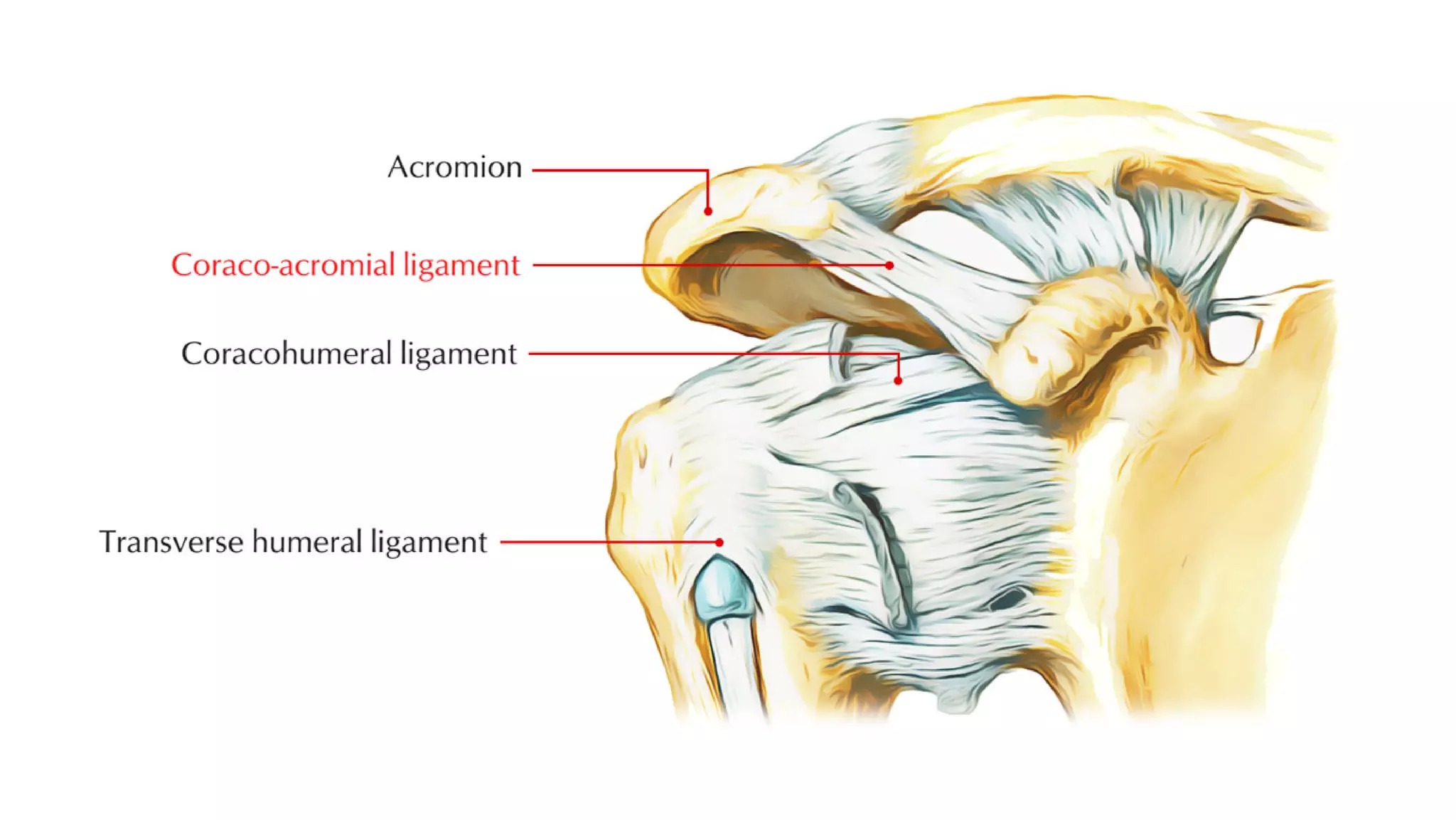
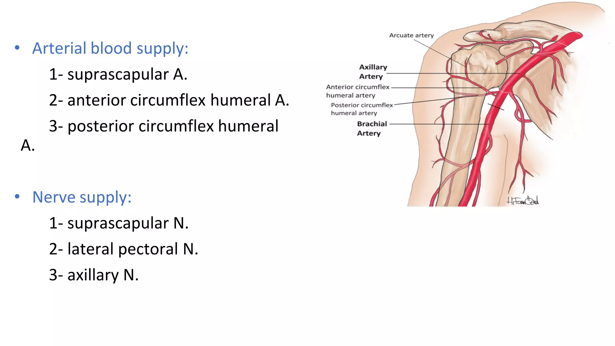
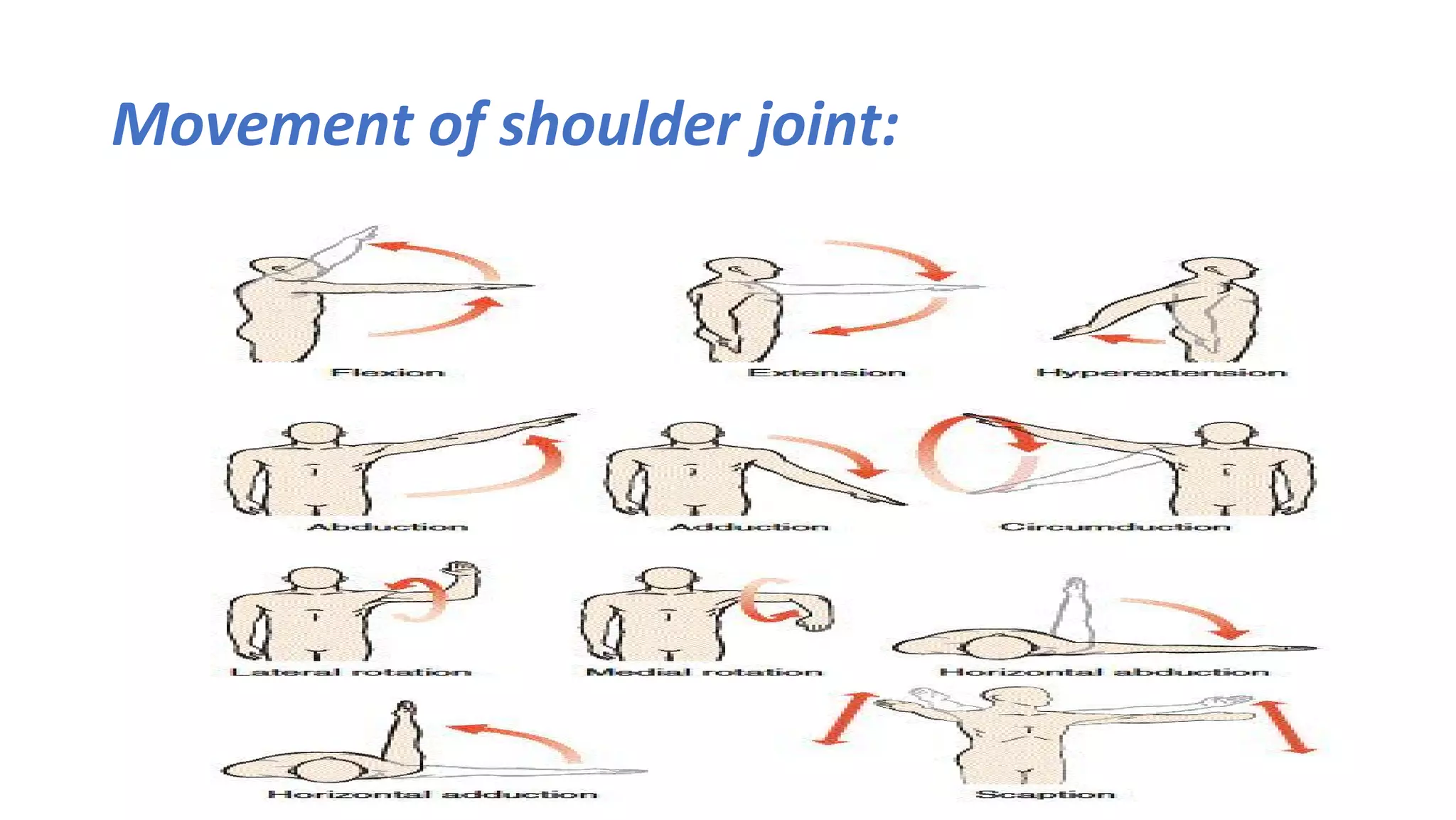
6. • Blood supply: 1- internal thoracic A.
2- suprascapular A.
• Nerve supply: 1- medial suprascapular N.
2- N. to subclavius.
Branches of subclavian A.
7. 2- Acromioclavicular joint:
• Type: plane synovial joint.
• Articulating bones: lateral (acromial) end of clavicle
+
clavicular facet of acromion process
• Fibrous capsule
• Synovial membrane: articular disc frequently present
8.
9. • Supporting ligaments:
1- Acromioclavicular ligament.
2- coracoclavicular ligament: it
has 2 parts
✔ Trapezoid part (quadrangular)
✔ Conoid part (cone shaped)
10. • Arterial blood supply: 1- suprascapular A.
2- Acromial br. Of thoracoacromial A.
• Nerve supply: 1- suprascapular N.
2- lateral pectoral N.
11. • Clavicle fracture: Most common site of fracture is the middle third.
• Acromial dislocation: leads to tear in the conoid & trapezoid lig.
Leads to elevation of the clavicle & shoulder drop.
• Sternal end dislocation: may be anterior or posterior (may compress
great vessels).
14. 3- shoulder (glenohumeral) joint:
• Type: ball & socket.
• Articulating bones: head of humerus
+
glenoid cavity of scapula
• Fibrous capsule: medially attached to the labrum glenoidal
laterally attached to the anatomical neck of humerus
• Synovial membrane: form a sheath for tendon of long head of biceps.
15.
16. • Supporting ligaments:
1- coracohumeral ligament: support the superior aspect of the joint
2- glenohumeral ligament: support the front of the capsule
3- transverse humeral ligament: keep tendon of long head of biceps
in position
17. Stability of the shoulder joint:
1- tendons of the rotator cuff
2- coracoacromial arch
3- long head of biceps
4- labrum glenoidal
24. - sternoclavicular ,
acromioclavicular ,shoulder joint ----->
suprascapular nerve and artery
sternoclavicular ----> internal thoracic artery
N. to sub scapularis
acromioclavicular ----> branch from thoracoacromial A
lateral pectoral nerve
shoulder joint -----> ant and post circumflex humaral A
- lateral pectoral N
- Axillary N