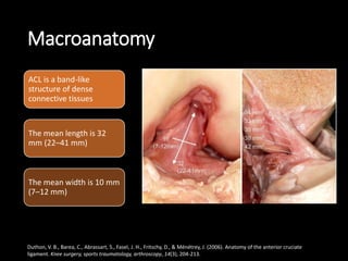
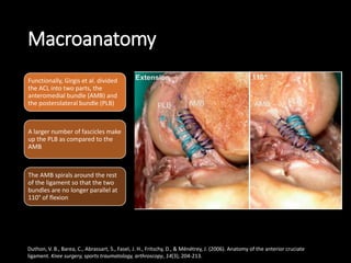
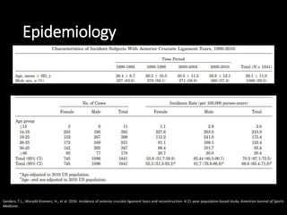
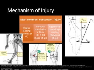
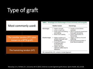
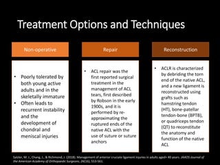
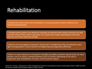
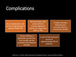
Anterior Cruciate
Ligament Injury
Introduction
ACL is a key structure in the knee joint, as it resists
anterior tibial translation and rotational loads
One of the most frequently injured structures during
high impact or sporting activities
ACL does not heal when torn, and surgical reconstruction
is the standard treatment in the field of sports medicine
Duthon, V. B., Barea, C., Abrassart, S., Fasel, J. H., Fritschy, D., & Ménétrey, J. (2006). Anatomy of the anterior cruciate
ligament. Knee surgery, sports traumatology, arthroscopy, 14(3), 204-213.
ANATOMY
ACL is composed of longitudinally oriented bundles of collagen
tissue
Inserts on the tibial plateau, medial to the insertion of the anterior
horn of the lateral meniscus in a depressed area anterolateral to
the anterior tibial spine
Posteromedial aspect of lateral femoral condyle to anterior tib ial
eminence
Azar, F.M., Canale, S.T. and Beaty, J.H., 2016. Campbell's operative orthopaedics e-book. Elsevier Health Sciences.
ANATOMY
Thompson, J.C., 2015. Netter's Concise Orthopaedic Anatomy E-Book. Elsevier Health Sciences.
Macroanatomy
ACL is a band-like
structure of dense
connective tissues
The mean length is 32
mm (22–41 mm)
The mean width is 10 mm
(7–12 mm)
Duthon, V. B., Barea, C., Abrassart, S., Fasel, J. H., Fritschy, D., & Ménétrey, J. (2006). Anatomy of the anterior cruciate
ligament. Knee surgery, sports traumatology, arthroscopy, 14(3), 204-213.
Macroanatomy
Functionally, Girgis et al. divided
the ACL into two parts, the
anteromedial bundle (AMB) and
the posterolateral bundle (PLB)
A larger number of fascicles make
up the PLB as compared to the
AMB
The AMB spirals around the rest
of the ligament so that the two
bundles are no longer parallel at
110° of flexion
Duthon, V. B., Barea, C., Abrassart, S., Fasel, J. H., Fritschy, D., & Ménétrey, J. (2006). Anatomy of the anterior cruciate
ligament. Knee surgery, sports traumatology, arthroscopy, 14(3), 204-213.
BIOMECHANICS
• The anterior cruciate ligament is the primary
restraint to anterior tibial displacement
• Anteromedial band is tight in flexion, providing the
primary restraint,
• whereas the Posterolateral bulky portion of
this ligament is tight in extension
• The posterolateral bundle provides the principal
resistance to hyperextension
• Secondary restraint on tibial rotation and varus-
valgus angulation at full extension
Azar, F.M., Canale, S.T. and Beaty, J.H., 2016. Campbell's operative orthopaedics e-book. Elsevier Health Sciences.
Vascularization
MGA
Popliteal artery
Infrapatellar fat pad
• The primary blood supply to the ligament is from the middle geniculate artery
• The distribution of blood vessels within the substance of the ligament is not
homogeneous. The proximal part of the ACL is better endowed with blood vessels
than the distal part
Duthon, V. B., Barea, C., Abrassart, S., Fasel, J. H., Fritschy, D., & Ménétrey, J. (2006). Anatomy of the anterior cruciate
ligament. Knee surgery, sports traumatology, arthroscopy, 14(3), 204-213.
Epidemiology
Sanders, T.L., Maradit Kremers, H., et al. 2016. Incidence of anterior cruciate ligament tears and reconstruction: A 21-year population-based study. American Journal of Sports
Medicine.
Mechanism of Injury
Most common: noncontact injury
Valgus
loading
(Hewett et
al.)
Femoral
translation
+ Tibial
IR → ER
(Koga et al.)
Aggressive
quadriceps
loading
(DeMorat
et al.)
DeMorat, G., Weinhold, P., Blackburn, T., Chudik, S. & Garrett, W. 2004. Aggressive Quadriceps Loading Can Induce Noncontact Anterior Cruciate Ligament Injury. American Journal of Sports Medicine.
Hewett, T.E., Myer, G.D., et al. 2005. Biomechanical measures of neuromuscular control and valgus loading of the knee predict anterior cruciate ligament injury risk in female athletes: A prospective study. American
Journal of Sports Medicine.
Koga, H., Muneta, T., Bahr, R., Engebretsen, L. & Krosshaug, T. 2016. ACL Injury Mechanisms: Lessons Learned from Video Analysis. In: Rotatory Knee Instability,.
Clinic Manifestation
An appropriate and detailed patient history can raise suspicion for
an ACL injury, especially having been hyperextended or popping out
of joint
2.The patient who sustains a knee injury during sports activity
that is followed by knee swelling within 1 to 6 hours should be
evaluated carefully for a possible ACL injury
2.Patients with a chronic ACL injury may report recurrent episodes
of knee instability, mechanical symptoms from a secondary
meniscal tear, or pain and swelling.
Lieberman, J. R. (2019). AAOS Comprehensive Orthopaedic Review 3. Lippincott Williams & Wilkins.
Clinical Findings
Anterior Drawer
Test
Lachman Test Pivot-Shift Test Lelli's Test
Kuroda, R., Matsushita, T. & Araki, D. 2016. Physical examinations and device measurements for ACL deficiency. In: ACL Injury and Its Treatment.
Radiologic Findings - MRI
Direct Signs
ACL discontinuity
Loss of parallel
orientation
Empty notch sign
Yonetani, Y. & Tanaka, Y. 2016. Diagnostics of ACL injury using magnetic resonance imaging (MRI). In: ACL Injury and Its Treatment.
Radiologic Findings – 3D CT
Multidetector row CT scanner
Depicting injured ACL in 3D
Cannot depict the status of ACL
attachment
Crain’s classification
• Type I, bridging between the
posterior cruciate ligament and
tibia
• Type II, bridging between roof of
the intercondylar notch and tibia
• Type III, bridging between the
lateral wall of the intercondylar
notch and tibia
• Type IV, no substantial ACL
remnants
Adachi, N. 2016. Diagnosis of injured ACL using three-dimensional computed tomography: Usefulness for preoperative decision making. In: ACL Injury and Its Treatment.
Treatment
• Partial injuries of the ACL
• Rehabilitation to strengthen the hamstrings
and quadriceps
• Proprioceptive training
• Activity modification
• ACL sports braces
Nonsurgical
• Reconstruction surgery
• The decision to reconstruct the ACL
generally is related to the patient’s activity
level.
• Patients with jobs that may involve physical
combat (active military, police officers), risk
(firefighters), or activity on unstable surfaces
(construction workers)
1.Surgical
Lieberman, J. R. (2019). AAOS Comprehensive Orthopaedic Review 3. Lippincott Williams & Wilkins.
PRINCIPLES OF ACL RECONSTRUCTION
Timing of repair and rehabilitation
Type of graft
Allograft versus Autograft
1
2
3
Lieberman, J. R. (2019). AAOS Comprehensive Orthopaedic Review 3. Lippincott Williams & Wilkins.
Timing of Repair and Rehabilitation
- increased incidence of meniscus and chondral injuries following delayed ACL
reconstruction
- risk of arthrofibrosis associated with early ACL reconstruction
- associated loss of muscle strength due to inactivity when surgery is delayed
the timing of ACL reconstruction and
autograft choice can also influence
rehabilitation strategy
Early ACL reconstruction has been linked
with delay in quadriceps recovery
Specifically, 80% of patients treated with delayed ACL reconstruction (mean 40 days)
reached a 65% quadriceps strength at two months versus 53% of patients treated with
early ACL reconstruction (mean 11 days), respectively. By six months, 73% of patients that
had delayed ACL reconstruction (mean time from injury of 40 days) exhibited 80% muscle
strength versus only 47% in those reconstructed early.
(Nikolaos et al, 2016)
Lieberman, J. R. (2019). AAOS Comprehensive Orthopaedic Review 3. Lippincott Williams & Wilkins.
Type of graft
Most commonly used:
The patellar tendon (PT) (also
known as a BPTB graft)
The hamstring tendon (HT)
Macaulay, A. A., Perfetti, D. C., & Levine, W. N. (2012). Anterior cruciate ligament graft choices. Sports Health, 4(1), 63-68.
Treatment Options and Techniques
ReconstructionRepairNon-operative
• Poorly tolerated by
both young active
adults and in the
skeletally immature
• Often leads to
recurrent instability
and the
development of
chondral and
meniscal injuries
• ACL repair was the
first reported surgical
treatment in the
management of ACL
tears, first described
by Robson in the early
1900s, and it is
performed by re-
approximating the
ruptured ends of the
native ACL with the
use of suture or suture
anchors
• ACLR is characterized
by debriding the torn
end of the native ACL,
and a new ligament is
reconstructed using
grafts such as
hamstring tendon
(HT), bone-patellar
tendon-bone (BPTB),
or quadriceps tendon
(QT) to reconstitute
the anatomy and
function of the native
ACL
Salzler, M. J., Chang, J., & Richmond, J. (2018). Management of anterior cruciate ligament injuries in adults aged> 40 years. JAAOS-Journal of
the American Academy of Orthopaedic Surgeons, 26(16), 553-561.
Outcome
Clinical studies comparing outcomes of combined extra- and intra-articular procedures
found mixed results.
Historically, poor outcomes have been associated with primary ACL repair.
The inherent morbidity associated with open approaches and prolonged postoperative
immobilization led to significant motion loss and patellofemoral issues.
Recently, there has been an increasing interest in ACL preservation as an option to
perhaps better restore native ACL anatomy, biomechanics, and neurosensory function.
Salzler, M. J., Chang, J., & Richmond, J. (2018). Management of anterior cruciate ligament injuries in adults aged> 40 years. JAAOS-Journal of
the American Academy of Orthopaedic Surgeons, 26(16), 553-561.
Rehabilitation
1.Studies have shown that a knee immobilizer or continuous passive motion machine is not
required postoperatively.
1.Postoperatively, patients are allowed to ambulate as tolerated with crutches until they can walk
normally without them, which is generally 1 to 2 weeks after surgery depending on their pain
tolerance and lower extremity strength.
3.Patients are encouraged to emphasize “closed-chain” strengthening. They are allowed to start
light running between 3 and 4 months if strengthening has progressed sufficiently.
3.Return to sports depends on the patient’s strength and ability to perform specific tasks. For most
patients, return to sports is allowed between 6 and 12 months, depending on the rate of
progression with rehabilitation, the sport, and the position played.
Salzler, M. J., Chang, J., & Richmond, J. (2018). Management of anterior cruciate ligament injuries in adults aged> 40 years. JAAOS-Journal of
the American Academy of Orthopaedic Surgeons, 26(16), 553-561.
Complications
ACL complication rate
across all age groups is
approximately 5%
Meniscal tears with
acute anterior
cruciate ligament
injuries to range from
50% to 70%
2.Deep infection
following ACL
reconstruction is
uncommon (0.8%)
2.Knee stiffness is
generally uncommon
and usually can be
resolved with physical
therapy
2.Lack of full extension
occurs in
approximately 4% of
patients.
Lieberman, J. R. (2019). AAOS Comprehensive Orthopaedic Review 3. Lippincott Williams & Wilkins.
THANK YOU

ACL disorders

  • 1.
  • 2.
    Introduction ACL is akey structure in the knee joint, as it resists anterior tibial translation and rotational loads One of the most frequently injured structures during high impact or sporting activities ACL does not heal when torn, and surgical reconstruction is the standard treatment in the field of sports medicine Duthon, V. B., Barea, C., Abrassart, S., Fasel, J. H., Fritschy, D., & Ménétrey, J. (2006). Anatomy of the anterior cruciate ligament. Knee surgery, sports traumatology, arthroscopy, 14(3), 204-213.
  • 3.
    ANATOMY ACL is composedof longitudinally oriented bundles of collagen tissue Inserts on the tibial plateau, medial to the insertion of the anterior horn of the lateral meniscus in a depressed area anterolateral to the anterior tibial spine Posteromedial aspect of lateral femoral condyle to anterior tib ial eminence Azar, F.M., Canale, S.T. and Beaty, J.H., 2016. Campbell's operative orthopaedics e-book. Elsevier Health Sciences.
  • 4.
    ANATOMY Thompson, J.C., 2015.Netter's Concise Orthopaedic Anatomy E-Book. Elsevier Health Sciences.
  • 5.
    Macroanatomy ACL is aband-like structure of dense connective tissues The mean length is 32 mm (22–41 mm) The mean width is 10 mm (7–12 mm) Duthon, V. B., Barea, C., Abrassart, S., Fasel, J. H., Fritschy, D., & Ménétrey, J. (2006). Anatomy of the anterior cruciate ligament. Knee surgery, sports traumatology, arthroscopy, 14(3), 204-213.
  • 6.
    Macroanatomy Functionally, Girgis etal. divided the ACL into two parts, the anteromedial bundle (AMB) and the posterolateral bundle (PLB) A larger number of fascicles make up the PLB as compared to the AMB The AMB spirals around the rest of the ligament so that the two bundles are no longer parallel at 110° of flexion Duthon, V. B., Barea, C., Abrassart, S., Fasel, J. H., Fritschy, D., & Ménétrey, J. (2006). Anatomy of the anterior cruciate ligament. Knee surgery, sports traumatology, arthroscopy, 14(3), 204-213.
  • 7.
    BIOMECHANICS • The anteriorcruciate ligament is the primary restraint to anterior tibial displacement • Anteromedial band is tight in flexion, providing the primary restraint, • whereas the Posterolateral bulky portion of this ligament is tight in extension • The posterolateral bundle provides the principal resistance to hyperextension • Secondary restraint on tibial rotation and varus- valgus angulation at full extension Azar, F.M., Canale, S.T. and Beaty, J.H., 2016. Campbell's operative orthopaedics e-book. Elsevier Health Sciences.
  • 8.
    Vascularization MGA Popliteal artery Infrapatellar fatpad • The primary blood supply to the ligament is from the middle geniculate artery • The distribution of blood vessels within the substance of the ligament is not homogeneous. The proximal part of the ACL is better endowed with blood vessels than the distal part Duthon, V. B., Barea, C., Abrassart, S., Fasel, J. H., Fritschy, D., & Ménétrey, J. (2006). Anatomy of the anterior cruciate ligament. Knee surgery, sports traumatology, arthroscopy, 14(3), 204-213.
  • 9.
    Epidemiology Sanders, T.L., MaraditKremers, H., et al. 2016. Incidence of anterior cruciate ligament tears and reconstruction: A 21-year population-based study. American Journal of Sports Medicine.
  • 10.
    Mechanism of Injury Mostcommon: noncontact injury Valgus loading (Hewett et al.) Femoral translation + Tibial IR → ER (Koga et al.) Aggressive quadriceps loading (DeMorat et al.) DeMorat, G., Weinhold, P., Blackburn, T., Chudik, S. & Garrett, W. 2004. Aggressive Quadriceps Loading Can Induce Noncontact Anterior Cruciate Ligament Injury. American Journal of Sports Medicine. Hewett, T.E., Myer, G.D., et al. 2005. Biomechanical measures of neuromuscular control and valgus loading of the knee predict anterior cruciate ligament injury risk in female athletes: A prospective study. American Journal of Sports Medicine. Koga, H., Muneta, T., Bahr, R., Engebretsen, L. & Krosshaug, T. 2016. ACL Injury Mechanisms: Lessons Learned from Video Analysis. In: Rotatory Knee Instability,.
  • 11.
    Clinic Manifestation An appropriateand detailed patient history can raise suspicion for an ACL injury, especially having been hyperextended or popping out of joint 2.The patient who sustains a knee injury during sports activity that is followed by knee swelling within 1 to 6 hours should be evaluated carefully for a possible ACL injury 2.Patients with a chronic ACL injury may report recurrent episodes of knee instability, mechanical symptoms from a secondary meniscal tear, or pain and swelling. Lieberman, J. R. (2019). AAOS Comprehensive Orthopaedic Review 3. Lippincott Williams & Wilkins.
  • 12.
    Clinical Findings Anterior Drawer Test LachmanTest Pivot-Shift Test Lelli's Test Kuroda, R., Matsushita, T. & Araki, D. 2016. Physical examinations and device measurements for ACL deficiency. In: ACL Injury and Its Treatment.
  • 13.
    Radiologic Findings -MRI Direct Signs ACL discontinuity Loss of parallel orientation Empty notch sign Yonetani, Y. & Tanaka, Y. 2016. Diagnostics of ACL injury using magnetic resonance imaging (MRI). In: ACL Injury and Its Treatment.
  • 14.
    Radiologic Findings –3D CT Multidetector row CT scanner Depicting injured ACL in 3D Cannot depict the status of ACL attachment Crain’s classification • Type I, bridging between the posterior cruciate ligament and tibia • Type II, bridging between roof of the intercondylar notch and tibia • Type III, bridging between the lateral wall of the intercondylar notch and tibia • Type IV, no substantial ACL remnants Adachi, N. 2016. Diagnosis of injured ACL using three-dimensional computed tomography: Usefulness for preoperative decision making. In: ACL Injury and Its Treatment.
  • 15.
    Treatment • Partial injuriesof the ACL • Rehabilitation to strengthen the hamstrings and quadriceps • Proprioceptive training • Activity modification • ACL sports braces Nonsurgical • Reconstruction surgery • The decision to reconstruct the ACL generally is related to the patient’s activity level. • Patients with jobs that may involve physical combat (active military, police officers), risk (firefighters), or activity on unstable surfaces (construction workers) 1.Surgical Lieberman, J. R. (2019). AAOS Comprehensive Orthopaedic Review 3. Lippincott Williams & Wilkins.
  • 16.
    PRINCIPLES OF ACLRECONSTRUCTION Timing of repair and rehabilitation Type of graft Allograft versus Autograft 1 2 3 Lieberman, J. R. (2019). AAOS Comprehensive Orthopaedic Review 3. Lippincott Williams & Wilkins.
  • 17.
    Timing of Repairand Rehabilitation - increased incidence of meniscus and chondral injuries following delayed ACL reconstruction - risk of arthrofibrosis associated with early ACL reconstruction - associated loss of muscle strength due to inactivity when surgery is delayed the timing of ACL reconstruction and autograft choice can also influence rehabilitation strategy Early ACL reconstruction has been linked with delay in quadriceps recovery Specifically, 80% of patients treated with delayed ACL reconstruction (mean 40 days) reached a 65% quadriceps strength at two months versus 53% of patients treated with early ACL reconstruction (mean 11 days), respectively. By six months, 73% of patients that had delayed ACL reconstruction (mean time from injury of 40 days) exhibited 80% muscle strength versus only 47% in those reconstructed early. (Nikolaos et al, 2016) Lieberman, J. R. (2019). AAOS Comprehensive Orthopaedic Review 3. Lippincott Williams & Wilkins.
  • 18.
    Type of graft Mostcommonly used: The patellar tendon (PT) (also known as a BPTB graft) The hamstring tendon (HT) Macaulay, A. A., Perfetti, D. C., & Levine, W. N. (2012). Anterior cruciate ligament graft choices. Sports Health, 4(1), 63-68.
  • 19.
    Treatment Options andTechniques ReconstructionRepairNon-operative • Poorly tolerated by both young active adults and in the skeletally immature • Often leads to recurrent instability and the development of chondral and meniscal injuries • ACL repair was the first reported surgical treatment in the management of ACL tears, first described by Robson in the early 1900s, and it is performed by re- approximating the ruptured ends of the native ACL with the use of suture or suture anchors • ACLR is characterized by debriding the torn end of the native ACL, and a new ligament is reconstructed using grafts such as hamstring tendon (HT), bone-patellar tendon-bone (BPTB), or quadriceps tendon (QT) to reconstitute the anatomy and function of the native ACL Salzler, M. J., Chang, J., & Richmond, J. (2018). Management of anterior cruciate ligament injuries in adults aged> 40 years. JAAOS-Journal of the American Academy of Orthopaedic Surgeons, 26(16), 553-561.
  • 20.
    Outcome Clinical studies comparingoutcomes of combined extra- and intra-articular procedures found mixed results. Historically, poor outcomes have been associated with primary ACL repair. The inherent morbidity associated with open approaches and prolonged postoperative immobilization led to significant motion loss and patellofemoral issues. Recently, there has been an increasing interest in ACL preservation as an option to perhaps better restore native ACL anatomy, biomechanics, and neurosensory function. Salzler, M. J., Chang, J., & Richmond, J. (2018). Management of anterior cruciate ligament injuries in adults aged> 40 years. JAAOS-Journal of the American Academy of Orthopaedic Surgeons, 26(16), 553-561.
  • 21.
    Rehabilitation 1.Studies have shownthat a knee immobilizer or continuous passive motion machine is not required postoperatively. 1.Postoperatively, patients are allowed to ambulate as tolerated with crutches until they can walk normally without them, which is generally 1 to 2 weeks after surgery depending on their pain tolerance and lower extremity strength. 3.Patients are encouraged to emphasize “closed-chain” strengthening. They are allowed to start light running between 3 and 4 months if strengthening has progressed sufficiently. 3.Return to sports depends on the patient’s strength and ability to perform specific tasks. For most patients, return to sports is allowed between 6 and 12 months, depending on the rate of progression with rehabilitation, the sport, and the position played. Salzler, M. J., Chang, J., & Richmond, J. (2018). Management of anterior cruciate ligament injuries in adults aged> 40 years. JAAOS-Journal of the American Academy of Orthopaedic Surgeons, 26(16), 553-561.
  • 22.
    Complications ACL complication rate acrossall age groups is approximately 5% Meniscal tears with acute anterior cruciate ligament injuries to range from 50% to 70% 2.Deep infection following ACL reconstruction is uncommon (0.8%) 2.Knee stiffness is generally uncommon and usually can be resolved with physical therapy 2.Lack of full extension occurs in approximately 4% of patients. Lieberman, J. R. (2019). AAOS Comprehensive Orthopaedic Review 3. Lippincott Williams & Wilkins.
  • 23.

Editor's Notes

  • #13 The Lachman test is the most useful in diagnosing ACL injuries in the acute setting