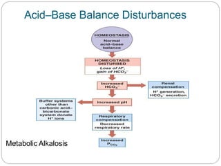
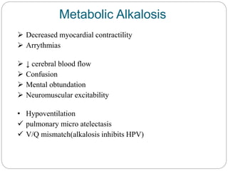
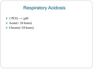
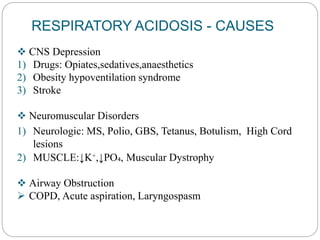
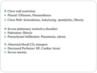
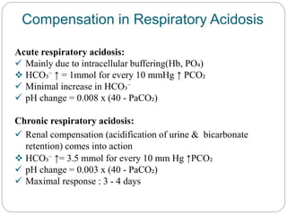
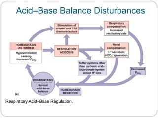
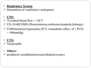
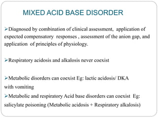
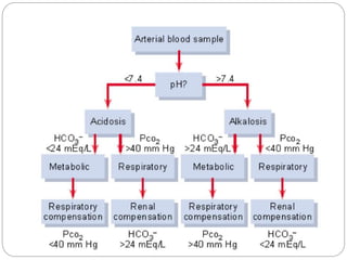
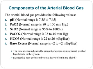
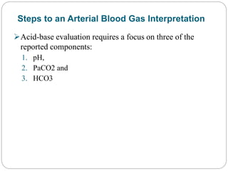
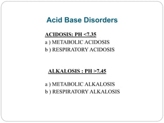
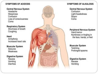
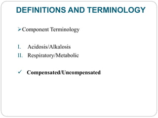
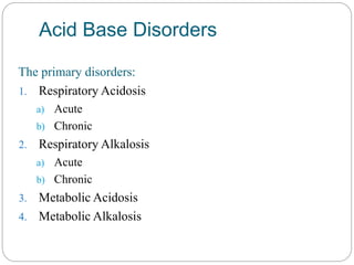
An arterial-blood gas test measures the amounts of arterial gases, such as oxygen and carbon dioxide. An ABG test requires that a small volume of blood be drawn from the radial artery with a syringe and a thin needle, but sometimes the femoral artery in the groin or another site is used.
The common indications for ABGs are:
Respiratory compromise, which leads to hypoxia or diminished ventilation.
Peri- or postcardiopulmonary arrest or collapse.
Medical conditions that cause significant metabolic derangement, such as sepsis, diabetic ketoacidosis, renal failure, heart failure, toxic substance ingestion, drug overdose, trauma, or burns.
Evaluating the effectiveness of therapies, monitoring the patient's clinical status, and determining treatment needs. For instance, clinicians often titrate oxygenation therapy, adjust the level of ventilator support, and make decisions about fluid and electrolyte therapy based on ABG results.
During the perioperative phase of major surgeries, which includes the preoperative, intraoperative, and postoperative care of the patient.
The components of an ABG analysis are PaO2, SaO2, hydrogen ion concentration (pH), PaCO2, HCO3-, base excess, and serum levels of hemoglobin, lactate, glucose, and electrolytes (sodium, potassium, calcium, and chloride).
ACID & BASE
Acid is a molecule or an ion that can function as a proton donor. Base is the molecule or an ion that can function as a proton acceptor.
pH
pH is negative log of H+ ion concentration.
Normal pH of arterial blood is 7.4 and that of venous blood and
ACID & BASE
Acid is a molecule or an ion that can function as a proton donor. Base is the molecule or an ion that can function as a proton acceptor.
pH
pH is negative log of H+ ion concentration.
Normal pH of arterial blood is 7.4 and that of venous blood and
Explain the chemical shifts of different types of protons for ethanol moleculeExplain the chemical shifts of different types of protons for ethanol moleculeExplain the chemical shifts of different types of protons for ethanol moleculeExplain the chemical shifts of different types of protons for ethanol moleculeExplain the chemical shifts of different types of protons for ethanol moleculeExplain the chemical shifts of different types of protons for ethanol moleculeExplain the chemical shifts of different types of protons for ethanol moleculeExplain the chemical shifts of different types of protons for ethanol moleculeExplain the chemical shifts of different types of protons for ethanol moleculeExplain the chemical shifts of different types of protons for ethanol moleculeExplain the chemical shifts of different types of protons for ethanol moleculeExplain the chemical shifts of different types of protons for ethanol moleculeExplain the chemical shifts of different types of protons for ethanol moleculeExplain the chemical shifts of different types of protons for ethanol moleculeExplain the chemical shifts of different types of protons for ethanol moleculeExplain the chemical shifts of different types of protons for ethanol moleculeExplain the chemical shifts of different types of protons for ethanol moleculeExplain the chemical shifts of different types of protons for ethanol moleculeExplain the chemical shifts of different types of protons for ethanol moleculeExplain the chemical shifts of different types of protons for ethanol moleculeExplain the chemical shifts of different types of protons for ethanol moleculeExplain the chemical shifts of different types of protons for ethanol moleculeExplain the chemical shifts of different types of protons for ethanol moleculeExplain the chemical shifts of different types of protons for ethanol moleculeExplain the chemical shifts of different types of protons for ethanol moleculeExplain the chemical shifts of different types of protons for ethanol moleculeExplain the chemical shifts of different types of protons for ethanol moleculeExplain the chemical shifts of different types of protons for ethanol moleculeExplain the chemical shifts of different types of protons for ethanol moleculeExplain the chemical shifts of different types of protons for ethanol moleculeExplain the chemical shifts of different types of protons for ethanol moleculeExplain the chemical shifts of different types of protons for ethanol moleculeExplain the chemical shifts of different types of protons for ethanol moleculeExplain the chemical shifts of different types of protons for ethanol moleculeExplain the chemical shifts of different types of protons for ethanol moleculeExplain the chemical shifts of different types of protons for ethanol moleculeExplain the chemical shifts of different types of protons for ethanol moleculeExplain the chemical shifts of different types of protons for ethanol moleculeExplain the chemical shifts of diffe
Non-Clinical Drug DevelopmentNon-Clinical Drug DevelopmentNon-Clinical Drug DevelopmentNon-Clinical Drug DevelopmentNon-Clinical Drug DevelopmentNon-Clinical Drug DevelopmentNon-Clinical Drug DevelopmentNon-Clinical Drug DevelopmentNon-Clinical Drug DevelopmentNon-Clinical Drug DevelopmentNon-Clinical Drug DevelopmentNon-Clinical Drug DevelopmentNon-Clinical Drug DevelopmentNon-Clinical Drug DevelopmentNon-Clinical Drug DevelopmentNon-Clinical Drug DevelopmentNon-Clinical Drug DevelopmentNon-Clinical Drug DevelopmentNon-Clinical Drug DevelopmentNon-Clinical Drug DevelopmentNon-Clinical Drug DevelopmentNon-Clinical Drug DevelopmentNon-Clinical Drug DevelopmentNon-Clinical Drug DevelopmentNon-Clinical Drug DevelopmentNon-Clinical Drug DevelopmentNon-Clinical Drug DevelopmentNon-Clinical Drug DevelopmentNon-Clinical Drug DevelopmentNon-Clinical Drug DevelopmentNon-Clinical Drug DevelopmentNon-Clinical Drug DevelopmentNon-Clinical Drug DevelopmentNon-Clinical Drug DevelopmentNon-Clinical Drug DevelopmentNon-Clinical Drug DevelopmentNon-Clinical Drug DevelopmentNon-Clinical Drug DevelopmentNon-Clinical Drug DevelopmentNon-Clinical Drug DevelopmentNon-Clinical Drug DevelopmentNon-Clinical Drug DevelopmentNon-Clinical Drug DevelopmentNon-Clinical Drug DevelopmentNon-Clinical Drug DevelopmentNon-Clinical Drug DevelopmentNon-Clinical Drug DevelopmentNon-Clinical Drug DevelopmentNon-Clinical Drug DevelopmentNon-Clinical Drug DevelopmentNon-Clinical Drug DevelopmentNon-Clinical Drug DevelopmentNon-Clinical Drug DevelopmentNon-Clinical Drug DevelopmentNon-Clinical Drug DevelopmentNon-Clinical Drug DevelopmentNon-Clinical Drug DevelopmentNon-Clinical Drug DevelopmentNon-Clinical Drug DevelopmentNon-Clinical Drug DevelopmentNon-Clinical Drug DevelopmentNon-Clinical Drug DevelopmentNon-Clinical Drug DevelopmentNon-Clinical Drug DevelopmentNon-Clinical Drug DevelopmentNon-Clinical Drug DevelopmentNon-Clinical Drug DevelopmentNon-Clinical Drug DevelopmentNon-Clinical Drug DevelopmentNon-Clinical Drug DevelopmentNon-Clinical Drug DevelopmentNon-Clinical Drug DevelopmentNon-Clinical Drug DevelopmentNon-Clinical Drug DevelopmentNon-Clinical Drug DevelopmentNon-Clinical Drug DevelopmentNon-Clinical Drug DevelopmentNon-Clinical Drug DevelopmentNon-Clinical Drug DevelopmentNon-Clinical Drug DevelopmentNon-Clinical Drug DevelopmentNon-Clinical Drug DevelopmentNon-Clinical Drug DevelopmentNon-Clinical Drug DevelopmentNon-Clinical Drug DevelopmentNon-Clinical Drug DevelopmentNon-Clinical Drug DevelopmentNon-Clinical Drug DevelopmentNon-Clinical Drug DevelopmentNon-Clinical Drug DevelopmentNon-Clinical Drug DevelopmentNon-Clinical Drug DevelopmentNon-Clinical Drug DevelopmentNon-Clinical Drug DevelopmentNon-Clinical Drug DevelopmentNon-Clinical Drug DevelopmentNon-Clinical Drug DevelopmentNon-Clinical Drug DevelopmentNon-Clinical Drug DevelopmentNon-Clinical Drug DevelopmentNon-Clinical Drug DevelopmentNon-Clinical Drug DevelopmentNon-Clinical Drug DevelopmentNon-Clinical
Synthetic Fiber Construction in lab .pptxPavel ( NSTU)
Synthetic fiber production is a fascinating and complex field that blends chemistry, engineering, and environmental science. By understanding these aspects, students can gain a comprehensive view of synthetic fiber production, its impact on society and the environment, and the potential for future innovations. Synthetic fibers play a crucial role in modern society, impacting various aspects of daily life, industry, and the environment. ynthetic fibers are integral to modern life, offering a range of benefits from cost-effectiveness and versatility to innovative applications and performance characteristics. While they pose environmental challenges, ongoing research and development aim to create more sustainable and eco-friendly alternatives. Understanding the importance of synthetic fibers helps in appreciating their role in the economy, industry, and daily life, while also emphasizing the need for sustainable practices and innovation.
Unit 8 - Information and Communication Technology (Paper I).pdfThiyagu K
This slides describes the basic concepts of ICT, basics of Email, Emerging Technology and Digital Initiatives in Education. This presentations aligns with the UGC Paper I syllabus.
A Strategic Approach: GenAI in EducationPeter Windle
Artificial Intelligence (AI) technologies such as Generative AI, Image Generators and Large Language Models have had a dramatic impact on teaching, learning and assessment over the past 18 months. The most immediate threat AI posed was to Academic Integrity with Higher Education Institutes (HEIs) focusing their efforts on combating the use of GenAI in assessment. Guidelines were developed for staff and students, policies put in place too. Innovative educators have forged paths in the use of Generative AI for teaching, learning and assessments leading to pockets of transformation springing up across HEIs, often with little or no top-down guidance, support or direction.
This Gasta posits a strategic approach to integrating AI into HEIs to prepare staff, students and the curriculum for an evolving world and workplace. We will highlight the advantages of working with these technologies beyond the realm of teaching, learning and assessment by considering prompt engineering skills, industry impact, curriculum changes, and the need for staff upskilling. In contrast, not engaging strategically with Generative AI poses risks, including falling behind peers, missed opportunities and failing to ensure our graduates remain employable. The rapid evolution of AI technologies necessitates a proactive and strategic approach if we are to remain relevant.
Instructions for Submissions thorugh G- Classroom.pptxJheel Barad
This presentation provides a briefing on how to upload submissions and documents in Google Classroom. It was prepared as part of an orientation for new Sainik School in-service teacher trainees. As a training officer, my goal is to ensure that you are comfortable and proficient with this essential tool for managing assignments and fostering student engagement.
Embracing GenAI - A Strategic ImperativePeter Windle
Artificial Intelligence (AI) technologies such as Generative AI, Image Generators and Large Language Models have had a dramatic impact on teaching, learning and assessment over the past 18 months. The most immediate threat AI posed was to Academic Integrity with Higher Education Institutes (HEIs) focusing their efforts on combating the use of GenAI in assessment. Guidelines were developed for staff and students, policies put in place too. Innovative educators have forged paths in the use of Generative AI for teaching, learning and assessments leading to pockets of transformation springing up across HEIs, often with little or no top-down guidance, support or direction.
This Gasta posits a strategic approach to integrating AI into HEIs to prepare staff, students and the curriculum for an evolving world and workplace. We will highlight the advantages of working with these technologies beyond the realm of teaching, learning and assessment by considering prompt engineering skills, industry impact, curriculum changes, and the need for staff upskilling. In contrast, not engaging strategically with Generative AI poses risks, including falling behind peers, missed opportunities and failing to ensure our graduates remain employable. The rapid evolution of AI technologies necessitates a proactive and strategic approach if we are to remain relevant.
The French Revolution, which began in 1789, was a period of radical social and political upheaval in France. It marked the decline of absolute monarchies, the rise of secular and democratic republics, and the eventual rise of Napoleon Bonaparte. This revolutionary period is crucial in understanding the transition from feudalism to modernity in Europe.
For more information, visit-www.vavaclasses.com
Introduction to AI for Nonprofits with Tapp NetworkTechSoup
Dive into the world of AI! Experts Jon Hill and Tareq Monaur will guide you through AI's role in enhancing nonprofit websites and basic marketing strategies, making it easy to understand and apply.
Francesca Gottschalk - How can education support child empowerment.pptxEduSkills OECD
Francesca Gottschalk from the OECD’s Centre for Educational Research and Innovation presents at the Ask an Expert Webinar: How can education support child empowerment?
Biological screening of herbal drugs: Introduction and Need for
Phyto-Pharmacological Screening, New Strategies for evaluating
Natural Products, In vitro evaluation techniques for Antioxidants, Antimicrobial and Anticancer drugs. In vivo evaluation techniques
for Anti-inflammatory, Antiulcer, Anticancer, Wound healing, Antidiabetic, Hepatoprotective, Cardio protective, Diuretics and
Antifertility, Toxicity studies as per OECD guidelines
2. 1. Introduction
2. Regulation of acid base balance
3. Blood buffers
4. Respiratory mechanism
5. Renal mechanism
6. Acid base disorders
7. ABG Analysis
Learning Objectives
3. Normal blood PH : 7.35-7.45
Maintenance of blood pH - important
homeostatic mechanism of the body.
PH less than 7.35 leads to acidosis and
pH more than 7.45 leads to alkalosis.
Introduction
4. Acid Base Balance
• pH : Signifies free hydrogen ion concentration. pH is
inversely related to H+ ion concentration.
• Acid: Substance that can donate H+ ion, i.e. lowers pH.
• Base: Substance that can accept H+ ion, i.e. raises pH.
• Anion: Ion with negative charge.
• Cation: Ion with positive charge.
• Acidemia: Blood pH< 7.35 with increased H+ concentration.
• Alkalemia: Blood pH>7.45 with decreased H+
concentration.
• Acidosis: Abnormal process or disease which reduces pH due
to increase in acid or decrease in alkali.
• Alkalosis: Abnormal process or disease which increases pH due
to decrease in acid or increase in alkali.
5. Acid Base Balance
The body produces acids daily
• 15,000 mmol CO2
• 50-100 mEq Nonvolatile acids
The primary source is from metabolism of sulfur containing
amino acids (cystine, methionine) and resultant formation
of sulfuric acid.
Other sources are non metabolized organic acids,
•Phosphoric acid, lactic acid, citric acid.
The lungs and kidneys attempt to maintain balance
6. Respiratory Regulation
10-12 mol/day CO2 is accumulated and is
transported to the lungs as Hb-generated
HCO3 and Hb-bound carbamino compounds
where it is freely excreted.
H2 O + CO2 ↔H2 CO3 ↔H+ + HCO3-
Accumulation/loss of Co2 changes pH within
minutes
7. Respiratory Regulation
Normally CO2 production and excretion are balanced which
maintain CO2 at 40 mm hg.
When rate of CO2 production increases it will stimulate
PaCO2 chemoreceptors at central medulla with resultant rise
in rate and depth of breathing.
This hyperventilation will maintain PaCO2 at normal range.
Response to alkalosis is biphasic. Initial hyperventilation to
remove excess pCO2 followed by suppression to increase
pCO2 to return pH to normal
8. Renal Regulation
Kidneys are the ultimate defense against the addition of
non-volatile acid/alkali
Kidneys play a role in the maintenance of this HCO3¯ by:
The kidneys regulate HCO3 by:
1. Excretion of H ions by tubular secretion.
2. Reabsorption of filtered bicarbonate ions.
3. Production of new HCO3 ions.
Kidneys balance nonvolatile acid generation during
metabolism by excreting acid.
9. Renal Regulation
Renal Excretion of acid –
combining hydrogen ions
with either urinary buffers to
form titrable acid. eg:
Phosphate, urate, ammonia
10. Acid Base Disorders
Acidosis/Alkalosis
Any process that tends to increase/decrease pH
Metabolic: Primarily affects Bicarbonate
Respiratory: Primarily affects PaCO2
Acidemia/Alkalemia
Net effect of all primary and compensatory changes
on arterial blood pH.
11. Response to ACID BASE challenge
1. Blood buffers : First line of defence
2. Respiratory regulation : Second line of defence
3. Renal regulation : Third line of defence
12. 1. Blood Buffer System
Can not remove H+ ions from the body.
Temporarily acts as a shock absorbant to reduce the free H+
ion.
3 buffer system :
Bicarbonate buffer
Phosphate buffer
Protein buffer
13. Blood buffer systems act instantaneously
Regulate pH by binding or releasing H⁺
Limitations of Buffer Systems
Provide only temporary solution to acid– base
imbalance
Do not eliminate H+ ions
Supply of buffer molecules is limited
14. 2. Respiratory Acid-Base Control Mechanisms
When chemical buffers alone cannot prevent changes in
blood pH, the respiratory system is the second line of defence
against changes.
Eliminate or Retain CO₂
Change in pH are RAPID
Occuring within minutes
PCO₂ ∞ VCO₂/VA
15. 3. Renal Acid-Base Control Mechanisms
The kidneys are the third line of defence against wide
changes in body fluid pH.
Movement of bicarbonate
Retention/Excretion of acids
Generating additional buffers
Long term regulator of ACID – BASE balance
May take hours to days for correction
16. Renal regulation of acid base balance
Role of kidneys is preservation of body’s bicarbonate stores.
Accomplished by:
Reabsorption of 99.9% of filtered bicarbonate
Regeneration of titrated bicarbonate by excretion of:
Titratable acidity (mainly phosphate)
Ammonium salts
17. Evaluation and investigations
History and examination :
•Careful history and examination can provide clue for underlying
clinical disorders.
Diarrhea or Ketoacidosis Metabolic acidosis
Presence of Kussmaul’s breathing Metabolic acidosis
18. Basic investigations are essential as they may provide
clue for underlying disorders.
Most useful investigations are serum sodium,
potassium, chloride, Hco3 and anion gap.
Other relevant investigations are CBC, urine
examination, urine electrolytes, blood sugar, renal
function test etc.
Primary investigations
19. Indications for ABG
1. Critical and unstable patients where significant acid base
disorder is suspected.
2. If history, examination and serum electrolytes suggest severe
progressive acid base disorders.
3. Sick patient with significant respiratory distress, secondary to
acute respiratory diseases or exacerbation of chronic
respiratory diseases
20.
21. Acid Base Disorders
ACIDOSIS: PH <7.35
a ) METABOLIC ACIDOSIS
b ) RESPIRATORY ACIDOSIS
ALKALOSIS : PH >7.45
a ) METABOLIC ALKALOSIS
b ) RESPIRATORY ALKALOSIS
24. Acid Base Disorders
The primary disorders:
1. Respiratory Acidosis
a) Acute
b) Chronic
2. Respiratory Alkalosis
a) Acute
b) Chronic
3. Metabolic Acidosis
4. Metabolic Alkalosis
26. The sum of cations and anions in ECF is always equal, so as
to maintain the electrical neutrality.
Commonly measured electrolytes in plasma are Na+, K+,Cl,
HCO3- .
Unmeasured anion in the plasma constitutes the anion gap.
Anion Gap
27. This is due to presence of protein anions, sulphate, phosphate
and organic acids.
Anion gap = (Na + k) - ( HCO3+ Cl- ) .
Normally anion gap is about 15 mEq/l
Normal range = 8-18 mEq/l.
28. High anion gap acidosis
I. Renal failure
II. Diabetic ketoacidosis
III. Lactic acidosis
IV. Starvation
Normal anion gap acidosis
I. Diarrhoea
II. Hyperchloremic acidosis
III. External pancreatic or small-bowel drainage
IV. Ureterosigmoidostomy, jejunal loop, ileal loop
Low anion gap
I. Multiple myeloma
29. Compensation for Metabolic acidosis
H+ buffered by ECF HCO - & Hb in RBC; Plasma
Hyperventilation – to reduce PCO₂
↓pH sensed by central and peripheral chemoreceptors
↑ in ventilation starts within minutes,well advanced at 2 hours
Maximal compensation takes 12 – 24 hours
31. Metabolic acidosis
Symptoms are specific and a result of the underlying pathology
I. Respiratory effects:
Hyperventilation
II. CVS:
↓ myocardial contractility
Sympathetic over activity
Resistant to catecholamines
III. CNS:
Lethargy,disorientation,stupor,muscle twitching,COMA, CN
palsies
Others : hyperkalemia
32. TREATMENT
• The treatment of metabolic acidosis consists of the
treatment of the primary pathophysiologic process, that is,
hypo- perfusion, hypoxia, and if pH is severely decreased,
administration of NaHCO -3
.
• Hyperventilation, although an important compensatory
response to metabolic acidosis, is not definitive therapy for
metabolic acidosis.
33. The initial dose of NaHCO3 can be calculated as:
NaHCO3 (mEq/L)=WT(kgs)x 0.3(24mEq/L-
actual HCO3) / 2
0.3 = the assumed distribution space for bicarbonate and 24
mEq/L is the normal value for [HCO 3-] on arterial blood gas
determination.
The calculation markedly underestimates dosage in severe
metabolic acidosis. In infants and children, a customary initial
dose is 1.0 to 2.0 mEq/kg of body weight.
34. Metabolic Alkalosis
↑ pH due to ↑HCO₃⁻ or ↓acid
Initiation process
↑in serum HCO₃⁻
Excessive secretion of net daily production of fixed acids
Maintenance
↓HCO₃⁻ excretion or ↑ HCO₃⁻ reclamation
Chloride depletion
Pottasium depletion
ECF volume depletion
Magnesium depletion
35. CAUSES OF METABOLIC ALKALOSIS
I. Exogenous HCO3 − loads
A.Acute alkali administration
B.Milk-alkali syndrome
II. Gastrointestinal origin
A. Vomiting
B. Gastric aspiration
C. Congenital chloridorrhea
D. Villous adenoma
III. Renal origin
1.Diuretics
2.Posthypercapnic state
3.Hypercalcemia/hypoparathyroidis
m
4.Recovery from lactic acidosis or
ketoacidosis
5.Nonreabsorbable anions including
penicillin, carbenicillin
6.Mg2+ deficiency
7.K+ depletion
37. Compensation for Metabolic Alkalosis
Respiratory compensation: HYPOVENTILATION
Maximal compensation: PCO₂ 55 – 60 mmHg
Hypoventilation not always found due to
Hyperventilation causes may be
Pain
Pulmonary congestion
Hypoxemia(PO₂ < 50mmHg)
38. TREATMENT
Etiologic therapy-
Expansion of intravascular volume or the administration of
potassium.
Infusion of 0.9% saline will dose-dependently increase
serum [Cl-] and decrease serum [HCO3-].
Nonetiologic therapy -
Acetazolamide (a carbonic anhydrase inhibitor that causes
renal bicarbonate wasting)
Infusion of [H+] in the form of ammonium chloride,
arginine hydrochloride, or 0.1 N hydrochloric acid
or dialysis against a high-chloride/low bicarbonate dialysate.
47. TREATMENT
The treatment of respiratory acidosis depends on whether the
process is acute or chronic.
Acute respiratory acidosis –
Require mechanical ventilation unless a simple etiologic factor
(i.e., narcotic overdosage or residual muscular blockade) can be
treated quickly.
Bicarbonate administration rarely indicated unless severe
metabolic acidosis or mechanical ventilation is ineffective in
reducing acute hypercarbia.
Chronic respiratory acidosis is rarely managed with
ventilation but rather with efforts to improve pulmonary
function.
48. Respiratory Alkalosis
Most common Acid base abnormality in critically ill
↓PCO₂ → ↑pH
Primary process: Hyperventilation
Acute: PaCO₂ ↓,pH-alkalemic
Chronic: PaCO₂↓,pH normal / near normal
49. CAUSES OF RESPIRATORY ALKALOSIS
A. Central nervous system
stimulation
1. Pain
2. Anxiety, psychosis
3. Fever
4. Cerebrovascular accident
5. Meningitis, encephalitis
6. Tumor
7. Trauma
B. Hypoxemia or
tissue hypoxia
1. High altitude
2. Septicemia
3. Hypotension
4. Severe anemia
50. C. Drugs or hormones
1. Pregnancy, progesterone
2. Salicylates
3. Cardiac failure
D. Stimulation of chest
receptors
1. Hemothorax
2. Flail chest
3. Cardiac failure
4. Pulmonary embolism
E. Miscellaneous
1. Septicemia
2. Hepatic failure
3. Mechanical ventilation
4. Heat exposure
5. Recovery from metabolic
acidosis
51. Compensation for respiratory Alkalosis
Acute respiratory alkalosis:
Intracellular buffering response-slight decrease in HCO₃⁻
Start within 10 mins ,maximal response 6 hrs
Chronic respiratory alkalosis:
Renal compensation (acid retention,HCO₃⁻ loss)
Starts after 6 hours, maximal response 2- 3 days
54. TREATMENT
Treatment of respiratory alkalosis per se is often not
required.
The most important steps are recognition and treatment
of the underlying cause.
For instance, correction of hypoxemia or hypoperfusion-
induced lactic acidosis should result in resolution of the
associated increases in respiratory drive.
55. MIXED ACID BASE DISORDER
Diagnosed by combination of clinical assessment, application of
expected compensatory responses , assessment of the anion gap, and
application of principles of physiology.
Respiratory acidosis and alkalosis never coexist
Metabolic disorders can coexist Eg: lactic acidosis/ DKA
with vomiting
Metabolic and respiratory Acid base disorders can coexist Eg:
salicylate poisoning (Metabolic acidosis + Respiratory alkalosis)
56.
57. Components of the Arterial Blood Gas
The arterial blood gas provides the following values:
1. pH (Normal range is 7.35 to 7.45)
2. PaO2 (Normal range is 80 to 100 mm Hg.)
3. SaO2 (Normal range is 95% to 100%.)
4. PaCO2 (Normal range is 35 to 45 mm Hg)
5. HCO3 (Normal range is 22 to 26 mEq/liter)
6. Base Excess (Normal range is –2 to +2 mEq/liter)
The base excess indicates the amount of excess or insufficient level of
bicarbonate in the system.
(A negative base excess indicates a base deficit in the blood.)
58. Steps to an Arterial Blood Gas Interpretation
Acid-base evaluation requires a focus on three of the
reported components:
1. pH,
2. PaCO2 and
3. HCO3
59. This process involves three steps.
Step 1
Assess the pH to determine if the blood is within normal
range, alkalotic or acidotic.
a) If it is above 7.45, the blood is alkalotic.
b) If it is below 7.35, the blood is acidotic.
60. Step 2
If the blood is alkalotic or acidotic, determine if it is caused
primarily by a
a) Respiratory or
b) Metabolic problem.
To do this, assess the PaCO2 level.
Remember that with a respiratory problem, as the pH decreases
below 7.35, the PaCO2 should rise.
If the pH rises above 7.45, the PaCO2 should fall. Compare the
pH and the PaCO2 values. If pH and PaCO2 are indeed moving
in opposite directions, then the problem is primarily respiratory
in nature.
61. Step 3
Finally, assess the HCO3 value.
With a metabolic problem, normally as the pH increases, the
HCO3 should also increase. Likewise, as the pH decreases, so
should the HCO3.
Compare the two values. If they are moving in the same
direction, then the problem is primarily metabolic in nature.
62. The following chart summarizes the relationships
between pH, PaCO2 and HCO3.