
Recommended
Recommended
More Related Content
What's hot
What's hot (20)
Similar to A RARE CASE PRESENTATION OF OVARIAN ECTOPIC PREGNANCY
Similar to A RARE CASE PRESENTATION OF OVARIAN ECTOPIC PREGNANCY (20)
More from Anu Manivannan
Recently uploaded
Recently uploaded (20)
A RARE CASE PRESENTATION OF OVARIAN ECTOPIC PREGNANCY
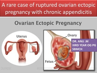
- 1. A rare case of ruptured ovarian ectopic pregnancy with chronic appendicitis DR. ANU .M IIIRD YEAR OG PG SBMCH
- 2. ABSTRACT • Ovarian pregnancy is a rare and dangerous form of ectopic pregnancy. • A little is known about its risk factors and incidence • We present a case of ruptured primary ovarian pregnancy which was a surprise intra operative finding • As assisted reproductive technology(ART) procedures are becoming popular, the incidence is likely to increase. • Clinicians should be well equipped to diagnose and treat this unusual form of ectopic pregnancy at the earliest
- 3. INTRODUCTION • Ovarian ectopic pregnancy is a rare diagnosis of exclusion and constitutes only 0.15 to 3 % of all ectopic pregnancies.
- 4. Incidence: 1:3000 to 1:7000 deliveries • Primary ovarian pregnancy result when an ovum not released from the ovary is fertilized or following a primary implantation of fertilized ovum in the ovary after reverse migration from the fallopian tube. • Intrafollicular – etiology is hormonal resulting in a rapped ovum inside the follicle • Thickened tunica albugenia of ovary • Inefficient sweeping of fimbria across the surface of ovary resulting in ineffective ovum pick up • Extrafollicular
- 5. • In secondary ovarian pregnancy, there is a tubal abortion or rupture with secondary implantation of gestational sac on the surface of ovary. • USG – Hyperdense chorial ring which moves with movement of ovary • Corpus luteum or hemorrhagic cyst of ovary
- 6. CASE REPORT • A 37 years, G 3 P 1 L 1 A1, with history of previous one LSCS, with regular cycles, presented to the casualty with history of 44 days amenorrhoea, and complaints of lower abdominal pain for past 4 days - acute in onset, - severe in intensity 2 days back - now dull aching and persistent in nature
- 7. MENSTRUAL HISTORY: Age of menarche – 14 years Regular 3-4 / 30 days cycle LMP 44 days back MARITAL HISTORY: Married since 4 years Non consanguinous marriage No history of contraceptives
- 8. • I – Male / 2.30kg/full term LSCS in view of PROM/ Oligohydramnios/ fetal distress/ 2 ½ years / A&H • II -One Spontanous abortion at 2 months amenorrhoea, 1 year back for which dilatation and curretage done, 1 Unit of blood transfused in view of anemia, at that time • III – present pregnancy • No history of ovulation induction drugs. OBSTETRIC HISTORY: G3P1L1A1
- 9. On examination She was severely anemic,afebrile, pulse rate- 102/min; BP= 100/60mmHg, CVS- S1,S2 heard; RS-normal vesicular breath sounds heard
- 10. • On abdominal examination, soft, non tender, mild distension noted, SPT scar healthy. • On speculum examination, cervix and vagina were healthy, no significant discharge • On per vaginum examination, cervix pointing downwards, soft, os closed uterus anteverted, normal size, fornices free, non tender, no cervical motion tenderness. Urinary beta human chorionic gonadotropin was positive
- 11. • Emergency ultrasound was done to confirm the diagnosis and showed no intrauterine pregnancy • ET regular , 6mm • Right ovary could not be visualized • Thick wall echogenic ring shaped lesion with irregular central anechoic area measuring 5.1 x 3.8 cm noted in the right adnexa. Periperal vascularity noted. • Free fluid in POD and right paracolic gutter noted • Thin rim of free fluid noted in morrison’s pouch.
- 12. Investigations • HB = 7.5 g/dl • Blood group = O Positive • Serology – NEG • After preoperative check up, patient was taken for exploratory laparotomy under general anesthesia, with a preoperative provisional diagnosis of ruptured right tubal ectopic pregnancy • Abdomen opened with suprapubic transverse incision
- 13. Intra op findings • Haemoperitoneum of 300 ml with blood clots • Uterus was normal in size • Right fallopian tube was congested • Left fallopian tube, and left ovary were normal • Right ovary was enlarged and showed a breech on the surface with an active bleeder on ruptured surface
- 14. Right ovary was enlarged and showed a breech on the surface with an active bleeder on ruptured surface
- 15. Uterus was normal in size Right fallopian tube was congested
- 16. • Right salphingo- oophrectomy done, as fallopian tube was congested, and the fresh bleeding vessels on the ovarian surface could not be secured after plication and electrocauterization. • Incidentally, appendix was found inflammed and edematous, proceeded with appendicectomy. Perfect hemostasis secured. Abdomen closed in layers
- 17. • Two units packed cells transfused in immediate postoperative period • Post operative period uneventful. • Serum beta HCG levels on • POD 1 = 2115mIU/ml • POD 3 = 378.14mIU/ml
- 18. HPE • Excised specimen – blood clot with few hyperplastic villi, synctiotrophoblast and cytotrophoblast • Right tube – numerous congested blood vessels in the wall • Right ovary – ovarian stroma with numerous corpora albicantes and hemorrhagic corpus luteal cyst. • IMP : RUPTURED OVARIAN PREGNANCY • Appendix – chronic appendicitis
- 19. Spiegelberg Criteria • The fallopian tube with its fimbriae should be intact and separate from the ovary • The gestational sac should occupy the normal position of the ovary • The gestational sac should be connected to the uterus by the ovarian ligament • A histologically proven ovarian tissue should be located in the sac wall. Distal tubal pregnancy VS primary ovarian pregnancy
- 20. DISCUSSION • There is often a delay in the diagnosis of ovarian pregnancy as the gestational sac mimics corpus luteum, hemorrhagic cyst, and endometriotic cyst of ovary • Ectopic pregnancy is responsible for 10% of maternal mortality. • Primary ovarian pregnancy is very uncommon among all types of extrauterine pregnancies, incidence of which is 0.15% - 3%.
- 21. • They pose a significant diagnostic and therapeutic challenge and carry a greater maternal mortality risk than tubal ampullary ectopic pregnancy • The developing chorionic villi may eventually erode into the blood vessels of ovary, causing severe hemorrhage • Significant maternal hemorrhage leading to hypovolemia and shock can rapidly result from ovarian pregnancy rupture
- 22. Causes of ovarian pregnancy remain obscure • PID • fibroids • Tubal diseases • IVF • Previous pelvic surgeries • IUCD • Favourable implantation surface as in endometriosis -Altered tubal motility- ↑progesterone – hyperstimulation -Excessive pressure on syringe during embryo transfer peri and intra tubal adhesions
- 23. • Mean age = 29 years • Mean gestational age = 45 days • Patient usually presents with • pain abdomen (100%) • Vaginal bleeding (33%) • Hypovolemic shock (8%)
- 24. DIAGNOSIS • Serum Beta HCG • TVS • Abdomino pelvic USG cannot always differentiate it from other types of ectopic pregnancies • Laparoscopy
- 25. TRANSVAGINAL ULTRASOUND CRITERIA FOR OVARIAN PREGNANCY • More echogenic white ring in the ovary compared with ovarian tissue • A yolk sac or fetal parts may be visualized but an embryonic pole is rarely seen • The corpus luteum has an anechoic texture and less wall echogenicity as compared to endometrium • 3D – Coronal plane of uterus – exact localization of GS relative to uterine tube and ovary
- 26. MEDICAL MANAGEMENT • Early diagnosis of ovarian pregnancy with TVS allows for first trimester conservative management with methotrexate • Serum beta HCG – 3000 IU/ml • Minimum symptoms
- 27. SURGICAL MANAGEMENT • LAPAROSCOPY • LAPARATOMY – • Unruptured - Ovarian wedge resection Ovarian cystectomy • Ruptured – Oophrectomy Salphingo - oophrectomy
- 28. • Ovarian pregnancy usually rupture in 91% of cases in first trimester • Laparoscopic visualisation is considered as gold standard of modern management of ovarian pregnancy – frozen section biopsy of gestational sac
- 29. CONCLUSION • Ovarian pregnancies are rare • Missed radiologically and intraoperatively • Difficult to differentiate between GS and corpus luteum/ hemorrhagic cyst in the ovary – 3D ultrasound • Frozen section biopsy • ART • In our patient, the ovarian gestation had ruptured and the only option was emergency salpingo - oophrectomy
- 30. THANK YOU
Editor's Notes
- A 37 years old female presented with dull aching lower abdominal pain at period of gestation 6weeks2days. Ultrasonography showed complex mass in right adnexa of size 5x4 cm. Emergency laparotomy was done under general anesthesia and ruptured right ovary found, with inflamed edematous appendix Right salphingo-oophrectomy and appendicectomy was done The histopathological examination confirmed the ovarian ectopic pregnancy and chronic appendicitis.
- Ectopic – gynec emergencies Ectopic 2% all pregnancies 95% fallopian tube We are presenting a case of ruptured ovarian ectopic pregnancy with chronic appendicitis, which presented for the emergency exploratory laparotomy
- MARITAL HISTORY: Married at 33 years of age Married since 4 years Non consanguinous marriage OBSTETRIC HISTORY: I – Male / 2.30kg/full term LSCS in view of PROM/ Oligohydramnios/ fetal distress/ 2 ½ years / A&H II -One Spontanous abortion at 2 months amenorrhoea, 1 year back for which dilatation and curretage done, certified, 1 Unit of blood transfused in view of anemia. III – Spontaneous conception Confirmed by UPT two days back H/O severe lower abdominal pain, 2 days back , for which USG was done showing Right Adnexal mass (74.7 x 48.2 mm) with free fluid in peritoneal cavity Suggestive of ? Ruptured ectopic pregnancy No H/O bleeding P/V.
- Haemoperitoneum – 300 ml Right ruptured adnexal mass 5x4 cm involving the ovarian tissue, associated with clots Right fallopian tube appeared congested and edematous Uterus normal size Left fallopian tubes and ovary – normal
- Ectopic pregnancy is responsible for 10% of maternal mortality. Primary ovarian pregnancy is very uncommon among all types of extrauterine pregnancies, incidence of which is 0.15% - 3%.