SlideShare a Scribd company logo
1 of 6
Download to read offline
IOSR Journal of Computer Engineering (IOSR-JCE)
e-ISSN: 2278-0661,p-ISSN: 2278-8727, Volume 17, Issue 3, Ver. V (May – Jun. 2015), PP 01-06
www.iosrjournals.org
DOI: 10.9790/0661-17350106 www.iosrjournals.org 1 | Page
Texture Analysis As An Aid In CAD And Computational Logic
Charu Sharma
Abstract: The thought to utilize composition examination in restorative imaging has been considered following
the mid-1970s. Then again, the energizing advancement of both surface investigation calculations and PC
innovation restored scientists' enthusiasm for applications for restorative imaging recently. Specialized
advancements in medicinal cross-sectional imaging have opened up new vistas for the investigation of the
human body, empowering both high spatial and temporal resolution. On Difficulties Caused by Missing
Reference Image Datasets, one of the fundamental issues in the improvement of frameworks for the investigation
of restorative pictures or division calculations is the situation of pictures that is utilized for both the
advancement and the testing of the framework. This paper proposes a symptomatic plan which takes after a well
known two-stage approach. At first, textural data is removed from the picture. Accordingly, the extricated data
is nourished into a decisional calculation that is intended to perform the analytic task.The talk covers new
routines for example, acknowledgment and PC helped determination in the field of MRI.Utilitarian MRI for
human cerebrum mapping, treatment control via programmed injury recognition in numerous sclerosis, and
new ways to deal with the diagnosis ofbreast cancer in MRI mammography. In the light of such creative systems
for example acknowledgment in biomedicine, the expanding interest for openly available approval stages is
underlined. With picture surface examination in the part of the visual perceptional capacity, the procedure of
highlight extraction and picture coding is accomplished. The separated elements can now be converted into a
finding by utilizing a decision making calculation, with decisions that range from the standard based models to
the customary factual examination to the more prominent and frequently more effective computerized reasoning
strategies, for example, neural systems and hereditary calculations. By utilizing picture texture analysis as the
preprocessing stride in CAD conspires, the information era procedure is mechanized and, hence, is
reproducible and vigorous. A few methods represent texture on the basis of the spectral properties of an image.
Others are model-based strategies that investigate surface by distinguishing a proper model that mirrors the
former convictions and information about the kind of pictures to be investigated. There are textural components
that depict nearby picture measurements and others that portray worldwide insights.
I. Introduction
Pattern recognition is "the demonstration of taking in crude information and making a move in view of
the class of the pattern". Most research in pattern acknowledgment is about techniques for regulated learning
and unsupervised learning. Pattern recognition plans to characterize information constructed either in light of the
earlier learning or on measurable data removed from the examples. The examples to be characterized are
typically gatherings of estimations or perceptions, characterizing focuses in a suitable multidimensional space.
This is as opposed to pattern coordinating, where the pattern is unbendingly indicated. A complete pattern
recognition framework comprises of a sensor that assembles the perceptions to be arranged or depicted, an
element extraction instrument that figures numeric or typical data from the perceptions, and a depiction strategy
that does the genuine occupation of characterizing or portraying perceptions, depending on the removed
elements.
The characterization or depiction plan is generally takes into account the accessibility of an
arrangement of patterns that have, as of now been grouped or depicted. This arrangement of patterns is termed
the preparation set or the training set, and the subsequent learning methodology is portrayed as managed
learning or in other terms, supervised learning. Learning can likewise be unsupervised in a manner, where the
system is not attached with earlier naming of examples, rather it itself sets up the classes in view of the inherent
regularities of the examples. The grouping or portrayal plan for the most part uses one of the accompanying
methodologies: factual or syntactic. Measurable pattern recognition is in light of factual portrayals of patterns,
accepting that the patterns are created by a probabilistic framework. Grammatical or basicpattern recognition is
in view of the auxiliary interrelationships of elements.
An extensive variety of calculations can be requisitioned for pattern recognition, from basic guileless
Bayes classifiers and neural systems to the intense k-closest neighbor calculation decisional algorithms. Design
acknowledgment is more perplexing when layouts are utilized to produce variations. To emphasize, in English,
sentences regularly take after the "N-VP" (thing - verb expression) design, however some learning of the
English dialect is obliged to identify the pattern. Pattern recognition is considered in numerous fields, including
brain science, ethology, intellectual science and software engineering. Holographic affiliated memory is another
Texture analysis as an aid in CAD and computational logic
DOI: 10.9790/0661-17350106 www.iosrjournals.org 2 | Page
sort of example coordinating where an extensive arrangement of scholarly examples in view of psychological
meta-weight is traversed for a little arrangement of target examples.
II. Specific Uses
Figure 1: Faces recognized distinctly among other elements.
The face was consequently recognized by unique programming set. In case of medicinal science,
pattern recognition is the premise for computer aided diagnostic (CAD) frameworks. CAD portrays a
methodology that backs up a specialist's translations and discoveries. Some specific applications are
programmed speech recognition software, characterization of content into a few classifications (e.g. spam/non-
spam email messages), the programmed acknowledgment of written by hand postal codes on postal envelopes,
or the programmed acknowledgment of pictures of human appearances. The last two cases shape the subtopic
picture examination of pattern recognition that makes arrangements with computerized pictures as information
to pattern recognition frameworks.
III. Role Of Image Texture Analysis
The concept of ‘computer aided diagnosis’ (which is also called CAD) refers to software that analyses
a radiographic finding to estimate the likelihood that the feature represents a specific disease process (e.g.
benign versus malignant). To my knowledge, this technology has not yet been approved for clinical use,
specifically depending more on biopsy tests done manually on sample tissues which usually is a very tedious
task. Currently, methods of image texture analysis are undergoing great development and utilization within the
field of medical imaging. Given the general interest and striking growth in computer-aided diagnosis (CAD), the
application of texture analysis in the diagnostic interpretation of radiologic images has become a rapidly
expanding field of research.
Proposed diagnostic scheme follows a familiar two-step approach. Initially, textural information is
extracted from the image. Subsequently, the extracted information is fed into a decisional algorithm (eg. an
artificial neural network) that is designed to perform the diagnostic task. However, from a methodological point
of view, the main attraction of this study is that the combination of image texture analysis and automated
decision making offers a promising approach to a clinical challenge. Successful applications of the above CAD
strategy have been reported for other types of medical images. While further development and testing is required
to establish the true clinical effect of such decisionsupport systems, texture analysis appears to open a new
exciting path in the journey toward CAD in radiology. Consequently, two questions are raised: What is the
realistic contribution of texture analysis in the computer-aided interpretation of medical images, and to what
extent can it be expected to improve interpretative accuracy?
Texture analysis as an aid in CAD and computational logic
DOI: 10.9790/0661-17350106 www.iosrjournals.org 3 | Page
IV. Formulation Of The Diagnosis
The symptomatic translation of therapeutic pictures is a multifaceted assignment. Its goal is the exact
recognition and exact portrayal of potential variations from the norm, animportant stride toward the foundation
of powerful treatment. Accomplishing this objective depends on the radiologists' effective mix of two particular
procedures: (a) the procedure of picture observation to perceive special picture patterns and (b) the procedure of
thinking to recognize connections between observed patterns and conceivable conclusions. Both procedures
depend intensely on the radiologists' exact learning, memory, instinct, and tirelessness.
Certainly, the radiologists approach the analytic undertaking with a level of insight, adaptability, and
an ability to think that is hard to copy with a computer brain. In spite of many years of concentrated research in
biological visual frameworks and perceptual insight, there is a constrained comprehension and a continuous
level headed discussion about the fundamental components that underlie visual observation. Interestingly, there
is general understanding that composition is a rich wellspring of visual data and is a key part in image
investigation and perception in people. Textureimportantly provides pieces of information about picturesque
profundity and texture introduction and, in that capacity, depicts the substance of both naturally produced and
manufactured images. Likewise, there is confirmation of perceptual adaption in composition coding systems and
in textural separation. In light of this, specialists have centered their consideration on creating calculations that
can evaluate the textural properties of a picture. Exact proof and analysts' innovativeness have prompted various
calculations for textureevaluation. The accessible calculations normally contrast in the sort of picture data that is
caught and in the coding instrument. The development and differences of accessible systems for surface
examination are a demonstration of the headway of this field.
A broad review of textural definitions, models, and diagnostic calculations can be discovered
somewhere else. Composition examination is at last concerned with robotized techniques that can get image
data from an absolutely computational perspective. As being what is indicated, it is just another kind of numeric
control of advanced or digitized pictures to get quantitative estimations. In any case, in opposition to the
segregation of morphologic data, there is proof that the human visual framework experiences issues in the
separation of textural data that is identified with higher-request insights or ghostly properties on a picture.
Hence, texture investigation can possibly increase the visual aptitudes of the radiologist by taking into
Texture analysis as an aid in CAD and computational logic
DOI: 10.9790/0661-17350106 www.iosrjournals.org 4 | Page
consideration the features of the image that may be applicable to the indicative issue however that are not so
much outwardly extractable.
In any case, surface examination is not a panacea for the symptomatic understanding of radiologic
pictures. The quest for composition investigation is taking into account the theory that the surface mark of a
picture is applicable to the indicative issue close by. Moreover, the adequacy of surface investigation is bound
by the kind of calculation that is utilized to concentrate significant components. A choice calculation that uses
consequently separated components has a superior possibility of creating a powerful CAD framework. A CAD
apparatus that uses naturally separated elements addresses a built up clinical shortcoming of the demonstrative
procedure furthermore supplements the radiologists' smart capacities. Texture investigation is appropriately
suited to this issue as the radiologists themselves depend on visual composition to distinguish and portray breast
injuries. The way image handling is utilized as a part of the study can be effortlessly performed with existing
programming bundles significantly streamlining the fuse of the proposed CAD device into the center. The
adequacy of any CAD instrument will dependably be contingent on two things: (a) how well the chosen
components portray the infection that need to be separated and (b) how well the study groupreflects the general
target patient population for the CAD apparatus.
V. Texture Analysis’ Clinical Implementation Along With Cad
Computer aided diagnostic (CAD) is an innovation intended to reduce observational oversights and in
this way decline the false negative rates of doctors deciphering restorative pictures. Planned clinical studies have
exhibited an increment in breast malignancy identification with CAD help. This review quickly depicts the
measurements that have been utilized to characterize CAD framework execution. PC projects have been created
and sanction for utilization in clinical practice that guide radiologists in identifying potential variations from the
norm on indicative radiology exams. This application has been termed PC aided location, usually alluded to as
CAD. The term 'computer aided diagnostic’ alludes to pattern recognition programming that recognizes
suspicious components on the picture and conveys them to the consideration of the radiologist, keeping in mind
the end goal to decline false negative readings.
As presently utilized, the radiologist first audits the exam, then initiates the CAD programming and re-
assesses the CAD-stamped regions of concern before issuing the last report. CAD is as of now FDA and CE
sanctioned for utilization with both film and advanced mammography, for both screening and analytic exams;
for midsection CT; and, for midsection radiographs. The essential objective of CAD is to build the discovery of
illness by diminishing the false negative rate because of observational oversights. The utilization of a PC as
opposed to a second human eyewitness has the benefit of not expanding the requests on the radiologist or
prepared spectator pool. An imperative part of either approach is to expand sickness recognition without a fix
effect on the review and work up rates.
At last, in a few applications CAD, with its related robotized programming instruments, can possibly
give work process efficiencies. CAD calculations are produced to scan for the same components that a
radiologist searches for amid case audit. Hence, for breast growth on mammograms, the CAD calculations hunt
down small scale calcifications and masses both hypothesized and non-theorized, design contortions and
asymmetries. On midsection radiographs and CT filters, flow CAD applications look for aspiratory densities
that have particular physical qualities, e.g. sphericity, that may speak to lung knobs. As anyone might expect,
CAD calculations will underline features that meet the calculation necessities, however which don't speak to
discoveries that the radiologist considers to warrant further examination, i.e. false CAD marks. Likewise, on
occasion a genuine positive CAD mark, upon survey by the radiologist, is released as not justifying further
examination. In this case, the false negative report would be the aftereffect of an interpretive—as opposed to a
discernment.
VI. Cad Methodology: Dealing With Textural Analysis
CAD is in a far-reaching way taking into account profoundly complex pattern recognition. X-ray
pictures are filtered for suspicious structures. Regularly a couple of thousand pictures are obliged to enhance the
calculation. Advanced picture information are replicated to a CAD server in a DICOM-format and are arranged
and investigated in a few stages.
6.1.1 Preprocessing for
 Decrease of relics (bugs in pictures)
 Picture commotion diminishment
 Leveling (harmonization) of picture quality for clearing the picture's distinctive fundamental conditions e.g.
diverse presentation parameter.
Texture analysis as an aid in CAD and computational logic
DOI: 10.9790/0661-17350106 www.iosrjournals.org 5 | Page
6.1.2 Segmentation for
 Separation of distinctive structures in the picture, e.g. heart, lung, ribcage , conceivable round
 Sores
 Coordinating with anatomic databank
6.1.3 Structure/ROI (Region of Interest) Analyze
Each distinguished area is examined exclusively for uncommon attributes:
 Closeness
 Frame, size and area
 Reference to near to structures/ ROIs
 Normal greylevel worth break down inside of a ROI
 Extent of greylevels to outskirt of the structure inside the ROI
6.1.4 Evaluation/ order
After the structure is broke down, every ROI is assessed exclusively (scoring) for the likelihood of a TP.
Subsequently the methodology are:
 Nearest-Neighbor Rule
 Minimum distance classifier
 Cascade Classifier
 Bayesian Classifier
 Multilayer perception
 Radial basis function network (RBF)
 SVM
In the event that the recognized structures have come to a certain edge level, they are highlighted in the
picture for the radiologist. Contingent upon the CAD framework these markings can be forever or provisional
spared. The latter's preference is that just the markings which are endorsed by the radiologist are spared. False
hits ought not to be spared, on the grounds that an examination at a later date turns out to be more troublesome
then.
VII. Implementation Of Cad In Accordance With Background Evaluation
The CAD calculations oblige an advanced information set of the picture for examination. In the event
that the picture is obtained on x-ray film, for example, a screen mammogram, the simple picture should first be
digitized. On the other hand, the CAD calculations can specifically investigate pictures obtained in
computerized arrangement, for example, with advanced mammography (FFDM) and CT.
In present practice (and as needed by the FDA), the exam ought to first be investigated and deciphered in the
typical manner. At exactly that point are the CAD imprints shown, taking after which the radiologist re-surveys
those ranges that are provoked by the CAD framework. Two vital standards must be held fast to:
 Current CAD frameworks don't check every noteworthy finding. Consequently, the nonappearance of a
CAD check on a discovering the radiologist was worried about on his/her pre CAD audit should not hinder
further assessment.
 Current CAD frameworks create numerous more false CAD marks than genuine CAD marks. Thus, it is the
obligation of the radiologist to figure out whether a CAD imprint warrants further assessment.
A CAD framework can be assessed in a few ways, which incorporates examination of information
produced in a research center or test setting, and by the effect of CAD on radiologist execution in a real clinical
work on setting. ‘Standalone' affectability and specificity, this data can be acquired by watching the execution of
a CAD framework on an arrangement of "truth" cases. The fact of the matter is by and large settled by
histological confirmation of the vicinity (e.g. disease) or nonattendance (e.g. clinical postliminary) of malady.
Affectability is dictated by the rate of positive cases in which the CAD framework puts an imprint on
the malady area. The quantity of false CAD marks per typical picture or case is generally utilized as a surrogate
for specificity. The aftereffects of this activity are, obviously, subject to the case accumulation. Inclination,
planned or not, in gathering positive cases that have more obvious discoveries will evidently bring about better
CAD execution analyzed than cases that are less prominent, despite the fact that the CAD calculation is the
same. Along these lines, the same CAD calculation will exhibit changing sensitivities and specificities (false
checks per case) contingent upon the case creation.
A favored technique to contrast CAD frameworks is to determine the sensitivity and false marker rates
on the same arrangement of "truth" cases. These cases must be "obscure" to the CAD framework, that is, they
Texture analysis as an aid in CAD and computational logic
DOI: 10.9790/0661-17350106 www.iosrjournals.org 6 | Page
ought not to have been utilized to prepare the CAD calculations. Adequate and regularly extensive quantities of
cases will be required so as to build up measurable noteworthiness of predominance or proportionality in
execution when contrasting CAD frameworks. "Research center" investigations of potential recognition change
initiate radiologists (or other 'pursuers') to assess an arrangement of "truth" cases to focus the affectability and
get back to rate of the unaided pursuer (pre CAD) with that of the pursuer with CAD help. Such studies are
valuable to survey the potential advantage of CAD and give evaluations of expected changes in ailment
discovery and workup/review rates. In any case, the test setting regularly bargains the execution of the pursuer,
in that the pursuers might either, over or under call the investigated cases in a test domain.
This should be possible in a 'consecutive read' clinical trial, in which the exam is first read before, and
later on taking after, CAD information. The adjustment in sickness recognition because of the CAD data, and
additionally the adjustment in the review/workup rates, will focus the commitment of CAD to patient
administration. Vitally, the rate increment in ailment discovery ought to be concordant with, or not exactly, the
rate increment in the review/workup rate.
VIII. Conclusion
The thought to utilize pattern recognition in restorative imaging has been considered following the mid
1970s. On Difficulties Caused by Missing Reference Image Datasets One of the fundamental issues in the
improvement of frameworks for the examination of medicinal pictures or division calculations is the situation of
pictures that is utilized for both the advancement and the testing of the framework. This paper depicts the system
with picture composition examination in the part of the visual perceptional capacity, the procedure of highlight
extraction and picture coding is accomplished. The extricated elements can now be converted into a conclusion
by utilizing a decisional calculation, with decisions that range from the tenet based models to the customary
factual examination to the more prevalent and regularly more fruitful, manmade brainpower systems, for
example, neural systems and genetic calculations.
By utilizing image texture analysis as the preprocessing stride in CAD schemes, the input generation
procedure is computerized and, accordingly, is reproducible and strong. Hitherto, discoveries have demonstrated
that CAD can improve the symptomatic execution of radiologists, in the event that it is utilized as a second
assessment. Be that as it may, it is still hard to foresee its acknowledgement in medicinal analysis, and its
acknowledgement won't be resolved until discoveries from broad clinical utilization exhibit quantifiable
advantages. In the meantime, business CAD items will need to face new difficulties identified with promoting,
specialized bolster, adjustments, and item life-cycles and practices in the human services environment are
always re-imagined.
References
[1]. Chen DR, Chang RF, Huang YL. Computer-aided diagnosis applied to US of solid breast nodules by using neural networks.
[2]. D R. Brunelli, Template Matching Techniques in Computer Vision: Theory and Practice, Wiley, ISBN
[3]. Fukunaga, Keinosuke. Introduction to Statistical Pattern Recognition (2nd ed.).
[4]. Bishop, Christopher. Pattern Recognition and Machine Learning.
[5]. Godfried T. Toussaint. Computational Morphology. Amsterdam: North-Holland Publishing Company.
[6]. Kulikowski, Casimir A.; Weiss, Sholom M. Computer Systems That Learn: Classification and Prediction Methods from Statistics,
Neural Nets, Machine Learning, and Expert Systems. Machine Learning.
[7]. Krupinski EA. The future of image perception in radiology.
[8]. Vittitoe NF, Baker JA, Floyd CE, Jr. Fractal texture analysis in computer-aided diagnosis of solitary pulmonary nodules.
[9]. S Wei D, Chan HP, Helvie MA, et al. Classification of mass and normal breast tissue on digital mammograms: multiresolution
texture analysis.
[10]. Chan HP, Sahiner B, Petrick N, et al. Computerized classification of malignant and benign microcalcifications on mammograms:
texture analysis using an artificial neural network.
[11]. Petrick N, Chan HP, Wei D, Sahiner B, Helvie MA, Adler DD. Automated detection of breast masses on mammograms using
adaptive contrast enhancement and texture classification.
[12]. McPherson DD, Aylward PE, Knosp BM, et al.
[13]. Ultrasound characterization of acute myocardial ischemia by quantitative texture analysis.
[14]. Lucht R, Brix G, Lorenz WJ. Texture analysis of differently reconstructed PET images.

More Related Content

What's hot

MR Image Segmentation of Patients’ Brain Using Disease Specific a priori Know...
MR Image Segmentation of Patients’ Brain Using Disease Specific a priori Know...MR Image Segmentation of Patients’ Brain Using Disease Specific a priori Know...
MR Image Segmentation of Patients’ Brain Using Disease Specific a priori Know...CSCJournals
 
deep learning applications in medical image analysis brain tumor
deep learning applications in medical image analysis brain tumordeep learning applications in medical image analysis brain tumor
deep learning applications in medical image analysis brain tumorVenkat Projects
 
A novel framework for efficient identification of brain cancer region from br...
A novel framework for efficient identification of brain cancer region from br...A novel framework for efficient identification of brain cancer region from br...
A novel framework for efficient identification of brain cancer region from br...IJECEIAES
 
IRJET- A Novel Segmentation Technique for MRI Brain Tumor Images
IRJET- A Novel Segmentation Technique for MRI Brain Tumor ImagesIRJET- A Novel Segmentation Technique for MRI Brain Tumor Images
IRJET- A Novel Segmentation Technique for MRI Brain Tumor ImagesIRJET Journal
 
Paper id 25201494
Paper id 25201494Paper id 25201494
Paper id 25201494IJRAT
 
Brain Image Fusion using DWT and Laplacian Pyramid Approach and Tumor Detecti...
Brain Image Fusion using DWT and Laplacian Pyramid Approach and Tumor Detecti...Brain Image Fusion using DWT and Laplacian Pyramid Approach and Tumor Detecti...
Brain Image Fusion using DWT and Laplacian Pyramid Approach and Tumor Detecti...INFOGAIN PUBLICATION
 
Classification of MR medical images Based Rough-Fuzzy KMeans
Classification of MR medical images Based Rough-Fuzzy KMeansClassification of MR medical images Based Rough-Fuzzy KMeans
Classification of MR medical images Based Rough-Fuzzy KMeansIOSRJM
 
ENHANCED SYSTEM FOR COMPUTER-AIDED DETECTION OF MRI BRAIN TUMORS
ENHANCED SYSTEM FOR COMPUTER-AIDED DETECTION OF MRI BRAIN TUMORSENHANCED SYSTEM FOR COMPUTER-AIDED DETECTION OF MRI BRAIN TUMORS
ENHANCED SYSTEM FOR COMPUTER-AIDED DETECTION OF MRI BRAIN TUMORSsipij
 
Automatic grading of diabetic retinopathy through machine learning
Automatic grading of diabetic retinopathy through machine learning   Automatic grading of diabetic retinopathy through machine learning
Automatic grading of diabetic retinopathy through machine learning SupriyaKamatgi
 
Glioblastomas brain tumour segmentation based on convolutional neural network...
Glioblastomas brain tumour segmentation based on convolutional neural network...Glioblastomas brain tumour segmentation based on convolutional neural network...
Glioblastomas brain tumour segmentation based on convolutional neural network...IJECEIAES
 
An Ameliorate Technique for Brain Lumps Detection Using Fuzzy C-Means Clustering
An Ameliorate Technique for Brain Lumps Detection Using Fuzzy C-Means ClusteringAn Ameliorate Technique for Brain Lumps Detection Using Fuzzy C-Means Clustering
An Ameliorate Technique for Brain Lumps Detection Using Fuzzy C-Means ClusteringIRJET Journal
 
Illustration of Medical Image Segmentation based on Clustering Algorithms
Illustration of Medical Image Segmentation based on Clustering AlgorithmsIllustration of Medical Image Segmentation based on Clustering Algorithms
Illustration of Medical Image Segmentation based on Clustering Algorithmsrahulmonikasharma
 
Image Binarization for the uses of Preprocessing to Detect Brain Abnormality ...
Image Binarization for the uses of Preprocessing to Detect Brain Abnormality ...Image Binarization for the uses of Preprocessing to Detect Brain Abnormality ...
Image Binarization for the uses of Preprocessing to Detect Brain Abnormality ...Journal For Research
 
IJCER (www.ijceronline.com) International Journal of computational Engineerin...
IJCER (www.ijceronline.com) International Journal of computational Engineerin...IJCER (www.ijceronline.com) International Journal of computational Engineerin...
IJCER (www.ijceronline.com) International Journal of computational Engineerin...ijceronline
 
BRAIN TUMOR MRIIMAGE CLASSIFICATION WITH FEATURE SELECTION AND EXTRACTION USI...
BRAIN TUMOR MRIIMAGE CLASSIFICATION WITH FEATURE SELECTION AND EXTRACTION USI...BRAIN TUMOR MRIIMAGE CLASSIFICATION WITH FEATURE SELECTION AND EXTRACTION USI...
BRAIN TUMOR MRIIMAGE CLASSIFICATION WITH FEATURE SELECTION AND EXTRACTION USI...ijistjournal
 
Comparative Study on Medical Image Classification Techniques
Comparative Study on Medical Image Classification TechniquesComparative Study on Medical Image Classification Techniques
Comparative Study on Medical Image Classification TechniquesINFOGAIN PUBLICATION
 
Development of Computational Tool for Lung Cancer Prediction Using Data Mining
Development of Computational Tool for Lung Cancer Prediction Using Data MiningDevelopment of Computational Tool for Lung Cancer Prediction Using Data Mining
Development of Computational Tool for Lung Cancer Prediction Using Data MiningEditor IJCATR
 
Automatic Brain Tumour Detection Using Symmetry Information
Automatic Brain Tumour Detection Using Symmetry InformationAutomatic Brain Tumour Detection Using Symmetry Information
Automatic Brain Tumour Detection Using Symmetry InformationIJERA Editor
 

What's hot (20)

MR Image Segmentation of Patients’ Brain Using Disease Specific a priori Know...
MR Image Segmentation of Patients’ Brain Using Disease Specific a priori Know...MR Image Segmentation of Patients’ Brain Using Disease Specific a priori Know...
MR Image Segmentation of Patients’ Brain Using Disease Specific a priori Know...
 
deep learning applications in medical image analysis brain tumor
deep learning applications in medical image analysis brain tumordeep learning applications in medical image analysis brain tumor
deep learning applications in medical image analysis brain tumor
 
A novel framework for efficient identification of brain cancer region from br...
A novel framework for efficient identification of brain cancer region from br...A novel framework for efficient identification of brain cancer region from br...
A novel framework for efficient identification of brain cancer region from br...
 
IRJET- A Novel Segmentation Technique for MRI Brain Tumor Images
IRJET- A Novel Segmentation Technique for MRI Brain Tumor ImagesIRJET- A Novel Segmentation Technique for MRI Brain Tumor Images
IRJET- A Novel Segmentation Technique for MRI Brain Tumor Images
 
Paper id 25201494
Paper id 25201494Paper id 25201494
Paper id 25201494
 
Brain Image Fusion using DWT and Laplacian Pyramid Approach and Tumor Detecti...
Brain Image Fusion using DWT and Laplacian Pyramid Approach and Tumor Detecti...Brain Image Fusion using DWT and Laplacian Pyramid Approach and Tumor Detecti...
Brain Image Fusion using DWT and Laplacian Pyramid Approach and Tumor Detecti...
 
Bt36430432
Bt36430432Bt36430432
Bt36430432
 
C1103041623
C1103041623C1103041623
C1103041623
 
Classification of MR medical images Based Rough-Fuzzy KMeans
Classification of MR medical images Based Rough-Fuzzy KMeansClassification of MR medical images Based Rough-Fuzzy KMeans
Classification of MR medical images Based Rough-Fuzzy KMeans
 
ENHANCED SYSTEM FOR COMPUTER-AIDED DETECTION OF MRI BRAIN TUMORS
ENHANCED SYSTEM FOR COMPUTER-AIDED DETECTION OF MRI BRAIN TUMORSENHANCED SYSTEM FOR COMPUTER-AIDED DETECTION OF MRI BRAIN TUMORS
ENHANCED SYSTEM FOR COMPUTER-AIDED DETECTION OF MRI BRAIN TUMORS
 
Automatic grading of diabetic retinopathy through machine learning
Automatic grading of diabetic retinopathy through machine learning   Automatic grading of diabetic retinopathy through machine learning
Automatic grading of diabetic retinopathy through machine learning
 
Glioblastomas brain tumour segmentation based on convolutional neural network...
Glioblastomas brain tumour segmentation based on convolutional neural network...Glioblastomas brain tumour segmentation based on convolutional neural network...
Glioblastomas brain tumour segmentation based on convolutional neural network...
 
An Ameliorate Technique for Brain Lumps Detection Using Fuzzy C-Means Clustering
An Ameliorate Technique for Brain Lumps Detection Using Fuzzy C-Means ClusteringAn Ameliorate Technique for Brain Lumps Detection Using Fuzzy C-Means Clustering
An Ameliorate Technique for Brain Lumps Detection Using Fuzzy C-Means Clustering
 
Illustration of Medical Image Segmentation based on Clustering Algorithms
Illustration of Medical Image Segmentation based on Clustering AlgorithmsIllustration of Medical Image Segmentation based on Clustering Algorithms
Illustration of Medical Image Segmentation based on Clustering Algorithms
 
Image Binarization for the uses of Preprocessing to Detect Brain Abnormality ...
Image Binarization for the uses of Preprocessing to Detect Brain Abnormality ...Image Binarization for the uses of Preprocessing to Detect Brain Abnormality ...
Image Binarization for the uses of Preprocessing to Detect Brain Abnormality ...
 
IJCER (www.ijceronline.com) International Journal of computational Engineerin...
IJCER (www.ijceronline.com) International Journal of computational Engineerin...IJCER (www.ijceronline.com) International Journal of computational Engineerin...
IJCER (www.ijceronline.com) International Journal of computational Engineerin...
 
BRAIN TUMOR MRIIMAGE CLASSIFICATION WITH FEATURE SELECTION AND EXTRACTION USI...
BRAIN TUMOR MRIIMAGE CLASSIFICATION WITH FEATURE SELECTION AND EXTRACTION USI...BRAIN TUMOR MRIIMAGE CLASSIFICATION WITH FEATURE SELECTION AND EXTRACTION USI...
BRAIN TUMOR MRIIMAGE CLASSIFICATION WITH FEATURE SELECTION AND EXTRACTION USI...
 
Comparative Study on Medical Image Classification Techniques
Comparative Study on Medical Image Classification TechniquesComparative Study on Medical Image Classification Techniques
Comparative Study on Medical Image Classification Techniques
 
Development of Computational Tool for Lung Cancer Prediction Using Data Mining
Development of Computational Tool for Lung Cancer Prediction Using Data MiningDevelopment of Computational Tool for Lung Cancer Prediction Using Data Mining
Development of Computational Tool for Lung Cancer Prediction Using Data Mining
 
Automatic Brain Tumour Detection Using Symmetry Information
Automatic Brain Tumour Detection Using Symmetry InformationAutomatic Brain Tumour Detection Using Symmetry Information
Automatic Brain Tumour Detection Using Symmetry Information
 

Viewers also liked

Designing and Performance Evaluation of 64 QAM OFDM System
Designing and Performance Evaluation of 64 QAM OFDM SystemDesigning and Performance Evaluation of 64 QAM OFDM System
Designing and Performance Evaluation of 64 QAM OFDM SystemIOSR Journals
 
Thermal conductivity Characterization of Bamboo fiber reinforced in Epoxy Resin
Thermal conductivity Characterization of Bamboo fiber reinforced in Epoxy ResinThermal conductivity Characterization of Bamboo fiber reinforced in Epoxy Resin
Thermal conductivity Characterization of Bamboo fiber reinforced in Epoxy ResinIOSR Journals
 
Brain Bridge: A Comparative Study between Database Querying and Human Memory ...
Brain Bridge: A Comparative Study between Database Querying and Human Memory ...Brain Bridge: A Comparative Study between Database Querying and Human Memory ...
Brain Bridge: A Comparative Study between Database Querying and Human Memory ...IOSR Journals
 
Enhanced Mobile Node Tracking With Received Signal Strength in Wireless Senso...
Enhanced Mobile Node Tracking With Received Signal Strength in Wireless Senso...Enhanced Mobile Node Tracking With Received Signal Strength in Wireless Senso...
Enhanced Mobile Node Tracking With Received Signal Strength in Wireless Senso...IOSR Journals
 

Viewers also liked (20)

H017545662
H017545662H017545662
H017545662
 
E017523439
E017523439E017523439
E017523439
 
G017375362
G017375362G017375362
G017375362
 
Designing and Performance Evaluation of 64 QAM OFDM System
Designing and Performance Evaluation of 64 QAM OFDM SystemDesigning and Performance Evaluation of 64 QAM OFDM System
Designing and Performance Evaluation of 64 QAM OFDM System
 
F018123136
F018123136F018123136
F018123136
 
Thermal conductivity Characterization of Bamboo fiber reinforced in Epoxy Resin
Thermal conductivity Characterization of Bamboo fiber reinforced in Epoxy ResinThermal conductivity Characterization of Bamboo fiber reinforced in Epoxy Resin
Thermal conductivity Characterization of Bamboo fiber reinforced in Epoxy Resin
 
H012125765
H012125765H012125765
H012125765
 
Brain Bridge: A Comparative Study between Database Querying and Human Memory ...
Brain Bridge: A Comparative Study between Database Querying and Human Memory ...Brain Bridge: A Comparative Study between Database Querying and Human Memory ...
Brain Bridge: A Comparative Study between Database Querying and Human Memory ...
 
C017631521
C017631521C017631521
C017631521
 
Enhanced Mobile Node Tracking With Received Signal Strength in Wireless Senso...
Enhanced Mobile Node Tracking With Received Signal Strength in Wireless Senso...Enhanced Mobile Node Tracking With Received Signal Strength in Wireless Senso...
Enhanced Mobile Node Tracking With Received Signal Strength in Wireless Senso...
 
E1804012536
E1804012536E1804012536
E1804012536
 
H018124360
H018124360H018124360
H018124360
 
B017140612
B017140612B017140612
B017140612
 
K010418290
K010418290K010418290
K010418290
 
K0111199101
K0111199101K0111199101
K0111199101
 
B013120712
B013120712B013120712
B013120712
 
A018120105
A018120105A018120105
A018120105
 
D011122934
D011122934D011122934
D011122934
 
H1304034853
H1304034853H1304034853
H1304034853
 
B012620814
B012620814B012620814
B012620814
 

Similar to A017350106

Pneumonia Detection Using Convolutional Neural Network Writers
Pneumonia Detection Using Convolutional Neural Network WritersPneumonia Detection Using Convolutional Neural Network Writers
Pneumonia Detection Using Convolutional Neural Network WritersIRJET Journal
 
DOJProposal7.doc
DOJProposal7.docDOJProposal7.doc
DOJProposal7.docbutest
 
Performance Analysis of SVM Classifier for Classification of MRI Image
Performance Analysis of SVM Classifier for Classification of MRI ImagePerformance Analysis of SVM Classifier for Classification of MRI Image
Performance Analysis of SVM Classifier for Classification of MRI ImageIRJET Journal
 
An Investigation into Brain Tumor Segmentation Techniques
An Investigation into Brain Tumor Segmentation Techniques An Investigation into Brain Tumor Segmentation Techniques
An Investigation into Brain Tumor Segmentation Techniques IIRindia
 
IMAGE SEGMENTATION USING FCM ALGORITM | J4RV3I12021
IMAGE SEGMENTATION USING FCM ALGORITM | J4RV3I12021IMAGE SEGMENTATION USING FCM ALGORITM | J4RV3I12021
IMAGE SEGMENTATION USING FCM ALGORITM | J4RV3I12021Journal For Research
 
Detection of Cancer in Pap smear Cytological Images Using Bag of Texture Feat...
Detection of Cancer in Pap smear Cytological Images Using Bag of Texture Feat...Detection of Cancer in Pap smear Cytological Images Using Bag of Texture Feat...
Detection of Cancer in Pap smear Cytological Images Using Bag of Texture Feat...IOSR Journals
 
A Novel Efficient Medical Image Segmentation Methodology
A Novel Efficient Medical Image Segmentation MethodologyA Novel Efficient Medical Image Segmentation Methodology
A Novel Efficient Medical Image Segmentation Methodologyaciijournal
 
An Introduction To Artificial Intelligence And Its Applications In Biomedical...
An Introduction To Artificial Intelligence And Its Applications In Biomedical...An Introduction To Artificial Intelligence And Its Applications In Biomedical...
An Introduction To Artificial Intelligence And Its Applications In Biomedical...Jill Brown
 
Survey on Segmentation Techniques for Spinal Cord Images
Survey on Segmentation Techniques for Spinal Cord ImagesSurvey on Segmentation Techniques for Spinal Cord Images
Survey on Segmentation Techniques for Spinal Cord ImagesIIRindia
 
Image Segmentation and Classification using Neural Network
Image Segmentation and Classification using Neural NetworkImage Segmentation and Classification using Neural Network
Image Segmentation and Classification using Neural NetworkAIRCC Publishing Corporation
 
Image Segmentation and Classification using Neural Network
Image Segmentation and Classification using Neural NetworkImage Segmentation and Classification using Neural Network
Image Segmentation and Classification using Neural NetworkAIRCC Publishing Corporation
 
An efficient convolutional neural network-based classifier for an imbalanced ...
An efficient convolutional neural network-based classifier for an imbalanced ...An efficient convolutional neural network-based classifier for an imbalanced ...
An efficient convolutional neural network-based classifier for an imbalanced ...IAESIJAI
 
Brain Tumor Extraction from T1- Weighted MRI using Co-clustering and Level Se...
Brain Tumor Extraction from T1- Weighted MRI using Co-clustering and Level Se...Brain Tumor Extraction from T1- Weighted MRI using Co-clustering and Level Se...
Brain Tumor Extraction from T1- Weighted MRI using Co-clustering and Level Se...CSCJournals
 
International Journal of Computational Engineering Research(IJCER)
 International Journal of Computational Engineering Research(IJCER)  International Journal of Computational Engineering Research(IJCER)
International Journal of Computational Engineering Research(IJCER) ijceronline
 
Automatic brain tumor detection using adaptive region growing with thresholdi...
Automatic brain tumor detection using adaptive region growing with thresholdi...Automatic brain tumor detection using adaptive region growing with thresholdi...
Automatic brain tumor detection using adaptive region growing with thresholdi...IAESIJAI
 
Image annotation - Segmentation & Annotation
Image annotation - Segmentation & AnnotationImage annotation - Segmentation & Annotation
Image annotation - Segmentation & AnnotationTaposh Roy
 
MRIIMAGE SEGMENTATION USING LEVEL SET METHOD AND IMPLEMENT AN MEDICAL DIAGNOS...
MRIIMAGE SEGMENTATION USING LEVEL SET METHOD AND IMPLEMENT AN MEDICAL DIAGNOS...MRIIMAGE SEGMENTATION USING LEVEL SET METHOD AND IMPLEMENT AN MEDICAL DIAGNOS...
MRIIMAGE SEGMENTATION USING LEVEL SET METHOD AND IMPLEMENT AN MEDICAL DIAGNOS...cseij
 

Similar to A017350106 (20)

Af044212215
Af044212215Af044212215
Af044212215
 
Pneumonia Detection Using Convolutional Neural Network Writers
Pneumonia Detection Using Convolutional Neural Network WritersPneumonia Detection Using Convolutional Neural Network Writers
Pneumonia Detection Using Convolutional Neural Network Writers
 
DOJProposal7.doc
DOJProposal7.docDOJProposal7.doc
DOJProposal7.doc
 
Performance Analysis of SVM Classifier for Classification of MRI Image
Performance Analysis of SVM Classifier for Classification of MRI ImagePerformance Analysis of SVM Classifier for Classification of MRI Image
Performance Analysis of SVM Classifier for Classification of MRI Image
 
An Investigation into Brain Tumor Segmentation Techniques
An Investigation into Brain Tumor Segmentation Techniques An Investigation into Brain Tumor Segmentation Techniques
An Investigation into Brain Tumor Segmentation Techniques
 
IMAGE SEGMENTATION USING FCM ALGORITM | J4RV3I12021
IMAGE SEGMENTATION USING FCM ALGORITM | J4RV3I12021IMAGE SEGMENTATION USING FCM ALGORITM | J4RV3I12021
IMAGE SEGMENTATION USING FCM ALGORITM | J4RV3I12021
 
Detection of Cancer in Pap smear Cytological Images Using Bag of Texture Feat...
Detection of Cancer in Pap smear Cytological Images Using Bag of Texture Feat...Detection of Cancer in Pap smear Cytological Images Using Bag of Texture Feat...
Detection of Cancer in Pap smear Cytological Images Using Bag of Texture Feat...
 
A01110107
A01110107A01110107
A01110107
 
A Novel Efficient Medical Image Segmentation Methodology
A Novel Efficient Medical Image Segmentation MethodologyA Novel Efficient Medical Image Segmentation Methodology
A Novel Efficient Medical Image Segmentation Methodology
 
An Introduction To Artificial Intelligence And Its Applications In Biomedical...
An Introduction To Artificial Intelligence And Its Applications In Biomedical...An Introduction To Artificial Intelligence And Its Applications In Biomedical...
An Introduction To Artificial Intelligence And Its Applications In Biomedical...
 
Survey on Segmentation Techniques for Spinal Cord Images
Survey on Segmentation Techniques for Spinal Cord ImagesSurvey on Segmentation Techniques for Spinal Cord Images
Survey on Segmentation Techniques for Spinal Cord Images
 
Image Segmentation and Classification using Neural Network
Image Segmentation and Classification using Neural NetworkImage Segmentation and Classification using Neural Network
Image Segmentation and Classification using Neural Network
 
Image Segmentation and Classification using Neural Network
Image Segmentation and Classification using Neural NetworkImage Segmentation and Classification using Neural Network
Image Segmentation and Classification using Neural Network
 
Ea4301770773
Ea4301770773Ea4301770773
Ea4301770773
 
An efficient convolutional neural network-based classifier for an imbalanced ...
An efficient convolutional neural network-based classifier for an imbalanced ...An efficient convolutional neural network-based classifier for an imbalanced ...
An efficient convolutional neural network-based classifier for an imbalanced ...
 
Brain Tumor Extraction from T1- Weighted MRI using Co-clustering and Level Se...
Brain Tumor Extraction from T1- Weighted MRI using Co-clustering and Level Se...Brain Tumor Extraction from T1- Weighted MRI using Co-clustering and Level Se...
Brain Tumor Extraction from T1- Weighted MRI using Co-clustering and Level Se...
 
International Journal of Computational Engineering Research(IJCER)
 International Journal of Computational Engineering Research(IJCER)  International Journal of Computational Engineering Research(IJCER)
International Journal of Computational Engineering Research(IJCER)
 
Automatic brain tumor detection using adaptive region growing with thresholdi...
Automatic brain tumor detection using adaptive region growing with thresholdi...Automatic brain tumor detection using adaptive region growing with thresholdi...
Automatic brain tumor detection using adaptive region growing with thresholdi...
 
Image annotation - Segmentation & Annotation
Image annotation - Segmentation & AnnotationImage annotation - Segmentation & Annotation
Image annotation - Segmentation & Annotation
 
MRIIMAGE SEGMENTATION USING LEVEL SET METHOD AND IMPLEMENT AN MEDICAL DIAGNOS...
MRIIMAGE SEGMENTATION USING LEVEL SET METHOD AND IMPLEMENT AN MEDICAL DIAGNOS...MRIIMAGE SEGMENTATION USING LEVEL SET METHOD AND IMPLEMENT AN MEDICAL DIAGNOS...
MRIIMAGE SEGMENTATION USING LEVEL SET METHOD AND IMPLEMENT AN MEDICAL DIAGNOS...
 

More from IOSR Journals (20)

A011140104
A011140104A011140104
A011140104
 
M0111397100
M0111397100M0111397100
M0111397100
 
L011138596
L011138596L011138596
L011138596
 
K011138084
K011138084K011138084
K011138084
 
J011137479
J011137479J011137479
J011137479
 
I011136673
I011136673I011136673
I011136673
 
G011134454
G011134454G011134454
G011134454
 
H011135565
H011135565H011135565
H011135565
 
F011134043
F011134043F011134043
F011134043
 
E011133639
E011133639E011133639
E011133639
 
D011132635
D011132635D011132635
D011132635
 
C011131925
C011131925C011131925
C011131925
 
B011130918
B011130918B011130918
B011130918
 
A011130108
A011130108A011130108
A011130108
 
I011125160
I011125160I011125160
I011125160
 
H011124050
H011124050H011124050
H011124050
 
G011123539
G011123539G011123539
G011123539
 
F011123134
F011123134F011123134
F011123134
 
E011122530
E011122530E011122530
E011122530
 
D011121524
D011121524D011121524
D011121524
 

Recently uploaded

Unleash Your Potential - Namagunga Girls Coding Club
Unleash Your Potential - Namagunga Girls Coding ClubUnleash Your Potential - Namagunga Girls Coding Club
Unleash Your Potential - Namagunga Girls Coding ClubKalema Edgar
 
Bluetooth Controlled Car with Arduino.pdf
Bluetooth Controlled Car with Arduino.pdfBluetooth Controlled Car with Arduino.pdf
Bluetooth Controlled Car with Arduino.pdfngoud9212
 
New from BookNet Canada for 2024: BNC BiblioShare - Tech Forum 2024
New from BookNet Canada for 2024: BNC BiblioShare - Tech Forum 2024New from BookNet Canada for 2024: BNC BiblioShare - Tech Forum 2024
New from BookNet Canada for 2024: BNC BiblioShare - Tech Forum 2024BookNet Canada
 
Build your next Gen AI Breakthrough - April 2024
Build your next Gen AI Breakthrough - April 2024Build your next Gen AI Breakthrough - April 2024
Build your next Gen AI Breakthrough - April 2024Neo4j
 
SIP trunking in Janus @ Kamailio World 2024
SIP trunking in Janus @ Kamailio World 2024SIP trunking in Janus @ Kamailio World 2024
SIP trunking in Janus @ Kamailio World 2024Lorenzo Miniero
 
Beyond Boundaries: Leveraging No-Code Solutions for Industry Innovation
Beyond Boundaries: Leveraging No-Code Solutions for Industry InnovationBeyond Boundaries: Leveraging No-Code Solutions for Industry Innovation
Beyond Boundaries: Leveraging No-Code Solutions for Industry InnovationSafe Software
 
Understanding the Laravel MVC Architecture
Understanding the Laravel MVC ArchitectureUnderstanding the Laravel MVC Architecture
Understanding the Laravel MVC ArchitecturePixlogix Infotech
 
Bun (KitWorks Team Study 노별마루 발표 2024.4.22)
Bun (KitWorks Team Study 노별마루 발표 2024.4.22)Bun (KitWorks Team Study 노별마루 발표 2024.4.22)
Bun (KitWorks Team Study 노별마루 발표 2024.4.22)Wonjun Hwang
 
"Federated learning: out of reach no matter how close",Oleksandr Lapshyn
"Federated learning: out of reach no matter how close",Oleksandr Lapshyn"Federated learning: out of reach no matter how close",Oleksandr Lapshyn
"Federated learning: out of reach no matter how close",Oleksandr LapshynFwdays
 
Unblocking The Main Thread Solving ANRs and Frozen Frames
Unblocking The Main Thread Solving ANRs and Frozen FramesUnblocking The Main Thread Solving ANRs and Frozen Frames
Unblocking The Main Thread Solving ANRs and Frozen FramesSinan KOZAK
 
CloudStudio User manual (basic edition):
CloudStudio User manual (basic edition):CloudStudio User manual (basic edition):
CloudStudio User manual (basic edition):comworks
 
AI as an Interface for Commercial Buildings
AI as an Interface for Commercial BuildingsAI as an Interface for Commercial Buildings
AI as an Interface for Commercial BuildingsMemoori
 
Key Features Of Token Development (1).pptx
Key  Features Of Token  Development (1).pptxKey  Features Of Token  Development (1).pptx
Key Features Of Token Development (1).pptxLBM Solutions
 
"LLMs for Python Engineers: Advanced Data Analysis and Semantic Kernel",Oleks...
"LLMs for Python Engineers: Advanced Data Analysis and Semantic Kernel",Oleks..."LLMs for Python Engineers: Advanced Data Analysis and Semantic Kernel",Oleks...
"LLMs for Python Engineers: Advanced Data Analysis and Semantic Kernel",Oleks...Fwdays
 
Streamlining Python Development: A Guide to a Modern Project Setup
Streamlining Python Development: A Guide to a Modern Project SetupStreamlining Python Development: A Guide to a Modern Project Setup
Streamlining Python Development: A Guide to a Modern Project SetupFlorian Wilhelm
 
Integration and Automation in Practice: CI/CD in Mule Integration and Automat...
Integration and Automation in Practice: CI/CD in Mule Integration and Automat...Integration and Automation in Practice: CI/CD in Mule Integration and Automat...
Integration and Automation in Practice: CI/CD in Mule Integration and Automat...Patryk Bandurski
 
Tech-Forward - Achieving Business Readiness For Copilot in Microsoft 365
Tech-Forward - Achieving Business Readiness For Copilot in Microsoft 365Tech-Forward - Achieving Business Readiness For Copilot in Microsoft 365
Tech-Forward - Achieving Business Readiness For Copilot in Microsoft 3652toLead Limited
 

Recently uploaded (20)

Unleash Your Potential - Namagunga Girls Coding Club
Unleash Your Potential - Namagunga Girls Coding ClubUnleash Your Potential - Namagunga Girls Coding Club
Unleash Your Potential - Namagunga Girls Coding Club
 
Bluetooth Controlled Car with Arduino.pdf
Bluetooth Controlled Car with Arduino.pdfBluetooth Controlled Car with Arduino.pdf
Bluetooth Controlled Car with Arduino.pdf
 
New from BookNet Canada for 2024: BNC BiblioShare - Tech Forum 2024
New from BookNet Canada for 2024: BNC BiblioShare - Tech Forum 2024New from BookNet Canada for 2024: BNC BiblioShare - Tech Forum 2024
New from BookNet Canada for 2024: BNC BiblioShare - Tech Forum 2024
 
DMCC Future of Trade Web3 - Special Edition
DMCC Future of Trade Web3 - Special EditionDMCC Future of Trade Web3 - Special Edition
DMCC Future of Trade Web3 - Special Edition
 
E-Vehicle_Hacking_by_Parul Sharma_null_owasp.pptx
E-Vehicle_Hacking_by_Parul Sharma_null_owasp.pptxE-Vehicle_Hacking_by_Parul Sharma_null_owasp.pptx
E-Vehicle_Hacking_by_Parul Sharma_null_owasp.pptx
 
Build your next Gen AI Breakthrough - April 2024
Build your next Gen AI Breakthrough - April 2024Build your next Gen AI Breakthrough - April 2024
Build your next Gen AI Breakthrough - April 2024
 
SIP trunking in Janus @ Kamailio World 2024
SIP trunking in Janus @ Kamailio World 2024SIP trunking in Janus @ Kamailio World 2024
SIP trunking in Janus @ Kamailio World 2024
 
Vulnerability_Management_GRC_by Sohang Sengupta.pptx
Vulnerability_Management_GRC_by Sohang Sengupta.pptxVulnerability_Management_GRC_by Sohang Sengupta.pptx
Vulnerability_Management_GRC_by Sohang Sengupta.pptx
 
Beyond Boundaries: Leveraging No-Code Solutions for Industry Innovation
Beyond Boundaries: Leveraging No-Code Solutions for Industry InnovationBeyond Boundaries: Leveraging No-Code Solutions for Industry Innovation
Beyond Boundaries: Leveraging No-Code Solutions for Industry Innovation
 
Understanding the Laravel MVC Architecture
Understanding the Laravel MVC ArchitectureUnderstanding the Laravel MVC Architecture
Understanding the Laravel MVC Architecture
 
Bun (KitWorks Team Study 노별마루 발표 2024.4.22)
Bun (KitWorks Team Study 노별마루 발표 2024.4.22)Bun (KitWorks Team Study 노별마루 발표 2024.4.22)
Bun (KitWorks Team Study 노별마루 발표 2024.4.22)
 
"Federated learning: out of reach no matter how close",Oleksandr Lapshyn
"Federated learning: out of reach no matter how close",Oleksandr Lapshyn"Federated learning: out of reach no matter how close",Oleksandr Lapshyn
"Federated learning: out of reach no matter how close",Oleksandr Lapshyn
 
Unblocking The Main Thread Solving ANRs and Frozen Frames
Unblocking The Main Thread Solving ANRs and Frozen FramesUnblocking The Main Thread Solving ANRs and Frozen Frames
Unblocking The Main Thread Solving ANRs and Frozen Frames
 
CloudStudio User manual (basic edition):
CloudStudio User manual (basic edition):CloudStudio User manual (basic edition):
CloudStudio User manual (basic edition):
 
AI as an Interface for Commercial Buildings
AI as an Interface for Commercial BuildingsAI as an Interface for Commercial Buildings
AI as an Interface for Commercial Buildings
 
Key Features Of Token Development (1).pptx
Key  Features Of Token  Development (1).pptxKey  Features Of Token  Development (1).pptx
Key Features Of Token Development (1).pptx
 
"LLMs for Python Engineers: Advanced Data Analysis and Semantic Kernel",Oleks...
"LLMs for Python Engineers: Advanced Data Analysis and Semantic Kernel",Oleks..."LLMs for Python Engineers: Advanced Data Analysis and Semantic Kernel",Oleks...
"LLMs for Python Engineers: Advanced Data Analysis and Semantic Kernel",Oleks...
 
Streamlining Python Development: A Guide to a Modern Project Setup
Streamlining Python Development: A Guide to a Modern Project SetupStreamlining Python Development: A Guide to a Modern Project Setup
Streamlining Python Development: A Guide to a Modern Project Setup
 
Integration and Automation in Practice: CI/CD in Mule Integration and Automat...
Integration and Automation in Practice: CI/CD in Mule Integration and Automat...Integration and Automation in Practice: CI/CD in Mule Integration and Automat...
Integration and Automation in Practice: CI/CD in Mule Integration and Automat...
 
Tech-Forward - Achieving Business Readiness For Copilot in Microsoft 365
Tech-Forward - Achieving Business Readiness For Copilot in Microsoft 365Tech-Forward - Achieving Business Readiness For Copilot in Microsoft 365
Tech-Forward - Achieving Business Readiness For Copilot in Microsoft 365
 

A017350106

  • 1. IOSR Journal of Computer Engineering (IOSR-JCE) e-ISSN: 2278-0661,p-ISSN: 2278-8727, Volume 17, Issue 3, Ver. V (May – Jun. 2015), PP 01-06 www.iosrjournals.org DOI: 10.9790/0661-17350106 www.iosrjournals.org 1 | Page Texture Analysis As An Aid In CAD And Computational Logic Charu Sharma Abstract: The thought to utilize composition examination in restorative imaging has been considered following the mid-1970s. Then again, the energizing advancement of both surface investigation calculations and PC innovation restored scientists' enthusiasm for applications for restorative imaging recently. Specialized advancements in medicinal cross-sectional imaging have opened up new vistas for the investigation of the human body, empowering both high spatial and temporal resolution. On Difficulties Caused by Missing Reference Image Datasets, one of the fundamental issues in the improvement of frameworks for the investigation of restorative pictures or division calculations is the situation of pictures that is utilized for both the advancement and the testing of the framework. This paper proposes a symptomatic plan which takes after a well known two-stage approach. At first, textural data is removed from the picture. Accordingly, the extricated data is nourished into a decisional calculation that is intended to perform the analytic task.The talk covers new routines for example, acknowledgment and PC helped determination in the field of MRI.Utilitarian MRI for human cerebrum mapping, treatment control via programmed injury recognition in numerous sclerosis, and new ways to deal with the diagnosis ofbreast cancer in MRI mammography. In the light of such creative systems for example acknowledgment in biomedicine, the expanding interest for openly available approval stages is underlined. With picture surface examination in the part of the visual perceptional capacity, the procedure of highlight extraction and picture coding is accomplished. The separated elements can now be converted into a finding by utilizing a decision making calculation, with decisions that range from the standard based models to the customary factual examination to the more prominent and frequently more effective computerized reasoning strategies, for example, neural systems and hereditary calculations. By utilizing picture texture analysis as the preprocessing stride in CAD conspires, the information era procedure is mechanized and, hence, is reproducible and vigorous. A few methods represent texture on the basis of the spectral properties of an image. Others are model-based strategies that investigate surface by distinguishing a proper model that mirrors the former convictions and information about the kind of pictures to be investigated. There are textural components that depict nearby picture measurements and others that portray worldwide insights. I. Introduction Pattern recognition is "the demonstration of taking in crude information and making a move in view of the class of the pattern". Most research in pattern acknowledgment is about techniques for regulated learning and unsupervised learning. Pattern recognition plans to characterize information constructed either in light of the earlier learning or on measurable data removed from the examples. The examples to be characterized are typically gatherings of estimations or perceptions, characterizing focuses in a suitable multidimensional space. This is as opposed to pattern coordinating, where the pattern is unbendingly indicated. A complete pattern recognition framework comprises of a sensor that assembles the perceptions to be arranged or depicted, an element extraction instrument that figures numeric or typical data from the perceptions, and a depiction strategy that does the genuine occupation of characterizing or portraying perceptions, depending on the removed elements. The characterization or depiction plan is generally takes into account the accessibility of an arrangement of patterns that have, as of now been grouped or depicted. This arrangement of patterns is termed the preparation set or the training set, and the subsequent learning methodology is portrayed as managed learning or in other terms, supervised learning. Learning can likewise be unsupervised in a manner, where the system is not attached with earlier naming of examples, rather it itself sets up the classes in view of the inherent regularities of the examples. The grouping or portrayal plan for the most part uses one of the accompanying methodologies: factual or syntactic. Measurable pattern recognition is in light of factual portrayals of patterns, accepting that the patterns are created by a probabilistic framework. Grammatical or basicpattern recognition is in view of the auxiliary interrelationships of elements. An extensive variety of calculations can be requisitioned for pattern recognition, from basic guileless Bayes classifiers and neural systems to the intense k-closest neighbor calculation decisional algorithms. Design acknowledgment is more perplexing when layouts are utilized to produce variations. To emphasize, in English, sentences regularly take after the "N-VP" (thing - verb expression) design, however some learning of the English dialect is obliged to identify the pattern. Pattern recognition is considered in numerous fields, including brain science, ethology, intellectual science and software engineering. Holographic affiliated memory is another
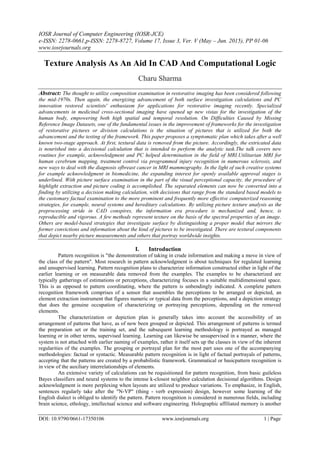
  • 2. Texture analysis as an aid in CAD and computational logic DOI: 10.9790/0661-17350106 www.iosrjournals.org 2 | Page sort of example coordinating where an extensive arrangement of scholarly examples in view of psychological meta-weight is traversed for a little arrangement of target examples. II. Specific Uses Figure 1: Faces recognized distinctly among other elements. The face was consequently recognized by unique programming set. In case of medicinal science, pattern recognition is the premise for computer aided diagnostic (CAD) frameworks. CAD portrays a methodology that backs up a specialist's translations and discoveries. Some specific applications are programmed speech recognition software, characterization of content into a few classifications (e.g. spam/non- spam email messages), the programmed acknowledgment of written by hand postal codes on postal envelopes, or the programmed acknowledgment of pictures of human appearances. The last two cases shape the subtopic picture examination of pattern recognition that makes arrangements with computerized pictures as information to pattern recognition frameworks. III. Role Of Image Texture Analysis The concept of ‘computer aided diagnosis’ (which is also called CAD) refers to software that analyses a radiographic finding to estimate the likelihood that the feature represents a specific disease process (e.g. benign versus malignant). To my knowledge, this technology has not yet been approved for clinical use, specifically depending more on biopsy tests done manually on sample tissues which usually is a very tedious task. Currently, methods of image texture analysis are undergoing great development and utilization within the field of medical imaging. Given the general interest and striking growth in computer-aided diagnosis (CAD), the application of texture analysis in the diagnostic interpretation of radiologic images has become a rapidly expanding field of research. Proposed diagnostic scheme follows a familiar two-step approach. Initially, textural information is extracted from the image. Subsequently, the extracted information is fed into a decisional algorithm (eg. an artificial neural network) that is designed to perform the diagnostic task. However, from a methodological point of view, the main attraction of this study is that the combination of image texture analysis and automated decision making offers a promising approach to a clinical challenge. Successful applications of the above CAD strategy have been reported for other types of medical images. While further development and testing is required to establish the true clinical effect of such decisionsupport systems, texture analysis appears to open a new exciting path in the journey toward CAD in radiology. Consequently, two questions are raised: What is the realistic contribution of texture analysis in the computer-aided interpretation of medical images, and to what extent can it be expected to improve interpretative accuracy?
  • 3. Texture analysis as an aid in CAD and computational logic DOI: 10.9790/0661-17350106 www.iosrjournals.org 3 | Page IV. Formulation Of The Diagnosis The symptomatic translation of therapeutic pictures is a multifaceted assignment. Its goal is the exact recognition and exact portrayal of potential variations from the norm, animportant stride toward the foundation of powerful treatment. Accomplishing this objective depends on the radiologists' effective mix of two particular procedures: (a) the procedure of picture observation to perceive special picture patterns and (b) the procedure of thinking to recognize connections between observed patterns and conceivable conclusions. Both procedures depend intensely on the radiologists' exact learning, memory, instinct, and tirelessness. Certainly, the radiologists approach the analytic undertaking with a level of insight, adaptability, and an ability to think that is hard to copy with a computer brain. In spite of many years of concentrated research in biological visual frameworks and perceptual insight, there is a constrained comprehension and a continuous level headed discussion about the fundamental components that underlie visual observation. Interestingly, there is general understanding that composition is a rich wellspring of visual data and is a key part in image investigation and perception in people. Textureimportantly provides pieces of information about picturesque profundity and texture introduction and, in that capacity, depicts the substance of both naturally produced and manufactured images. Likewise, there is confirmation of perceptual adaption in composition coding systems and in textural separation. In light of this, specialists have centered their consideration on creating calculations that can evaluate the textural properties of a picture. Exact proof and analysts' innovativeness have prompted various calculations for textureevaluation. The accessible calculations normally contrast in the sort of picture data that is caught and in the coding instrument. The development and differences of accessible systems for surface examination are a demonstration of the headway of this field. A broad review of textural definitions, models, and diagnostic calculations can be discovered somewhere else. Composition examination is at last concerned with robotized techniques that can get image data from an absolutely computational perspective. As being what is indicated, it is just another kind of numeric control of advanced or digitized pictures to get quantitative estimations. In any case, in opposition to the segregation of morphologic data, there is proof that the human visual framework experiences issues in the separation of textural data that is identified with higher-request insights or ghostly properties on a picture. Hence, texture investigation can possibly increase the visual aptitudes of the radiologist by taking into
  • 4. Texture analysis as an aid in CAD and computational logic DOI: 10.9790/0661-17350106 www.iosrjournals.org 4 | Page consideration the features of the image that may be applicable to the indicative issue however that are not so much outwardly extractable. In any case, surface examination is not a panacea for the symptomatic understanding of radiologic pictures. The quest for composition investigation is taking into account the theory that the surface mark of a picture is applicable to the indicative issue close by. Moreover, the adequacy of surface investigation is bound by the kind of calculation that is utilized to concentrate significant components. A choice calculation that uses consequently separated components has a superior possibility of creating a powerful CAD framework. A CAD apparatus that uses naturally separated elements addresses a built up clinical shortcoming of the demonstrative procedure furthermore supplements the radiologists' smart capacities. Texture investigation is appropriately suited to this issue as the radiologists themselves depend on visual composition to distinguish and portray breast injuries. The way image handling is utilized as a part of the study can be effortlessly performed with existing programming bundles significantly streamlining the fuse of the proposed CAD device into the center. The adequacy of any CAD instrument will dependably be contingent on two things: (a) how well the chosen components portray the infection that need to be separated and (b) how well the study groupreflects the general target patient population for the CAD apparatus. V. Texture Analysis’ Clinical Implementation Along With Cad Computer aided diagnostic (CAD) is an innovation intended to reduce observational oversights and in this way decline the false negative rates of doctors deciphering restorative pictures. Planned clinical studies have exhibited an increment in breast malignancy identification with CAD help. This review quickly depicts the measurements that have been utilized to characterize CAD framework execution. PC projects have been created and sanction for utilization in clinical practice that guide radiologists in identifying potential variations from the norm on indicative radiology exams. This application has been termed PC aided location, usually alluded to as CAD. The term 'computer aided diagnostic’ alludes to pattern recognition programming that recognizes suspicious components on the picture and conveys them to the consideration of the radiologist, keeping in mind the end goal to decline false negative readings. As presently utilized, the radiologist first audits the exam, then initiates the CAD programming and re- assesses the CAD-stamped regions of concern before issuing the last report. CAD is as of now FDA and CE sanctioned for utilization with both film and advanced mammography, for both screening and analytic exams; for midsection CT; and, for midsection radiographs. The essential objective of CAD is to build the discovery of illness by diminishing the false negative rate because of observational oversights. The utilization of a PC as opposed to a second human eyewitness has the benefit of not expanding the requests on the radiologist or prepared spectator pool. An imperative part of either approach is to expand sickness recognition without a fix effect on the review and work up rates. At last, in a few applications CAD, with its related robotized programming instruments, can possibly give work process efficiencies. CAD calculations are produced to scan for the same components that a radiologist searches for amid case audit. Hence, for breast growth on mammograms, the CAD calculations hunt down small scale calcifications and masses both hypothesized and non-theorized, design contortions and asymmetries. On midsection radiographs and CT filters, flow CAD applications look for aspiratory densities that have particular physical qualities, e.g. sphericity, that may speak to lung knobs. As anyone might expect, CAD calculations will underline features that meet the calculation necessities, however which don't speak to discoveries that the radiologist considers to warrant further examination, i.e. false CAD marks. Likewise, on occasion a genuine positive CAD mark, upon survey by the radiologist, is released as not justifying further examination. In this case, the false negative report would be the aftereffect of an interpretive—as opposed to a discernment. VI. Cad Methodology: Dealing With Textural Analysis CAD is in a far-reaching way taking into account profoundly complex pattern recognition. X-ray pictures are filtered for suspicious structures. Regularly a couple of thousand pictures are obliged to enhance the calculation. Advanced picture information are replicated to a CAD server in a DICOM-format and are arranged and investigated in a few stages. 6.1.1 Preprocessing for  Decrease of relics (bugs in pictures)  Picture commotion diminishment  Leveling (harmonization) of picture quality for clearing the picture's distinctive fundamental conditions e.g. diverse presentation parameter.
  • 5. Texture analysis as an aid in CAD and computational logic DOI: 10.9790/0661-17350106 www.iosrjournals.org 5 | Page 6.1.2 Segmentation for  Separation of distinctive structures in the picture, e.g. heart, lung, ribcage , conceivable round  Sores  Coordinating with anatomic databank 6.1.3 Structure/ROI (Region of Interest) Analyze Each distinguished area is examined exclusively for uncommon attributes:  Closeness  Frame, size and area  Reference to near to structures/ ROIs  Normal greylevel worth break down inside of a ROI  Extent of greylevels to outskirt of the structure inside the ROI 6.1.4 Evaluation/ order After the structure is broke down, every ROI is assessed exclusively (scoring) for the likelihood of a TP. Subsequently the methodology are:  Nearest-Neighbor Rule  Minimum distance classifier  Cascade Classifier  Bayesian Classifier  Multilayer perception  Radial basis function network (RBF)  SVM In the event that the recognized structures have come to a certain edge level, they are highlighted in the picture for the radiologist. Contingent upon the CAD framework these markings can be forever or provisional spared. The latter's preference is that just the markings which are endorsed by the radiologist are spared. False hits ought not to be spared, on the grounds that an examination at a later date turns out to be more troublesome then. VII. Implementation Of Cad In Accordance With Background Evaluation The CAD calculations oblige an advanced information set of the picture for examination. In the event that the picture is obtained on x-ray film, for example, a screen mammogram, the simple picture should first be digitized. On the other hand, the CAD calculations can specifically investigate pictures obtained in computerized arrangement, for example, with advanced mammography (FFDM) and CT. In present practice (and as needed by the FDA), the exam ought to first be investigated and deciphered in the typical manner. At exactly that point are the CAD imprints shown, taking after which the radiologist re-surveys those ranges that are provoked by the CAD framework. Two vital standards must be held fast to:  Current CAD frameworks don't check every noteworthy finding. Consequently, the nonappearance of a CAD check on a discovering the radiologist was worried about on his/her pre CAD audit should not hinder further assessment.  Current CAD frameworks create numerous more false CAD marks than genuine CAD marks. Thus, it is the obligation of the radiologist to figure out whether a CAD imprint warrants further assessment. A CAD framework can be assessed in a few ways, which incorporates examination of information produced in a research center or test setting, and by the effect of CAD on radiologist execution in a real clinical work on setting. ‘Standalone' affectability and specificity, this data can be acquired by watching the execution of a CAD framework on an arrangement of "truth" cases. The fact of the matter is by and large settled by histological confirmation of the vicinity (e.g. disease) or nonattendance (e.g. clinical postliminary) of malady. Affectability is dictated by the rate of positive cases in which the CAD framework puts an imprint on the malady area. The quantity of false CAD marks per typical picture or case is generally utilized as a surrogate for specificity. The aftereffects of this activity are, obviously, subject to the case accumulation. Inclination, planned or not, in gathering positive cases that have more obvious discoveries will evidently bring about better CAD execution analyzed than cases that are less prominent, despite the fact that the CAD calculation is the same. Along these lines, the same CAD calculation will exhibit changing sensitivities and specificities (false checks per case) contingent upon the case creation. A favored technique to contrast CAD frameworks is to determine the sensitivity and false marker rates on the same arrangement of "truth" cases. These cases must be "obscure" to the CAD framework, that is, they
  • 6. Texture analysis as an aid in CAD and computational logic DOI: 10.9790/0661-17350106 www.iosrjournals.org 6 | Page ought not to have been utilized to prepare the CAD calculations. Adequate and regularly extensive quantities of cases will be required so as to build up measurable noteworthiness of predominance or proportionality in execution when contrasting CAD frameworks. "Research center" investigations of potential recognition change initiate radiologists (or other 'pursuers') to assess an arrangement of "truth" cases to focus the affectability and get back to rate of the unaided pursuer (pre CAD) with that of the pursuer with CAD help. Such studies are valuable to survey the potential advantage of CAD and give evaluations of expected changes in ailment discovery and workup/review rates. In any case, the test setting regularly bargains the execution of the pursuer, in that the pursuers might either, over or under call the investigated cases in a test domain. This should be possible in a 'consecutive read' clinical trial, in which the exam is first read before, and later on taking after, CAD information. The adjustment in sickness recognition because of the CAD data, and additionally the adjustment in the review/workup rates, will focus the commitment of CAD to patient administration. Vitally, the rate increment in ailment discovery ought to be concordant with, or not exactly, the rate increment in the review/workup rate. VIII. Conclusion The thought to utilize pattern recognition in restorative imaging has been considered following the mid 1970s. On Difficulties Caused by Missing Reference Image Datasets One of the fundamental issues in the improvement of frameworks for the examination of medicinal pictures or division calculations is the situation of pictures that is utilized for both the advancement and the testing of the framework. This paper depicts the system with picture composition examination in the part of the visual perceptional capacity, the procedure of highlight extraction and picture coding is accomplished. The extricated elements can now be converted into a conclusion by utilizing a decisional calculation, with decisions that range from the tenet based models to the customary factual examination to the more prevalent and regularly more fruitful, manmade brainpower systems, for example, neural systems and genetic calculations. By utilizing image texture analysis as the preprocessing stride in CAD schemes, the input generation procedure is computerized and, accordingly, is reproducible and strong. Hitherto, discoveries have demonstrated that CAD can improve the symptomatic execution of radiologists, in the event that it is utilized as a second assessment. Be that as it may, it is still hard to foresee its acknowledgement in medicinal analysis, and its acknowledgement won't be resolved until discoveries from broad clinical utilization exhibit quantifiable advantages. In the meantime, business CAD items will need to face new difficulties identified with promoting, specialized bolster, adjustments, and item life-cycles and practices in the human services environment are always re-imagined. References [1]. Chen DR, Chang RF, Huang YL. Computer-aided diagnosis applied to US of solid breast nodules by using neural networks. [2]. D R. Brunelli, Template Matching Techniques in Computer Vision: Theory and Practice, Wiley, ISBN [3]. Fukunaga, Keinosuke. Introduction to Statistical Pattern Recognition (2nd ed.). [4]. Bishop, Christopher. Pattern Recognition and Machine Learning. [5]. Godfried T. Toussaint. Computational Morphology. Amsterdam: North-Holland Publishing Company. [6]. Kulikowski, Casimir A.; Weiss, Sholom M. Computer Systems That Learn: Classification and Prediction Methods from Statistics, Neural Nets, Machine Learning, and Expert Systems. Machine Learning. [7]. Krupinski EA. The future of image perception in radiology. [8]. Vittitoe NF, Baker JA, Floyd CE, Jr. Fractal texture analysis in computer-aided diagnosis of solitary pulmonary nodules. [9]. S Wei D, Chan HP, Helvie MA, et al. Classification of mass and normal breast tissue on digital mammograms: multiresolution texture analysis. [10]. Chan HP, Sahiner B, Petrick N, et al. Computerized classification of malignant and benign microcalcifications on mammograms: texture analysis using an artificial neural network. [11]. Petrick N, Chan HP, Wei D, Sahiner B, Helvie MA, Adler DD. Automated detection of breast masses on mammograms using adaptive contrast enhancement and texture classification. [12]. McPherson DD, Aylward PE, Knosp BM, et al. [13]. Ultrasound characterization of acute myocardial ischemia by quantitative texture analysis. [14]. Lucht R, Brix G, Lorenz WJ. Texture analysis of differently reconstructed PET images.