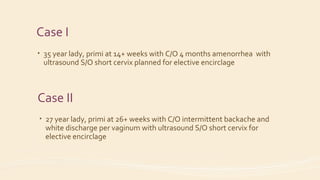
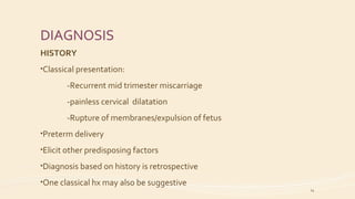
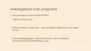
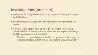
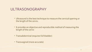
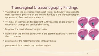
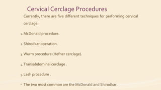
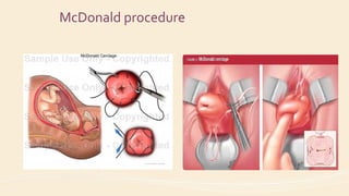
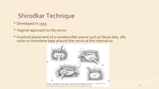
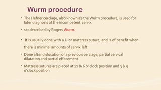
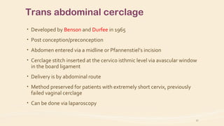
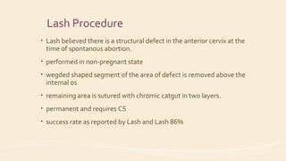
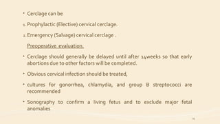
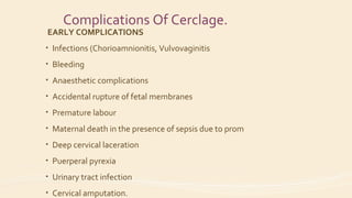
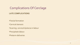
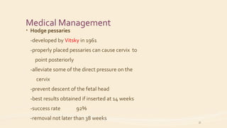
Cervical Incompetence
By Dr. Indraneel Jadhav
Case I
• 35 year lady, primi at 14+ weeks with C/O 4 months amenorrhea with
ultrasound S/O short cervix planned for elective encirclage
Case II
• 27 year lady, primi at 26+ weeks with C/O intermittent backache and
white discharge per vaginum with ultrasound S/O short cervix for
elective encirclage
Case I
• Spontaneous conception. Excellent dates. Taking Folic Acid
• Blood investigations done were normal
• At two and half month recorded high BP recording, persistent on
subsequent ANC visit, Hence started on Labetolol 50mg 1-0-1.
• At 3rd
month was admitted for threatened abortion due to episode of
bleeding – managed conservatively.
• Scan done was normal along with blood test S/O low risk.
• Wanted to continue further ANC’s at Mumbai hence scan done S/O short
cervix.
• N/H/O Excessive vomiting, fever with rash, burning micturation, white
discharge per vaginum, radiation exposure
• Medical/ surgical history: k/c/o Hypothyroidism on Thyronorm 100 mcg
1-0-0 .H/O hospitalisation for fever on and off 1 year back – told to have
urinary infection .
• Menstrual history: Cycles regular; LMP: 13/3/17; EDD: 20/12/17;
• Obstetric history: Married since 2 years (non – consanguineous )
,voluntary sub fertility, primigravida
• Family history: Father –DM + HTN , Mother – HTN and thyroid
• Personal History : Not significant
• Dietary history : (sedentary – 2400 kcal) calorie defecit -650 kcal
Protein defecit – 7 gm
Examination
• GC Fair
• Height: 154 cm ; Weight: 38Kg BMI : 24 kg/m2
• Vitals stable ,No pallor, icterus, pedal edema
• Cardiovascular system: S1 and S2 heard, no added sounds
• Respiratory system: B/L NVBS heard, no added sounds
• P/A:Inspection : linea nigra seen, No scars, sinuses or dilated veins
Palpation: Uterus just palpable ,No tenderness ,organomegaly, ascites
Case II
• Spontaneous conception. Excellent dates. Folic acid taken
• Blood investigations done S/O Thyroid abnormality, started on
thyronorm 25 mcg 1-0-0
• Scan done was normal at 3rd
month along with blood test S/O low risk.
Started on Iron/Calcium
• 5th
month scan done found to be normal.
• At 6th
month came with complaints of low intermittent backache and
white discharge per vaginum,hence ultrasound done S/O short cervix
• N/H/O Excessive vomiting, fever with rash, burning micturation, white
discharge/bleeding per vaginum, radiation exposure,high BP recordings
or high sugars.
• Medical/ surgical history:.H/O appendicectomy 4 years back
• Menstrual history: Cycles regular; LMP: 28/12/16; EDD: 02/10/17;
• Obstetric history: Married since 6 months (non – consanguineous )
primigravida
• Family history: Father –DM + HTN , Mother – HTN and thyroid
• Personal History : Not significant
• Dietary history : (sedentary – 2400 kcal) calorie defecit -478 kcal
Protein defecit – 6 gm
Examination
• GC Fair
• Height: 140 cm ; Weight: 41Kg BMI : 20.4 kg/m2
• Vitals stable ,No pallor, icterus, pedal edema
• Cardiovascular system: S1 and S2 heard, no added sounds
• Respiratory system: B/L NVBS heard, no added sounds
• P/A:Inspection : linea nigra seen, Appendicectomy scar seen, sinuses or
dilated veins
Palpation: Uterus 24-26 wks, EB +,No tenderness ,organomegaly, ascites
Definition
Cervical incompetence is the inability
for the cervix to retain an intra-
uterine pregnancy till term as a result
of structural and functional defects of
the cervix.
9
Anatomy
10
-Internal os: the opening of the
cervix into the body of uterus.
-External os – its size and shape
varies widely with age, hormonal
state, and whether the woman has
had a vaginal birth. In nonparous
women it appears as a small,
circular opening. Versus being
fish mouthed (meaning wider,
more slit-like and gaping) in
parous women.
In pregnancy, the normal cervical
length is
4 + 1cm.
Pathogenesis/ Aetiology
The cervical competence is a specific entity involving not just an
abnormality or defect of cervical collagen, but is also due to either:
1. Absence of the usual cervical musculature in cases of congenital
cervical incompetence, or
2. Injury or damage to the cervical musculature caused by previous
trauma.
11
Aetiology
- Unknown, Congenital and Acquired.
- Congenital causes include:
- congenital mullerian duct abnormalities (e.g. septate uterus,
bicornuate uterus)
- Connective tissue disorder (Ehlers-Danlos syndrome)
- DES exposure in utero
12
• Acquired causes include:
Surgical trauma
 Cone biopsy or loop electrosurgical excision procedures
 Repeated forced cervical dilatation asso. with 2nd
trimester D&C
 Previous Manchester repair with cervical amputation.
Obstetric injuries
 Compression necrosis of the cervix due to prolonged 2nd
stage of
labor
 Spontaneous and iatrogenic cervical laceration (e.g. Duhrssen’s
incision)
 Extension of the uterine incision into the cervix during CS
13
DIAGNOSIS
HISTORY
•Classical presentation:
-Recurrent mid trimester miscarriage
-painless cervical dilatation
-Rupture of membranes/expulsion of fetus
•Preterm delivery
•Elicit other predisposing factors
•Diagnosis based on history is retrospective
•One classical hx may also be suggestive
14
Investigations (non-pregnant)
• Easy passage of a size 8 Hegar’s/Pratt’s
dilator number 15-17
• Foley’s catheter traction test - size 16 F balloon filled with 1ml of water
(6mm)
• Hysterosalpingography- dilated internal os > 8mm /widened
isthmus( funnel/ Inverted Bishop’s cap)
15
Investigations (pregnant)
• Weekly or forthnightly cervical assessment- softening effacement
and dilatation.
• Serial ultrasound assessment of the lower uterine segment and
cervix.
• Cervical stress test usually done at 15 –24 weeks (increasing
transfundal intrauterine pressure while monitoring cervical length
and the appearance of funnelling).
• This test is currently recommended for patients with suspected
features of incompetent cervix, undergoing ultrasonography.
16
ULTRASONOGRAPHY
• Ultrasound is the best technique to measure the cervical opening or
the length of the cervix.
• It provides an objective and reproducible method of measuring the
length of the cervix
• Transabdominal (requires full bladder)
• Transvaginal (more accurate)
17
Transvaginal Ultrasonography Findings
• ‘Funneling’ of the internal cervical os (at rest or particularly in respond to
transabdominal pressure on the uterine fundus) is the ultrasonographic
appearance of cervical incompetence.
• Y= initial effacement and subsequent V, U visualized on progressive
endocervial change and cervical shortening.
• length of the cervical canal: <2.5cm – risk
• diameter of the internal os:>15 mm in the 1st trimester and > 20mm in
the 2nd
trimester
• protrusion of the fetal membranes through the os
• presence of fetal parts in the cervix or vagina
18
Transvaginal Sonography
• TVS in contrast to HSG, is
 Non-invasive
 Repeatable over time
 Can be performed during pregnancy
• Negative USS can not exclude CI
• Positive USS during routine screening in pregnant women without
history of pregnancy loss do not necessarily place them at risk, but close
follow up is required.
19
Investigations (Cont’d)
• In some situation it may be necessary to conduct test to exclude other
causes of recurrent pregnancy losses-
-Diabetes mellitus
-Thyroid dysfuntion
-Lupus anticoagulant
-Chromosonal anomalies
-Cervical infection
• The role of magnetic resonance imaging- cost prohibitive
20
CERVICAL CERCLAGE
• Evidence-based procedure that reduces perinatal and maternal
morbidity and mortality arising from cervical incompetence.
• The success rate can be high (80-90%) when done early in pregnancy.
• Usually done prior to 18 weeks, but after 13 weeks. Many say between
14-16 weeks.
21
Cervical Cerclage
INDICATIONS FOR CERCLAGE
•Suspected/confirmed cervical incompetence remains the only acceptable
indication for cervical cerclage.
CONTRA-INDICATIONS
• Uterine contractions
• Uterine bleeding
• Chorioamnionitis
• Premature rupture of membranes.
• Fetal anomaly incompatible with life.
22
Cervical Cerclage Procedures
Currently, there are five different techniques for performing cervical
cerclage:
1. McDonald procedure.
2. Shirodkar operation.
3. Wurm procedure (Hefner cerclage).
4. Transabdominal cerclage .
5. Lash procedure .
• The two most common are the McDonald and Shirodkar.
McDonald procedure
Shirodkar Technique
 Developed in 1955
 Vaginal approach to the cervix
 Involved placement of a nonabsorble suture such as fascia lata, silk,
nylon or mersilene tape around the cervix at the internal os.
25
Wurm procedure
• The Hefner cerclage, also known as the Wurm procedure, is used for
later diagnosis of the incompetent cervix.
• 1st described by Rogers Wurm.
• It is usually done with a U or mattress suture, and is of benefit when
there is minimal amounts of cervix left.
• Done after dislocation of a previous cerclage, partial cervical
dilatation and partial effacement
• Mattress sutures are placed at 12 & 6 o’ clock position and 3 & 9
o’clock position
Trans abdominal cerclage
• Developed by Benson and Durfee in 1965
• Post conception/preconception
• Abdomen entered via a midline or Pfannenstiel’s incision
• Cerclage stitch inserted at the cervico isthmic level via avascular window
in the board ligament
• Delivery is by abdominal route
• Method preserved for patients with extremely short cervix, previously
failed vaginal cerclage
• Can be done via laparoscopy
27
Lash Procedure
• Lash believed there is a structural defect in the anterior cervix at the
time of spontanous abortion.
• performed in non-pregnant state
• wegded shaped segment of the area of defect is removed above the
internal os
• remaining area is sutured with chromic catgut in two layers.
• permanent and requires CS
• success rate as reported by Lash and Lash 86%
• Cerclage can be
1. Prophylactic (Elective) cervical cerclage.
2. Emergency (Salvage) cervical cerclage .
Preoperative evaluation.
• Cerclage should generally be delayed until after 14weeks so that early
abortions due to other factors will be completed.
• Obvious cervical infection should be treated,
• cultures for gonorrhea, chlamydia, and group B streptococci are
recommended
• Sonography to confirm a living fetus and to exclude major fetal
anomalies
29
A. Prophylactic/Elective Cerclage
• A planned cerclage placement after history, examination and
investigations have been done.
• It may be placed prior to pregnancy, but is more commonly
placed between 14 -16 weeks’ gestation.
• The stitch is usually removed around 37 weeks or at the onset of
labour.
B. Emergency Cerclage (cont’d)
 Refers to placement of a cerclage in the setting of significant cervical
dilatation and/or effacement prior to 28 weeks’ gestation and in the
absence of labor.
 Preserved for patients without classical features incompetence.
 Patient experiencing features of incompetence in an index
pregnancy: prolapse of membranes, cervical dilatation and
effacement
 Success rate lower than that of elective.
 Higher incidence of infection
 Prolonged hospital stay
 The prognosis is influenced by the gestational age at the time when
the suture is placed.
The bulging membrane during emergency
cerclage
• Management options
• Insertion of a foley’s catheter with 20ml balloon with the distal cut
end inserted into the cervical canal and inflated.
• Pre cerclage amniocentesis to remove sufficient fluid to reduce the
bulging membranes can be helpful.
• Overfilling the bladder with 1,000 ml of saline may help by elevating
the membranes out of the operative field.
• Use of 6-10 stay stitches attached to the edges of the cervix with the
patient in deep trendelenburg position. Traction pushes back the
membrane
32
Post-operatively,
• Antibiotics
• Tocolytics
-controversial except for patients with uterine irritability.
• Bed rest advised for the 1st
24 hours followed by mobilization and
activity
• Discharge after a couple of days advised - studies
have found no benefit for staying more than one week.
Removal of cerclage
• Timing: usually b/w 37-38 weeks
• Earlier removal
-excessive vaginal bleeding
-intrauterine fetal death
-persistent uterine contraction
-Rupture of fetal membranes
-chorioamnionitis
34
Complications Of Cerclage.
EARLY COMPLICATIONS
• Infections (Chorioamnionitis, Vulvovaginitis
• Bleeding
• Anaesthetic complications
• Accidental rupture of fetal membranes
• Premature labour
• Maternal death in the presence of sepsis due to prom
• Deep cervical laceration
• Puerperal pyrexia
• Urinary tract infection
• Cervical amputation.
Complications Of Cerclage
LATE COMPLICATIONS
•Fistula formation
•Cervical stenosis
•Scarring- cervical dystocia in labour
•Precipitate labour.
•Preterm deliveries
36
Medical Management
• Hodge pessaries
-developed by Vitsky in 1961
-properly placed pessaries can cause cervix to
point posteriorly
-alleviate some of the direct pressure on the
cervix
-prevent descent of the fetal head
-best results obtained if inserted at 14 weeks
-success rate 92%
-removal not later than 38 weeks
37
Medical Management
• Baylor Balloon
-Proposed in 1972
-Double silicon plastic cuff inserted on
cervix to act as cuff.
• Progesterone
-Reduces uterine tone
38

Cervical insufficiency

  • 1.
  • 2.
    Case I • 35year lady, primi at 14+ weeks with C/O 4 months amenorrhea with ultrasound S/O short cervix planned for elective encirclage Case II • 27 year lady, primi at 26+ weeks with C/O intermittent backache and white discharge per vaginum with ultrasound S/O short cervix for elective encirclage
  • 3.
    Case I • Spontaneousconception. Excellent dates. Taking Folic Acid • Blood investigations done were normal • At two and half month recorded high BP recording, persistent on subsequent ANC visit, Hence started on Labetolol 50mg 1-0-1. • At 3rd month was admitted for threatened abortion due to episode of bleeding – managed conservatively. • Scan done was normal along with blood test S/O low risk. • Wanted to continue further ANC’s at Mumbai hence scan done S/O short cervix. • N/H/O Excessive vomiting, fever with rash, burning micturation, white discharge per vaginum, radiation exposure
  • 4.
    • Medical/ surgicalhistory: k/c/o Hypothyroidism on Thyronorm 100 mcg 1-0-0 .H/O hospitalisation for fever on and off 1 year back – told to have urinary infection . • Menstrual history: Cycles regular; LMP: 13/3/17; EDD: 20/12/17; • Obstetric history: Married since 2 years (non – consanguineous ) ,voluntary sub fertility, primigravida • Family history: Father –DM + HTN , Mother – HTN and thyroid • Personal History : Not significant • Dietary history : (sedentary – 2400 kcal) calorie defecit -650 kcal Protein defecit – 7 gm
  • 5.
    Examination • GC Fair •Height: 154 cm ; Weight: 38Kg BMI : 24 kg/m2 • Vitals stable ,No pallor, icterus, pedal edema • Cardiovascular system: S1 and S2 heard, no added sounds • Respiratory system: B/L NVBS heard, no added sounds • P/A:Inspection : linea nigra seen, No scars, sinuses or dilated veins Palpation: Uterus just palpable ,No tenderness ,organomegaly, ascites
  • 6.
    Case II • Spontaneousconception. Excellent dates. Folic acid taken • Blood investigations done S/O Thyroid abnormality, started on thyronorm 25 mcg 1-0-0 • Scan done was normal at 3rd month along with blood test S/O low risk. Started on Iron/Calcium • 5th month scan done found to be normal. • At 6th month came with complaints of low intermittent backache and white discharge per vaginum,hence ultrasound done S/O short cervix • N/H/O Excessive vomiting, fever with rash, burning micturation, white discharge/bleeding per vaginum, radiation exposure,high BP recordings or high sugars.
  • 7.
    • Medical/ surgicalhistory:.H/O appendicectomy 4 years back • Menstrual history: Cycles regular; LMP: 28/12/16; EDD: 02/10/17; • Obstetric history: Married since 6 months (non – consanguineous ) primigravida • Family history: Father –DM + HTN , Mother – HTN and thyroid • Personal History : Not significant • Dietary history : (sedentary – 2400 kcal) calorie defecit -478 kcal Protein defecit – 6 gm
  • 8.
    Examination • GC Fair •Height: 140 cm ; Weight: 41Kg BMI : 20.4 kg/m2 • Vitals stable ,No pallor, icterus, pedal edema • Cardiovascular system: S1 and S2 heard, no added sounds • Respiratory system: B/L NVBS heard, no added sounds • P/A:Inspection : linea nigra seen, Appendicectomy scar seen, sinuses or dilated veins Palpation: Uterus 24-26 wks, EB +,No tenderness ,organomegaly, ascites
  • 9.
    Definition Cervical incompetence isthe inability for the cervix to retain an intra- uterine pregnancy till term as a result of structural and functional defects of the cervix. 9
  • 10.
    Anatomy 10 -Internal os: theopening of the cervix into the body of uterus. -External os – its size and shape varies widely with age, hormonal state, and whether the woman has had a vaginal birth. In nonparous women it appears as a small, circular opening. Versus being fish mouthed (meaning wider, more slit-like and gaping) in parous women. In pregnancy, the normal cervical length is 4 + 1cm.
  • 11.
    Pathogenesis/ Aetiology The cervicalcompetence is a specific entity involving not just an abnormality or defect of cervical collagen, but is also due to either: 1. Absence of the usual cervical musculature in cases of congenital cervical incompetence, or 2. Injury or damage to the cervical musculature caused by previous trauma. 11
  • 12.
    Aetiology - Unknown, Congenitaland Acquired. - Congenital causes include: - congenital mullerian duct abnormalities (e.g. septate uterus, bicornuate uterus) - Connective tissue disorder (Ehlers-Danlos syndrome) - DES exposure in utero 12
  • 13.
    • Acquired causesinclude: Surgical trauma  Cone biopsy or loop electrosurgical excision procedures  Repeated forced cervical dilatation asso. with 2nd trimester D&C  Previous Manchester repair with cervical amputation. Obstetric injuries  Compression necrosis of the cervix due to prolonged 2nd stage of labor  Spontaneous and iatrogenic cervical laceration (e.g. Duhrssen’s incision)  Extension of the uterine incision into the cervix during CS 13
  • 14.
    DIAGNOSIS HISTORY •Classical presentation: -Recurrent midtrimester miscarriage -painless cervical dilatation -Rupture of membranes/expulsion of fetus •Preterm delivery •Elicit other predisposing factors •Diagnosis based on history is retrospective •One classical hx may also be suggestive 14
  • 15.
    Investigations (non-pregnant) • Easypassage of a size 8 Hegar’s/Pratt’s dilator number 15-17 • Foley’s catheter traction test - size 16 F balloon filled with 1ml of water (6mm) • Hysterosalpingography- dilated internal os > 8mm /widened isthmus( funnel/ Inverted Bishop’s cap) 15
  • 16.
    Investigations (pregnant) • Weeklyor forthnightly cervical assessment- softening effacement and dilatation. • Serial ultrasound assessment of the lower uterine segment and cervix. • Cervical stress test usually done at 15 –24 weeks (increasing transfundal intrauterine pressure while monitoring cervical length and the appearance of funnelling). • This test is currently recommended for patients with suspected features of incompetent cervix, undergoing ultrasonography. 16
  • 17.
    ULTRASONOGRAPHY • Ultrasound isthe best technique to measure the cervical opening or the length of the cervix. • It provides an objective and reproducible method of measuring the length of the cervix • Transabdominal (requires full bladder) • Transvaginal (more accurate) 17
  • 18.
    Transvaginal Ultrasonography Findings •‘Funneling’ of the internal cervical os (at rest or particularly in respond to transabdominal pressure on the uterine fundus) is the ultrasonographic appearance of cervical incompetence. • Y= initial effacement and subsequent V, U visualized on progressive endocervial change and cervical shortening. • length of the cervical canal: <2.5cm – risk • diameter of the internal os:>15 mm in the 1st trimester and > 20mm in the 2nd trimester • protrusion of the fetal membranes through the os • presence of fetal parts in the cervix or vagina 18
  • 19.
    Transvaginal Sonography • TVSin contrast to HSG, is  Non-invasive  Repeatable over time  Can be performed during pregnancy • Negative USS can not exclude CI • Positive USS during routine screening in pregnant women without history of pregnancy loss do not necessarily place them at risk, but close follow up is required. 19
  • 20.
    Investigations (Cont’d) • Insome situation it may be necessary to conduct test to exclude other causes of recurrent pregnancy losses- -Diabetes mellitus -Thyroid dysfuntion -Lupus anticoagulant -Chromosonal anomalies -Cervical infection • The role of magnetic resonance imaging- cost prohibitive 20
  • 21.
    CERVICAL CERCLAGE • Evidence-basedprocedure that reduces perinatal and maternal morbidity and mortality arising from cervical incompetence. • The success rate can be high (80-90%) when done early in pregnancy. • Usually done prior to 18 weeks, but after 13 weeks. Many say between 14-16 weeks. 21
  • 22.
    Cervical Cerclage INDICATIONS FORCERCLAGE •Suspected/confirmed cervical incompetence remains the only acceptable indication for cervical cerclage. CONTRA-INDICATIONS • Uterine contractions • Uterine bleeding • Chorioamnionitis • Premature rupture of membranes. • Fetal anomaly incompatible with life. 22
  • 23.
    Cervical Cerclage Procedures Currently,there are five different techniques for performing cervical cerclage: 1. McDonald procedure. 2. Shirodkar operation. 3. Wurm procedure (Hefner cerclage). 4. Transabdominal cerclage . 5. Lash procedure . • The two most common are the McDonald and Shirodkar.
  • 24.
  • 25.
    Shirodkar Technique  Developedin 1955  Vaginal approach to the cervix  Involved placement of a nonabsorble suture such as fascia lata, silk, nylon or mersilene tape around the cervix at the internal os. 25
  • 26.
    Wurm procedure • TheHefner cerclage, also known as the Wurm procedure, is used for later diagnosis of the incompetent cervix. • 1st described by Rogers Wurm. • It is usually done with a U or mattress suture, and is of benefit when there is minimal amounts of cervix left. • Done after dislocation of a previous cerclage, partial cervical dilatation and partial effacement • Mattress sutures are placed at 12 & 6 o’ clock position and 3 & 9 o’clock position
  • 27.
    Trans abdominal cerclage •Developed by Benson and Durfee in 1965 • Post conception/preconception • Abdomen entered via a midline or Pfannenstiel’s incision • Cerclage stitch inserted at the cervico isthmic level via avascular window in the board ligament • Delivery is by abdominal route • Method preserved for patients with extremely short cervix, previously failed vaginal cerclage • Can be done via laparoscopy 27
  • 28.
    Lash Procedure • Lashbelieved there is a structural defect in the anterior cervix at the time of spontanous abortion. • performed in non-pregnant state • wegded shaped segment of the area of defect is removed above the internal os • remaining area is sutured with chromic catgut in two layers. • permanent and requires CS • success rate as reported by Lash and Lash 86%
  • 29.
    • Cerclage canbe 1. Prophylactic (Elective) cervical cerclage. 2. Emergency (Salvage) cervical cerclage . Preoperative evaluation. • Cerclage should generally be delayed until after 14weeks so that early abortions due to other factors will be completed. • Obvious cervical infection should be treated, • cultures for gonorrhea, chlamydia, and group B streptococci are recommended • Sonography to confirm a living fetus and to exclude major fetal anomalies 29
  • 30.
    A. Prophylactic/Elective Cerclage •A planned cerclage placement after history, examination and investigations have been done. • It may be placed prior to pregnancy, but is more commonly placed between 14 -16 weeks’ gestation. • The stitch is usually removed around 37 weeks or at the onset of labour.
  • 31.
    B. Emergency Cerclage(cont’d)  Refers to placement of a cerclage in the setting of significant cervical dilatation and/or effacement prior to 28 weeks’ gestation and in the absence of labor.  Preserved for patients without classical features incompetence.  Patient experiencing features of incompetence in an index pregnancy: prolapse of membranes, cervical dilatation and effacement  Success rate lower than that of elective.  Higher incidence of infection  Prolonged hospital stay  The prognosis is influenced by the gestational age at the time when the suture is placed.
  • 32.
    The bulging membraneduring emergency cerclage • Management options • Insertion of a foley’s catheter with 20ml balloon with the distal cut end inserted into the cervical canal and inflated. • Pre cerclage amniocentesis to remove sufficient fluid to reduce the bulging membranes can be helpful. • Overfilling the bladder with 1,000 ml of saline may help by elevating the membranes out of the operative field. • Use of 6-10 stay stitches attached to the edges of the cervix with the patient in deep trendelenburg position. Traction pushes back the membrane 32
  • 33.
    Post-operatively, • Antibiotics • Tocolytics -controversialexcept for patients with uterine irritability. • Bed rest advised for the 1st 24 hours followed by mobilization and activity • Discharge after a couple of days advised - studies have found no benefit for staying more than one week.
  • 34.
    Removal of cerclage •Timing: usually b/w 37-38 weeks • Earlier removal -excessive vaginal bleeding -intrauterine fetal death -persistent uterine contraction -Rupture of fetal membranes -chorioamnionitis 34
  • 35.
    Complications Of Cerclage. EARLYCOMPLICATIONS • Infections (Chorioamnionitis, Vulvovaginitis • Bleeding • Anaesthetic complications • Accidental rupture of fetal membranes • Premature labour • Maternal death in the presence of sepsis due to prom • Deep cervical laceration • Puerperal pyrexia • Urinary tract infection • Cervical amputation.
  • 36.
    Complications Of Cerclage LATECOMPLICATIONS •Fistula formation •Cervical stenosis •Scarring- cervical dystocia in labour •Precipitate labour. •Preterm deliveries 36
  • 37.
    Medical Management • Hodgepessaries -developed by Vitsky in 1961 -properly placed pessaries can cause cervix to point posteriorly -alleviate some of the direct pressure on the cervix -prevent descent of the fetal head -best results obtained if inserted at 14 weeks -success rate 92% -removal not later than 38 weeks 37
  • 38.
    Medical Management • BaylorBalloon -Proposed in 1972 -Double silicon plastic cuff inserted on cervix to act as cuff. • Progesterone -Reduces uterine tone 38