SlideShare a Scribd company logo
1 of 18
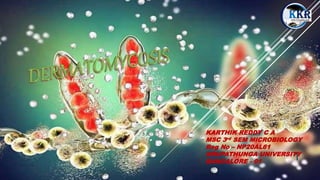
KARTHIK REDDY C A
MSC 3rd SEM MICROBIOLOGY
Reg No – NP20AL61
NRUPATHUNGA UNIVERSITY
BANGALORE - 01
KKR1116
1
INTRODUCTION
Dermatomycosis is one of the most frequent fungal
infections of skin and skin appendages, which
encompass nails and hair.
It is a mycotic diseases of skin caused by a few mycetes :
Dermatophytes and some opportunistic fungi.
These fungal infections impair superficial layers of the
skin, hair and nails.
Dermatomycosis mainly caused by filamentous fungi.
KKR1116
2
DERMATOPHYTES
• Dermatophytes are a common label for a group of fungus of arthrodermaceae
that commonly causes skin disease in animals and humans.
• These anamorphic mold genera are : Microsporum, Epidermophyton and
trichophyton.
• These cause infections of skin, hair, and nails, obtaining nutrients from
keratinized material.
• The organisms colonize the keratin tissues causing inflammation as the host
responds to metabolic byproducts.
KKR1116
3
EPIDERMIOLOGY
Dermatophyte fungi are the most common fungal infections world wide.
Dermatophytosis was first described by David Gruby, a Hungarian
physician in 1841.
Before Gruby, various scientist described lesions which were ring like and
were thought to be infective.
The description of lesions dates back to the Roman era.
Around 1890, Raimond sabouraud advance knowledge of
dermatomycology by studying extensively into the taxonomy, morphology
and treatment of dermatophytes even classifying these fungal agents into 4
genera.
Dermatophytosis has been prevalent since early 1900’S at which time
ringworm was treated with compounds of mercury or sometimes sulfur or
iodine.
KKR1116
4
MORPHOLOGY
• Hyphae of dermatophytes are long, undulant and branching.
• Many septa are present along the length of hyphae.
• Hyphae break at the septa into barrel shaped arthrospores.
CULTURALCHARACTERS
• In culture, dermatophytes form conidiophores with resulting microconidia and macroconidia.
• Genera and Species identification is based on gross characteristic of colony and microscopic
morphology of conidia.
KKR1116
5
CHARACTERISTICS
• Dermatophytes are a group of about 40 related fungi that belongs to three genera:
1. Microsporum
2. Trichophyton
3. Epidermophyton
• They are restricted to Non- viable skin because most are unable to grow at 37°C or in the presence of
Serum.
• Many species have particular keratinase, Elastase and other enzymes which make them host specific.
• Several are capable of sexual reproduction – produce ascospores. Thus belongs to genus
Arthroderma.
• In skin, they produce hyaline, septate, branching hyphae or chains of arthroconidia.
• Epidermophyton Flucosum is the only pathogen in this genus which produces macroconidia.
• They are highly contagious and frequently transmitted by exposure to shed scale, nails, hairs
containing hyphae and conidia.
• They remain viable for long periods on fomites.
KKR1116
6
SYMPTOMS
o The first symptom is a distinctive skin rash on the face, eyelids,
chest, nails, cuticle areas, knees or elbows.
o The rash is patchy and usually a bluish to purple colour.
o May also get muscle weakness that gets work over weeks or
months. This muscle weakness usually starts in neck, arms, or
hips and can be felt on both sides of body.
o Other symptoms are –
o Muscle pain and tenderness
o Swallowing problems and lung problems
o Hard calcium deposits underneath, the skin which is mostly seen
in children.
o Fatigue, unintentional weight loss, fever.
There is a subtype of dermatomyositis that includes the rash but
not muscle weakness. This is know as amyopathic dermatomyositis.
KKR1116
7
LABORATORYDIAGNOSIS
• MRI is used to observe abnormal muscles.
• Electromyography ( EMG ) to record electrical impulses that control
muscles.
• Blood analysis to check the levels of muscle enzymes and
autoantibodies, which are antibodies that attack normal cells.
• A muscle biopsy to look for inflammation and other problems
associated with the disease in a sample of muscle tissue.
• A skin biopsy to look for changes caused by the disease in a skin
sample.
KKR1116
8
TREATMENT
• Tinea corpora ( body ), tinea manus (hands), tinea cruris ( groin ), tinea pedis ( foot ) and tinea
facie ( face ) can be treated topically.
• Tinea unguiem ( nails ) usually will require oral treatment with terbinafine, itraconazole, or
griscofulvin.
• Tinea capitis ( scalp ) must be treated orally, as the medication must be present deep in the hair
follicles to eradicate the fungus. Griseofulvin is given orally for 2 to 3 months.
• Tinea pedis is usually treated with topical medicines, like ketoconazole or terbinafine and pills
or with medicine that contains miconazole, clotrimazole or tolnaftate.
• Antibiotics may be necessary to treat secondary bacterial infections that occur in addition to the
fungus.
KKR1116
9
Epidermophyton
• Epidermophyton floccosum is a filamentous dermatophytic fungus known to cause
skin and nail infections in humans.
• It is an anthropophilic dermatophyte hence it is transmissible from one individual to
another.
• Common infections caused by dermatophytes include tinea pedis (athlete’s foot),
tinea crusis, tinea corporis, and onychomycosis.
• It is the third most common cause of Tinea pedis (athletes foot).
KKR1116
10
Habitat of Epidermophyton floccosum
•It is an ascomycete and therefore it has a worldwide distribution.
•It commonly occurs in tropical and subtropical regions.
•It does not live in the soil.
•It is common in North America and Asian countries.
KKR1116
11
Morphological and Cultural Characteristics of E. Floccosum
o Growth in basic mycological medium, Epidermophyton floccosum grows slowly, producing greenish-
brown or khaki-colored colonies, with a suede-like surface.
o The colonies have a centralized occurrence that is raised and folded, a flat periphery, and submerged
fringe growth.
o Mature cultures produce white pleomorphic tufts of mycelium with a deep yellowish-brown reverse
pigment.
o Microscopic observation shows smooth, thin-walled macroconidia occurring in clusters on the hyphal
threads.
o They are also observed to be filamentous fungus, with septate and hyaline hyphae.
o The hyphae contain smooth, whin-walled, clavate, club-shaped, clusters macroconidia.
o Chlamydospores are also formed in mature/old cultures.
o They do not form microconidia.
o The distinction of Epidermophytes floccosum from the other dermatophytes
(Microsporum and Trichophyton)is that they do not form microconidia and they form macroconidia
which are shorter, wide, and smooth.
KKR1116
12
Figure: Pleomorphism of E. floccosum. A: Overview of the Wild-Type surface; B: Revise
side of wild-type; C: Overview of pleomorphic E. floccosum; D: Revise side of
pleomorphic E. floccosum.
KKR1116
13
Pathogenesis and clinical manifestations of Epidermophyton floccosum
o Epidermophyton floccosum is transmitted from person-to-person via skin contact, causing cutaneous
and subcutaneous infections on the skin, and nails.
o As a dermatophyte, it causes dermatophytosis affecting the keratinized regions in the body including
hair, skin, and nails.
o Symptoms vary depending on the site of the body affected, such as:
o Hair infections are Tinea capitis, Tinea barbae characterized by invasion of hair follicles (without
perforation), hair loss (ectothrix), and/or hair breakage (endothrix).
o On the skin, the formation of lesions is commonly characterized by circular or annular ringworm
infections (tinea corporis).
o As an anthropophilic fungus, it does not cause inflammations or hypersensitivity.
o They have also been associated with causing keratitis of the eye.
o Infection of the nails is commonly characterized by discoloration, dystrophy, hyperkeratosis, and
onycholysis.
o Manifestations occur several weeks after contracting the fungus.
o Disseminated and invasive disease infections are rare.
o However, invasive Epidermophyton floccosum infection has been documented in persons with Behcet’s
syndrome.
KKR1116
14
Laboratory Diagnosis of Epidermophytonfloccosum
Specimen: Skin scrapings, pus/lesion biopsies, hair scarpings
Microscopic Examination
10-20% KOH Wet mount for observation of smooth, thin-walled, wide macroconidia.
Cultural Examination
 Growth in Sabouraud Dextrose Agar with cycloheximide and chloramphenicol to suppress mold and
bacterial growth and observation of growth in 3 weeks. The colonies formed are greenish-brown or
khaki-colored with a suede-like surface.
 The colonies also appear raised and folded at the center with a flattened periphery.
 Extended growth produces white colonies showing pleomorphism in mycelial growth, producing deep
yellow to brown mycelium on reverse pigmentation.
KKR1116
15
Molecularcharacterization and differentiation
 Use of PCR-Restriction Fragment Length Plormophism (PCR-RFLP) is used to distinguish the 12
species that cause dermatophytosis infections.
 Real-time PCR is also used to identify Epidermophyton floccosum after fungal lysis
Treatment of Epidermophytonfloccosuminfections
 Tinea capitis is tretaed with griseofulvin or oral azoles (ketoconazole, itraconazole, and
voriconazole).
 Tinea corporis, tinea crisis, tinea pedis, and tinea manuun are treated tropically using naftifine,
terbinafine, butenafine, clotrimazole, econazole, ketoconazole, miconazole, oxiconazole,
sulconazole, cyclopyrox, and tolnaftate.
 Nail infections, topical therapy is normally unsuccessful, and therefore, the use of systemic oral
therapy for a prolonged period is advised
KKR1116
16
 A Text book of Microbes and Diseases of Man – W C DEB.
 Text book of Medical microbiology – H L CHOPRA.
 Fritz H. Kayser (2005). Medical microbiology. Thiemeverlag.
 https://www.healthline.com/health/dermatomycosis.
 https://microbnotes.com/Epidermophyton-floccosum.
 https:www.slideshare,net/AnkurVashishtha4/dermatophytes.
KKR1116
17
KKR1116
18

More Related Content

Similar to DERMATOMYCOSIS Epidermophyton KKR.pptx

Introduction to Myco and viro.pptx viruses and microb
Introduction to Myco and viro.pptx viruses and microbIntroduction to Myco and viro.pptx viruses and microb
Introduction to Myco and viro.pptx viruses and microbNathanDanielgashahun
 
100004568.ppt
100004568.ppt100004568.ppt
100004568.pptAvaB3
 
Overview of Skin infections- July 2022.pdf
Overview of Skin infections- July 2022.pdfOverview of Skin infections- July 2022.pdf
Overview of Skin infections- July 2022.pdfAdamu Mohammad
 
Superficial Fungal Infections
Superficial Fungal InfectionsSuperficial Fungal Infections
Superficial Fungal InfectionsNishkarshChugh
 
‫‫Mycology-main_-Medicine_2023.ppt
‫‫Mycology-main_-Medicine_2023.ppt‫‫Mycology-main_-Medicine_2023.ppt
‫‫Mycology-main_-Medicine_2023.pptssusera40762
 
2-Superficial, Cutanous &systemic.ppt
2-Superficial, Cutanous &systemic.ppt2-Superficial, Cutanous &systemic.ppt
2-Superficial, Cutanous &systemic.pptahmedmohammed358733
 
SUBCUTANEOUS AND FUNGAL INFECTIONS
SUBCUTANEOUS AND FUNGAL INFECTIONSSUBCUTANEOUS AND FUNGAL INFECTIONS
SUBCUTANEOUS AND FUNGAL INFECTIONSAjeet Singh
 
DEEP MYCOSES.pptx
DEEP MYCOSES.pptxDEEP MYCOSES.pptx
DEEP MYCOSES.pptxsriramck2
 
mycology 12345.pptx development of mmmmmbbbbbsssssssss
mycology 12345.pptx development of mmmmmbbbbbsssssssssmycology 12345.pptx development of mmmmmbbbbbsssssssss
mycology 12345.pptx development of mmmmmbbbbbsssssssssAnuragKumarKumar4
 
mycology ppt good for seminar12334₹fxvcxgcgcczg
mycology ppt good for seminar12334₹fxvcxgcgcczgmycology ppt good for seminar12334₹fxvcxgcgcczg
mycology ppt good for seminar12334₹fxvcxgcgcczgAnuragKumarKumar4
 
Superficial Mycosis.pptx
Superficial Mycosis.pptxSuperficial Mycosis.pptx
Superficial Mycosis.pptxmicroarunkumar
 
Dermatophytes undergraduates
Dermatophytes undergraduatesDermatophytes undergraduates
Dermatophytes undergraduateselsadekamr
 

Similar to DERMATOMYCOSIS Epidermophyton KKR.pptx (20)

Introduction to Myco and viro.pptx viruses and microb
Introduction to Myco and viro.pptx viruses and microbIntroduction to Myco and viro.pptx viruses and microb
Introduction to Myco and viro.pptx viruses and microb
 
100004568.ppt
100004568.ppt100004568.ppt
100004568.ppt
 
Overview of Skin infections- July 2022.pdf
Overview of Skin infections- July 2022.pdfOverview of Skin infections- July 2022.pdf
Overview of Skin infections- July 2022.pdf
 
Superficial Fungal Infections
Superficial Fungal InfectionsSuperficial Fungal Infections
Superficial Fungal Infections
 
‫‫Mycology-main_-Medicine_2023.ppt
‫‫Mycology-main_-Medicine_2023.ppt‫‫Mycology-main_-Medicine_2023.ppt
‫‫Mycology-main_-Medicine_2023.ppt
 
2-Superficial, Cutanous &systemic.ppt
2-Superficial, Cutanous &systemic.ppt2-Superficial, Cutanous &systemic.ppt
2-Superficial, Cutanous &systemic.ppt
 
SUBCUTANEOUS AND FUNGAL INFECTIONS
SUBCUTANEOUS AND FUNGAL INFECTIONSSUBCUTANEOUS AND FUNGAL INFECTIONS
SUBCUTANEOUS AND FUNGAL INFECTIONS
 
7. Fungal infections
7. Fungal infections7. Fungal infections
7. Fungal infections
 
Dermatophyte malassezia
Dermatophyte malasseziaDermatophyte malassezia
Dermatophyte malassezia
 
DEEP MYCOSES.pptx
DEEP MYCOSES.pptxDEEP MYCOSES.pptx
DEEP MYCOSES.pptx
 
mycology 12345.pptx development of mmmmmbbbbbsssssssss
mycology 12345.pptx development of mmmmmbbbbbsssssssssmycology 12345.pptx development of mmmmmbbbbbsssssssss
mycology 12345.pptx development of mmmmmbbbbbsssssssss
 
mycology ppt good for seminar12334₹fxvcxgcgcczg
mycology ppt good for seminar12334₹fxvcxgcgcczgmycology ppt good for seminar12334₹fxvcxgcgcczg
mycology ppt good for seminar12334₹fxvcxgcgcczg
 
Superficial mycosis
Superficial mycosisSuperficial mycosis
Superficial mycosis
 
Mycology
MycologyMycology
Mycology
 
Fungal skin infection
Fungal skin infectionFungal skin infection
Fungal skin infection
 
Dermatomycosis
DermatomycosisDermatomycosis
Dermatomycosis
 
Fungal infections lec
Fungal infections lecFungal infections lec
Fungal infections lec
 
6 cutaneus myco
6 cutaneus myco6 cutaneus myco
6 cutaneus myco
 
Superficial Mycosis.pptx
Superficial Mycosis.pptxSuperficial Mycosis.pptx
Superficial Mycosis.pptx
 
Dermatophytes undergraduates
Dermatophytes undergraduatesDermatophytes undergraduates
Dermatophytes undergraduates
 

More from KARTHIK REDDY C A

Types of Food packaging materials .pptx
Types of Food  packaging  materials .pptxTypes of Food  packaging  materials .pptx
Types of Food packaging materials .pptxKARTHIK REDDY C A
 
Solid-waste-management .pptx
Solid-waste-management               .pptxSolid-waste-management               .pptx
Solid-waste-management .pptxKARTHIK REDDY C A
 
QUALITY-ASSURANCE-AND-FOOD -SAFETY .pptx
QUALITY-ASSURANCE-AND-FOOD -SAFETY .pptxQUALITY-ASSURANCE-AND-FOOD -SAFETY .pptx
QUALITY-ASSURANCE-AND-FOOD -SAFETY .pptxKARTHIK REDDY C A
 
Preservation of Food and feed using irradiation .pptx
Preservation of Food and feed using irradiation .pptxPreservation of Food and feed using irradiation .pptx
Preservation of Food and feed using irradiation .pptxKARTHIK REDDY C A
 
Food Safety And Quality Assurance K R.pptx
Food Safety And Quality Assurance K R.pptxFood Safety And Quality Assurance K R.pptx
Food Safety And Quality Assurance K R.pptxKARTHIK REDDY C A
 
Food preservation by chemicals .pptx
Food preservation by chemicals      .pptxFood preservation by chemicals      .pptx
Food preservation by chemicals .pptxKARTHIK REDDY C A
 
Food Packaging and materials .pptx
Food Packaging and materials       .pptxFood Packaging and materials       .pptx
Food Packaging and materials .pptxKARTHIK REDDY C A
 
Systemic lupus Erytromatosis and Graves’ Disease .pptx
Systemic lupus Erytromatosis and Graves’ Disease .pptxSystemic lupus Erytromatosis and Graves’ Disease .pptx
Systemic lupus Erytromatosis and Graves’ Disease .pptxKARTHIK REDDY C A
 
Subcutaneous mycoses K R.pptx
Subcutaneous mycoses            K R.pptxSubcutaneous mycoses            K R.pptx
Subcutaneous mycoses K R.pptxKARTHIK REDDY C A
 
SPIROCHAETES TREPONEMA K R .pptx
SPIROCHAETES TREPONEMA     K R     .pptxSPIROCHAETES TREPONEMA     K R     .pptx
SPIROCHAETES TREPONEMA K R .pptxKARTHIK REDDY C A
 

More from KARTHIK REDDY C A (20)

Types of Food packaging materials .pptx
Types of Food  packaging  materials .pptxTypes of Food  packaging  materials .pptx
Types of Food packaging materials .pptx
 
Solid-waste-management .pptx
Solid-waste-management               .pptxSolid-waste-management               .pptx
Solid-waste-management .pptx
 
QUALITY-ASSURANCE-AND-FOOD -SAFETY .pptx
QUALITY-ASSURANCE-AND-FOOD -SAFETY .pptxQUALITY-ASSURANCE-AND-FOOD -SAFETY .pptx
QUALITY-ASSURANCE-AND-FOOD -SAFETY .pptx
 
Preservation of Food and feed using irradiation .pptx
Preservation of Food and feed using irradiation .pptxPreservation of Food and feed using irradiation .pptx
Preservation of Food and feed using irradiation .pptx
 
Food sanitation .pptx
Food sanitation                      .pptxFood sanitation                      .pptx
Food sanitation .pptx
 
Food Safety And Quality Assurance K R.pptx
Food Safety And Quality Assurance K R.pptxFood Safety And Quality Assurance K R.pptx
Food Safety And Quality Assurance K R.pptx
 
Food Preservation .pptx
Food Preservation                   .pptxFood Preservation                   .pptx
Food Preservation .pptx
 
Food preservation by chemicals .pptx
Food preservation by chemicals      .pptxFood preservation by chemicals      .pptx
Food preservation by chemicals .pptx
 
Food Packaging and materials .pptx
Food Packaging and materials       .pptxFood Packaging and materials       .pptx
Food Packaging and materials .pptx
 
Food Adulteration pptx
Food Adulteration                   pptxFood Adulteration                   pptx
Food Adulteration pptx
 
Biopreservation .pptx
Biopreservation                    .pptxBiopreservation                    .pptx
Biopreservation .pptx
 
YERSINIA .pptx
YERSINIA                           .pptxYERSINIA                           .pptx
YERSINIA .pptx
 
Vibrio cholera .pptx
Vibrio cholera                     .pptxVibrio cholera                     .pptx
Vibrio cholera .pptx
 
Vibrio Cholera .ppt
Vibrio Cholera                      .pptVibrio Cholera                      .ppt
Vibrio Cholera .ppt
 
Vaccines .pptx
Vaccines                            .pptxVaccines                            .pptx
Vaccines .pptx
 
TISSUE TYPING .pptx
TISSUE TYPING                       .pptxTISSUE TYPING                       .pptx
TISSUE TYPING .pptx
 
SYSTEMIC MYCOSIS .pptx
SYSTEMIC MYCOSIS                   .pptxSYSTEMIC MYCOSIS                   .pptx
SYSTEMIC MYCOSIS .pptx
 
Systemic lupus Erytromatosis and Graves’ Disease .pptx
Systemic lupus Erytromatosis and Graves’ Disease .pptxSystemic lupus Erytromatosis and Graves’ Disease .pptx
Systemic lupus Erytromatosis and Graves’ Disease .pptx
 
Subcutaneous mycoses K R.pptx
Subcutaneous mycoses            K R.pptxSubcutaneous mycoses            K R.pptx
Subcutaneous mycoses K R.pptx
 
SPIROCHAETES TREPONEMA K R .pptx
SPIROCHAETES TREPONEMA     K R     .pptxSPIROCHAETES TREPONEMA     K R     .pptx
SPIROCHAETES TREPONEMA K R .pptx
 

Recently uploaded

Forest laws, Indian forest laws, why they are important
Forest laws, Indian forest laws, why they are importantForest laws, Indian forest laws, why they are important
Forest laws, Indian forest laws, why they are importantadityabhardwaj282
 
Dashanga agada a formulation of Agada tantra dealt in 3 Rd year bams agada tanta
Dashanga agada a formulation of Agada tantra dealt in 3 Rd year bams agada tantaDashanga agada a formulation of Agada tantra dealt in 3 Rd year bams agada tanta
Dashanga agada a formulation of Agada tantra dealt in 3 Rd year bams agada tantaPraksha3
 
Bentham & Hooker's Classification. along with the merits and demerits of the ...
Bentham & Hooker's Classification. along with the merits and demerits of the ...Bentham & Hooker's Classification. along with the merits and demerits of the ...
Bentham & Hooker's Classification. along with the merits and demerits of the ...Nistarini College, Purulia (W.B) India
 
Analytical Profile of Coleus Forskohlii | Forskolin .pptx
Analytical Profile of Coleus Forskohlii | Forskolin .pptxAnalytical Profile of Coleus Forskohlii | Forskolin .pptx
Analytical Profile of Coleus Forskohlii | Forskolin .pptxSwapnil Therkar
 
Vision and reflection on Mining Software Repositories research in 2024
Vision and reflection on Mining Software Repositories research in 2024Vision and reflection on Mining Software Repositories research in 2024
Vision and reflection on Mining Software Repositories research in 2024AyushiRastogi48
 
Temporomandibular joint Muscles of Mastication
Temporomandibular joint Muscles of MasticationTemporomandibular joint Muscles of Mastication
Temporomandibular joint Muscles of Masticationvidulajaib
 
Evidences of Evolution General Biology 2
Evidences of Evolution General Biology 2Evidences of Evolution General Biology 2
Evidences of Evolution General Biology 2John Carlo Rollon
 
THE ROLE OF PHARMACOGNOSY IN TRADITIONAL AND MODERN SYSTEM OF MEDICINE.pptx
THE ROLE OF PHARMACOGNOSY IN TRADITIONAL AND MODERN SYSTEM OF MEDICINE.pptxTHE ROLE OF PHARMACOGNOSY IN TRADITIONAL AND MODERN SYSTEM OF MEDICINE.pptx
THE ROLE OF PHARMACOGNOSY IN TRADITIONAL AND MODERN SYSTEM OF MEDICINE.pptxNandakishor Bhaurao Deshmukh
 
Module 4: Mendelian Genetics and Punnett Square
Module 4:  Mendelian Genetics and Punnett SquareModule 4:  Mendelian Genetics and Punnett Square
Module 4: Mendelian Genetics and Punnett SquareIsiahStephanRadaza
 
Call Girls In Nihal Vihar Delhi ❤️8860477959 Looking Escorts In 24/7 Delhi NCR
Call Girls In Nihal Vihar Delhi ❤️8860477959 Looking Escorts In 24/7 Delhi NCRCall Girls In Nihal Vihar Delhi ❤️8860477959 Looking Escorts In 24/7 Delhi NCR
Call Girls In Nihal Vihar Delhi ❤️8860477959 Looking Escorts In 24/7 Delhi NCRlizamodels9
 
zoogeography of pakistan.pptx fauna of Pakistan
zoogeography of pakistan.pptx fauna of Pakistanzoogeography of pakistan.pptx fauna of Pakistan
zoogeography of pakistan.pptx fauna of Pakistanzohaibmir069
 
SOLUBLE PATTERN RECOGNITION RECEPTORS.pptx
SOLUBLE PATTERN RECOGNITION RECEPTORS.pptxSOLUBLE PATTERN RECOGNITION RECEPTORS.pptx
SOLUBLE PATTERN RECOGNITION RECEPTORS.pptxkessiyaTpeter
 
Microphone- characteristics,carbon microphone, dynamic microphone.pptx
Microphone- characteristics,carbon microphone, dynamic microphone.pptxMicrophone- characteristics,carbon microphone, dynamic microphone.pptx
Microphone- characteristics,carbon microphone, dynamic microphone.pptxpriyankatabhane
 
Grafana in space: Monitoring Japan's SLIM moon lander in real time
Grafana in space: Monitoring Japan's SLIM moon lander  in real timeGrafana in space: Monitoring Japan's SLIM moon lander  in real time
Grafana in space: Monitoring Japan's SLIM moon lander in real timeSatoshi NAKAHIRA
 
Call Girls in Hauz Khas Delhi 💯Call Us 🔝9953322196🔝 💯Escort.
Call Girls in Hauz Khas Delhi 💯Call Us 🔝9953322196🔝 💯Escort.Call Girls in Hauz Khas Delhi 💯Call Us 🔝9953322196🔝 💯Escort.
Call Girls in Hauz Khas Delhi 💯Call Us 🔝9953322196🔝 💯Escort.aasikanpl
 
Call Us ≽ 9953322196 ≼ Call Girls In Mukherjee Nagar(Delhi) |
Call Us ≽ 9953322196 ≼ Call Girls In Mukherjee Nagar(Delhi) |Call Us ≽ 9953322196 ≼ Call Girls In Mukherjee Nagar(Delhi) |
Call Us ≽ 9953322196 ≼ Call Girls In Mukherjee Nagar(Delhi) |aasikanpl
 
Scheme-of-Work-Science-Stage-4 cambridge science.docx
Scheme-of-Work-Science-Stage-4 cambridge science.docxScheme-of-Work-Science-Stage-4 cambridge science.docx
Scheme-of-Work-Science-Stage-4 cambridge science.docxyaramohamed343013
 
Call Girls in Mayapuri Delhi 💯Call Us 🔝9953322196🔝 💯Escort.
Call Girls in Mayapuri Delhi 💯Call Us 🔝9953322196🔝 💯Escort.Call Girls in Mayapuri Delhi 💯Call Us 🔝9953322196🔝 💯Escort.
Call Girls in Mayapuri Delhi 💯Call Us 🔝9953322196🔝 💯Escort.aasikanpl
 

Recently uploaded (20)

Forest laws, Indian forest laws, why they are important
Forest laws, Indian forest laws, why they are importantForest laws, Indian forest laws, why they are important
Forest laws, Indian forest laws, why they are important
 
Dashanga agada a formulation of Agada tantra dealt in 3 Rd year bams agada tanta
Dashanga agada a formulation of Agada tantra dealt in 3 Rd year bams agada tantaDashanga agada a formulation of Agada tantra dealt in 3 Rd year bams agada tanta
Dashanga agada a formulation of Agada tantra dealt in 3 Rd year bams agada tanta
 
Bentham & Hooker's Classification. along with the merits and demerits of the ...
Bentham & Hooker's Classification. along with the merits and demerits of the ...Bentham & Hooker's Classification. along with the merits and demerits of the ...
Bentham & Hooker's Classification. along with the merits and demerits of the ...
 
Analytical Profile of Coleus Forskohlii | Forskolin .pptx
Analytical Profile of Coleus Forskohlii | Forskolin .pptxAnalytical Profile of Coleus Forskohlii | Forskolin .pptx
Analytical Profile of Coleus Forskohlii | Forskolin .pptx
 
Vision and reflection on Mining Software Repositories research in 2024
Vision and reflection on Mining Software Repositories research in 2024Vision and reflection on Mining Software Repositories research in 2024
Vision and reflection on Mining Software Repositories research in 2024
 
Temporomandibular joint Muscles of Mastication
Temporomandibular joint Muscles of MasticationTemporomandibular joint Muscles of Mastication
Temporomandibular joint Muscles of Mastication
 
Evidences of Evolution General Biology 2
Evidences of Evolution General Biology 2Evidences of Evolution General Biology 2
Evidences of Evolution General Biology 2
 
THE ROLE OF PHARMACOGNOSY IN TRADITIONAL AND MODERN SYSTEM OF MEDICINE.pptx
THE ROLE OF PHARMACOGNOSY IN TRADITIONAL AND MODERN SYSTEM OF MEDICINE.pptxTHE ROLE OF PHARMACOGNOSY IN TRADITIONAL AND MODERN SYSTEM OF MEDICINE.pptx
THE ROLE OF PHARMACOGNOSY IN TRADITIONAL AND MODERN SYSTEM OF MEDICINE.pptx
 
Volatile Oils Pharmacognosy And Phytochemistry -I
Volatile Oils Pharmacognosy And Phytochemistry -IVolatile Oils Pharmacognosy And Phytochemistry -I
Volatile Oils Pharmacognosy And Phytochemistry -I
 
Module 4: Mendelian Genetics and Punnett Square
Module 4:  Mendelian Genetics and Punnett SquareModule 4:  Mendelian Genetics and Punnett Square
Module 4: Mendelian Genetics and Punnett Square
 
Call Girls In Nihal Vihar Delhi ❤️8860477959 Looking Escorts In 24/7 Delhi NCR
Call Girls In Nihal Vihar Delhi ❤️8860477959 Looking Escorts In 24/7 Delhi NCRCall Girls In Nihal Vihar Delhi ❤️8860477959 Looking Escorts In 24/7 Delhi NCR
Call Girls In Nihal Vihar Delhi ❤️8860477959 Looking Escorts In 24/7 Delhi NCR
 
zoogeography of pakistan.pptx fauna of Pakistan
zoogeography of pakistan.pptx fauna of Pakistanzoogeography of pakistan.pptx fauna of Pakistan
zoogeography of pakistan.pptx fauna of Pakistan
 
Engler and Prantl system of classification in plant taxonomy
Engler and Prantl system of classification in plant taxonomyEngler and Prantl system of classification in plant taxonomy
Engler and Prantl system of classification in plant taxonomy
 
SOLUBLE PATTERN RECOGNITION RECEPTORS.pptx
SOLUBLE PATTERN RECOGNITION RECEPTORS.pptxSOLUBLE PATTERN RECOGNITION RECEPTORS.pptx
SOLUBLE PATTERN RECOGNITION RECEPTORS.pptx
 
Microphone- characteristics,carbon microphone, dynamic microphone.pptx
Microphone- characteristics,carbon microphone, dynamic microphone.pptxMicrophone- characteristics,carbon microphone, dynamic microphone.pptx
Microphone- characteristics,carbon microphone, dynamic microphone.pptx
 
Grafana in space: Monitoring Japan's SLIM moon lander in real time
Grafana in space: Monitoring Japan's SLIM moon lander  in real timeGrafana in space: Monitoring Japan's SLIM moon lander  in real time
Grafana in space: Monitoring Japan's SLIM moon lander in real time
 
Call Girls in Hauz Khas Delhi 💯Call Us 🔝9953322196🔝 💯Escort.
Call Girls in Hauz Khas Delhi 💯Call Us 🔝9953322196🔝 💯Escort.Call Girls in Hauz Khas Delhi 💯Call Us 🔝9953322196🔝 💯Escort.
Call Girls in Hauz Khas Delhi 💯Call Us 🔝9953322196🔝 💯Escort.
 
Call Us ≽ 9953322196 ≼ Call Girls In Mukherjee Nagar(Delhi) |
Call Us ≽ 9953322196 ≼ Call Girls In Mukherjee Nagar(Delhi) |Call Us ≽ 9953322196 ≼ Call Girls In Mukherjee Nagar(Delhi) |
Call Us ≽ 9953322196 ≼ Call Girls In Mukherjee Nagar(Delhi) |
 
Scheme-of-Work-Science-Stage-4 cambridge science.docx
Scheme-of-Work-Science-Stage-4 cambridge science.docxScheme-of-Work-Science-Stage-4 cambridge science.docx
Scheme-of-Work-Science-Stage-4 cambridge science.docx
 
Call Girls in Mayapuri Delhi 💯Call Us 🔝9953322196🔝 💯Escort.
Call Girls in Mayapuri Delhi 💯Call Us 🔝9953322196🔝 💯Escort.Call Girls in Mayapuri Delhi 💯Call Us 🔝9953322196🔝 💯Escort.
Call Girls in Mayapuri Delhi 💯Call Us 🔝9953322196🔝 💯Escort.
 

DERMATOMYCOSIS Epidermophyton KKR.pptx

  • 1. KARTHIK REDDY C A MSC 3rd SEM MICROBIOLOGY Reg No – NP20AL61 NRUPATHUNGA UNIVERSITY BANGALORE - 01 KKR1116 1
  • 2. INTRODUCTION Dermatomycosis is one of the most frequent fungal infections of skin and skin appendages, which encompass nails and hair. It is a mycotic diseases of skin caused by a few mycetes : Dermatophytes and some opportunistic fungi. These fungal infections impair superficial layers of the skin, hair and nails. Dermatomycosis mainly caused by filamentous fungi. KKR1116 2
  • 3. DERMATOPHYTES • Dermatophytes are a common label for a group of fungus of arthrodermaceae that commonly causes skin disease in animals and humans. • These anamorphic mold genera are : Microsporum, Epidermophyton and trichophyton. • These cause infections of skin, hair, and nails, obtaining nutrients from keratinized material. • The organisms colonize the keratin tissues causing inflammation as the host responds to metabolic byproducts. KKR1116 3
  • 4. EPIDERMIOLOGY Dermatophyte fungi are the most common fungal infections world wide. Dermatophytosis was first described by David Gruby, a Hungarian physician in 1841. Before Gruby, various scientist described lesions which were ring like and were thought to be infective. The description of lesions dates back to the Roman era. Around 1890, Raimond sabouraud advance knowledge of dermatomycology by studying extensively into the taxonomy, morphology and treatment of dermatophytes even classifying these fungal agents into 4 genera. Dermatophytosis has been prevalent since early 1900’S at which time ringworm was treated with compounds of mercury or sometimes sulfur or iodine. KKR1116 4
  • 5. MORPHOLOGY • Hyphae of dermatophytes are long, undulant and branching. • Many septa are present along the length of hyphae. • Hyphae break at the septa into barrel shaped arthrospores. CULTURALCHARACTERS • In culture, dermatophytes form conidiophores with resulting microconidia and macroconidia. • Genera and Species identification is based on gross characteristic of colony and microscopic morphology of conidia. KKR1116 5
  • 6. CHARACTERISTICS • Dermatophytes are a group of about 40 related fungi that belongs to three genera: 1. Microsporum 2. Trichophyton 3. Epidermophyton • They are restricted to Non- viable skin because most are unable to grow at 37°C or in the presence of Serum. • Many species have particular keratinase, Elastase and other enzymes which make them host specific. • Several are capable of sexual reproduction – produce ascospores. Thus belongs to genus Arthroderma. • In skin, they produce hyaline, septate, branching hyphae or chains of arthroconidia. • Epidermophyton Flucosum is the only pathogen in this genus which produces macroconidia. • They are highly contagious and frequently transmitted by exposure to shed scale, nails, hairs containing hyphae and conidia. • They remain viable for long periods on fomites. KKR1116 6
  • 7. SYMPTOMS o The first symptom is a distinctive skin rash on the face, eyelids, chest, nails, cuticle areas, knees or elbows. o The rash is patchy and usually a bluish to purple colour. o May also get muscle weakness that gets work over weeks or months. This muscle weakness usually starts in neck, arms, or hips and can be felt on both sides of body. o Other symptoms are – o Muscle pain and tenderness o Swallowing problems and lung problems o Hard calcium deposits underneath, the skin which is mostly seen in children. o Fatigue, unintentional weight loss, fever. There is a subtype of dermatomyositis that includes the rash but not muscle weakness. This is know as amyopathic dermatomyositis. KKR1116 7
  • 8. LABORATORYDIAGNOSIS • MRI is used to observe abnormal muscles. • Electromyography ( EMG ) to record electrical impulses that control muscles. • Blood analysis to check the levels of muscle enzymes and autoantibodies, which are antibodies that attack normal cells. • A muscle biopsy to look for inflammation and other problems associated with the disease in a sample of muscle tissue. • A skin biopsy to look for changes caused by the disease in a skin sample. KKR1116 8
  • 9. TREATMENT • Tinea corpora ( body ), tinea manus (hands), tinea cruris ( groin ), tinea pedis ( foot ) and tinea facie ( face ) can be treated topically. • Tinea unguiem ( nails ) usually will require oral treatment with terbinafine, itraconazole, or griscofulvin. • Tinea capitis ( scalp ) must be treated orally, as the medication must be present deep in the hair follicles to eradicate the fungus. Griseofulvin is given orally for 2 to 3 months. • Tinea pedis is usually treated with topical medicines, like ketoconazole or terbinafine and pills or with medicine that contains miconazole, clotrimazole or tolnaftate. • Antibiotics may be necessary to treat secondary bacterial infections that occur in addition to the fungus. KKR1116 9
  • 10. Epidermophyton • Epidermophyton floccosum is a filamentous dermatophytic fungus known to cause skin and nail infections in humans. • It is an anthropophilic dermatophyte hence it is transmissible from one individual to another. • Common infections caused by dermatophytes include tinea pedis (athlete’s foot), tinea crusis, tinea corporis, and onychomycosis. • It is the third most common cause of Tinea pedis (athletes foot). KKR1116 10
  • 11. Habitat of Epidermophyton floccosum •It is an ascomycete and therefore it has a worldwide distribution. •It commonly occurs in tropical and subtropical regions. •It does not live in the soil. •It is common in North America and Asian countries. KKR1116 11
  • 12. Morphological and Cultural Characteristics of E. Floccosum o Growth in basic mycological medium, Epidermophyton floccosum grows slowly, producing greenish- brown or khaki-colored colonies, with a suede-like surface. o The colonies have a centralized occurrence that is raised and folded, a flat periphery, and submerged fringe growth. o Mature cultures produce white pleomorphic tufts of mycelium with a deep yellowish-brown reverse pigment. o Microscopic observation shows smooth, thin-walled macroconidia occurring in clusters on the hyphal threads. o They are also observed to be filamentous fungus, with septate and hyaline hyphae. o The hyphae contain smooth, whin-walled, clavate, club-shaped, clusters macroconidia. o Chlamydospores are also formed in mature/old cultures. o They do not form microconidia. o The distinction of Epidermophytes floccosum from the other dermatophytes (Microsporum and Trichophyton)is that they do not form microconidia and they form macroconidia which are shorter, wide, and smooth. KKR1116 12
  • 13. Figure: Pleomorphism of E. floccosum. A: Overview of the Wild-Type surface; B: Revise side of wild-type; C: Overview of pleomorphic E. floccosum; D: Revise side of pleomorphic E. floccosum. KKR1116 13
  • 14. Pathogenesis and clinical manifestations of Epidermophyton floccosum o Epidermophyton floccosum is transmitted from person-to-person via skin contact, causing cutaneous and subcutaneous infections on the skin, and nails. o As a dermatophyte, it causes dermatophytosis affecting the keratinized regions in the body including hair, skin, and nails. o Symptoms vary depending on the site of the body affected, such as: o Hair infections are Tinea capitis, Tinea barbae characterized by invasion of hair follicles (without perforation), hair loss (ectothrix), and/or hair breakage (endothrix). o On the skin, the formation of lesions is commonly characterized by circular or annular ringworm infections (tinea corporis). o As an anthropophilic fungus, it does not cause inflammations or hypersensitivity. o They have also been associated with causing keratitis of the eye. o Infection of the nails is commonly characterized by discoloration, dystrophy, hyperkeratosis, and onycholysis. o Manifestations occur several weeks after contracting the fungus. o Disseminated and invasive disease infections are rare. o However, invasive Epidermophyton floccosum infection has been documented in persons with Behcet’s syndrome. KKR1116 14
  • 15. Laboratory Diagnosis of Epidermophytonfloccosum Specimen: Skin scrapings, pus/lesion biopsies, hair scarpings Microscopic Examination 10-20% KOH Wet mount for observation of smooth, thin-walled, wide macroconidia. Cultural Examination  Growth in Sabouraud Dextrose Agar with cycloheximide and chloramphenicol to suppress mold and bacterial growth and observation of growth in 3 weeks. The colonies formed are greenish-brown or khaki-colored with a suede-like surface.  The colonies also appear raised and folded at the center with a flattened periphery.  Extended growth produces white colonies showing pleomorphism in mycelial growth, producing deep yellow to brown mycelium on reverse pigmentation. KKR1116 15
  • 16. Molecularcharacterization and differentiation  Use of PCR-Restriction Fragment Length Plormophism (PCR-RFLP) is used to distinguish the 12 species that cause dermatophytosis infections.  Real-time PCR is also used to identify Epidermophyton floccosum after fungal lysis Treatment of Epidermophytonfloccosuminfections  Tinea capitis is tretaed with griseofulvin or oral azoles (ketoconazole, itraconazole, and voriconazole).  Tinea corporis, tinea crisis, tinea pedis, and tinea manuun are treated tropically using naftifine, terbinafine, butenafine, clotrimazole, econazole, ketoconazole, miconazole, oxiconazole, sulconazole, cyclopyrox, and tolnaftate.  Nail infections, topical therapy is normally unsuccessful, and therefore, the use of systemic oral therapy for a prolonged period is advised KKR1116 16
  • 17.  A Text book of Microbes and Diseases of Man – W C DEB.  Text book of Medical microbiology – H L CHOPRA.  Fritz H. Kayser (2005). Medical microbiology. Thiemeverlag.  https://www.healthline.com/health/dermatomycosis.  https://microbnotes.com/Epidermophyton-floccosum.  https:www.slideshare,net/AnkurVashishtha4/dermatophytes. KKR1116 17