SlideShare a Scribd company logo
1 of 530
MCQ Cases for MD Radiology Doctors.
Dr/ ABD ALLAH NAZEER. MD.
X-Ray left leg: In the medial part of the calf, there is a
heterogeneous soft tissue lesion which contains numerous small
spherical calcifications characteristic of phleboliths. The bones appear
normal.
MRI left leg: In the medial gastrocnemius muscle all sequences show
a heterogeneous mass. T1-weighted images show an increase in fat
around the circumference of the lesion and multiple interlacing
vascular channels in the lesion, giving a “bag of worms” appearance.
STIR axial images through the hemangioma at the same level show the
vascular component of the lesion to be markedly hyperintense to
muscle. On T2-weighted images, the central portion of the mass shows
high signal intensity. (High-signal intralesional fat is abundant in many
hemangiomas.)Axial T1 post-contrast images show enhancement of
the lesion, which is composed of a cluster of tubular structures that
represent discrete vessels.
Soft tissue hemangioma: intramuscular, leg:
X-Ray: There is a well-defined deep soft tissue
swelling of the anterior aspect of the lower thigh
with no calcification or cortical erosion of the
femoral bone.
MRI Images: There is a relatively well-circumscribed
lobulated lesion centered on the vastus medialis
muscle, isointense to the muscles on T1WI,
hyperintense on T2WI, and STIR with areas of high
signal on T1WI, low signal on T2W, and STIR
(hemorrhagic areas) and peripheral rim of fatty signal
well-visualized on T1WI sequences. The postcontrast
sequences show a prominent enhancement.
Diagnosis: Intramuscular hemangioma:
A 33-year-old man with history of swelling of
his forearm for 6 years. Anteroposterior
radiograph of the radius and ulna
demonstrates multiple phleboliths in soft
tissue, suggesting intramuscular hemangioma.
A 33-year-old woman with a history of swelling
of her right lower extremity.
Coronal T2 weighted MR image (TR/TE
3500/80) of the right lower extremity shows a
soft tissue mass, which is homogeneous and
very bright in appearance with slightly
lobulated margins.
Hemangioma, soft tissue. There is
a soft tissue mass (white arrows)
which contains numerous spherical
calcifications characteristic of
phleboliths (red oval). The
combination is characteristic of
a hemangioma of the soft tissue,
most likely of the cavernous variety.
Hemangioma, soft tissue. There is
a huge soft tissue mass containing
innumerable phleboliths in the soft
tissue of the thigh (white oval) that
is seen to contain numerous
enlarged and disordered arterial and
venous channels (red arrows) on the
angiogram done on the right.
Conventional radiograph of the bilateral lower limbs shows disparity in limb length.
The right limb appears relatively longer and shows soft tissue hypertrophy, periosteal
and endosteal thickening (involving middle 1/3rd shaft of the tibia), scalloping of
planter surface of right calcaneum and erosion of articular surfaces of the right
talocalcaneal joint.
Plain computed tomography (bone window) demonstrates a large periosteal defect
giving way to the infiltrating mass lesion, periosteal/ endosteal thickening (axial and
coronal image-arrows) and cortical erosions at talocalcaneal joint (sagittal image-
arrows).
T1W sagittal and coronal sequences show marked atrophy and poor differentiation
of individual muscles and variegated appearance of masses, ranging from nodular to
thick irregular cords, few of them showing a branching pattern. The lesion shows
signal intensity similar to as well as slightly higher than that of muscle.
On T2W/STIR (sagittal and coronal) sequences, the lesion demonstrates a
characteristic “bag of worms” appearance of plexiform neurofibromatosis.
Postcontrast T1W fat saturation image reveals marked but slight inhomogeneous
enhancement of the soft tissue (asterisk) and subperiosteal component (arrow) of the
mass. The subperiosteal component also shows a central area of cystic necrosis.
Dynamic 3D postcontrast computed tomography angiography (arterial phase) reveals
hypertrophy of the right lower limb main arteries, which otherwise appear to be
patent. Multiple tortuous collateral branches infiltrating soft tissues of the calf are
also seen.
There is resorption of a portion of the ulnar shaft
at the junction of the middle and distal thirds.
There is a narrow-sharpened pencil-like
appearance of the ulnar distal to the absent
section with cupping of the ulnar proximal to this,
mimicking the appearance of a joint.
This abnormality is rare and has been termed ulnar
pseudoarthrosis and is usually only seen in patients
with neurofibromatosis type 1 (NF1) which was
true in this case. Involvement of the ulna is
uncommon with tibial pseudoarthrosis being many
times more prevalent in patients with NF1.
Right sphenoid wing hypoplasia causing deformity of the
right orbit which is flattened posteriorly.
Slightly asymmetry dilated CSF spaces at the right middle
cranial fossa.
Right upper eyelid subcutaneous soft tissue lesion, that
elicits intermediate signal at T1 & T2 WI showing post
contrast enhancement.
Right parasellar/cavernous intensely enhancing soft tissue
mass lesion is also noted.
The brain parenchyma demonstrates no abnormality.
Sphenoid wing dysplasia, right orbital plexiform
neurofibroma, & cavernous sinus mass that could be;
plexiform neurofibroma or schwannoma or less likely
meningioma (No biopsy was obtained)
Features impressive of Neurofibromatosis type 1,
Large confluent masses of low attenuation are seen within the superior, middle
and posterior mediastinum.
Diffuse abdominal and retroperitoneal plexiform neurofibromatosis with large
confluent hypodense masses almost totally occupying the abdominal and pelvic
cavities. They are seen within the celiac region, extending to the periportal region,
para-aortic region, small bowel mesentery, greater omentum, and retrocrural
space.
They also encase the patent splanchnic branches without narrowing, displacing
and compressing the adjacent organs and bowel loops without invasion.
Multiple diffuse and scattered subcutaneous and muscular nodules and masses
(plexiform neurofibromas).
Multiple low-density well-defined hypodense neurofibromas are seen along
lumbosacral spinal canal, in the neural foramina and paravertebral regions
bilaterally, along the sacral plexus in the pelvis on both sides and along the course
of the nerves in both thighs with large right anterior thigh mass. The patient is
known to have neurofibromatosis 1 and presented complaining of increased
abdominal girth and recurrent attacks of intestinal obstruction.
This is a case of huge abdominal and retroperitoneal plexiform neurofibromas
in a known case of neurofibromatosis type 1 (NF1).
He underwent abdominal surgical intervention and biopsy proved neurofibromas.
NF1, also known as von Recklinghausen disease
or peripheral neurofibromatosis, is characterized by a
progressive disease that affects the growth of neural
tissues and has varied manifestations, such as numerous
tumors of peripheral nerves, skin manifestations, ocular
disorders, gliomas, and dysplastic skeletal lesions.
Skin tumors can take on sessile or pedunculated forms.
The second form can reach great dimensions, as in this
case.
Axial (Fig. 2A) and sagittal (Fig. 2B) T1-weighted MR
images of the brain showed multiple masses with
cystic degeneration in bilateral CP-angles as low
signal intensity. Axial FLAIR image showed high signal
intensity of the lesions (Fig. 2C). Axial (Fig. 2D),
coronal (Fig. 2E) and sagittal T1-weighted with
contrast MR images (Fig. 2F) showed heterogeneously
contrast-enhancement of the lesions. The masses
were proved to be multiple acoustic neurinomas. One
of them extended into the right internal acoustic canal
as is shown on axial FLAIR image (Fig. 2C). Bilateral
acoustic neurinomas are diagnostic for
neurofibromatosis type 2
Axial post contrast CT scan image of the brain showed
multiple extra-axial coarse calcifications which were
proved to be meningiomas including intraventricular
meningiomas (Figs. 6A & B). Coronal post-contrast
T1-weighted MR image showed the same large
calcified mass on CT which is contrast enhanced and
well defined with a mass effect in this image. Coronal
post Gd injection MR image showed bilateral acoustic
schwannomas as enhancement lesions in CP angles.
Bilateral acoustic neurinomas and multiple
meningiomas are diagnostic for NF2.
CT scan of the abdominopelvis with IV and oral contrast
revealed a large retroperitoneal lobulated nonenhanced
and homogeneous mass with encasement of the aorta
and extension into the neural foramina. There is also
neural foraminal widening of the lumbar vertebra. The
para-spinal muscles contained multiple small non-
enhanced, homogeneous masses. The masses were
proved to be neurofibromas from the neural foramina to
the retroperitoneal space (plexiform neurofibromatosis-
type one) and from the para-spinal muscles (Fig. 7A). On
the lower slice of the same CT scan, multiple
subcutaneous neurofibromas and the extension of the
retroperitoneal neurofibroma to the pelvic cavity were
demonstrated (Fig. 7B).
Axial contrast-enhanced computed
tomography scan (a) showing plexiform
neurofibroma with calcifications (arrow)
in left parapharyngeal and carotid space
(b, c) axial magnetic resonance T1- and
T2- weighted images showing
heterogeneous signal intensity lesion
(arrows) with flow voids within the lesion.
X-ray of the right wrist: (a) lateral, (b) oblique, and (c) AP—soft
tissue swelling overlying ulnar aspect of wrist with remodeling
of triquetrum (white arrow), hamate (black arrow), and 4th
proximal metacarpal (arrowhead). *posterior subluxation of ulna.
Axial T2-weighted wrist MRI with fat suppression—
multilobulated right wrist mass (anterior/ulnar aspect).
Individual lesions (arrows) show target signs: rim of high-signal
intensity surrounding central low-signal intensity. *usual location
of ulnar nerve. DR: distal radius; DU: distal ulna; FD: flexor
digitorum longus; FCU: flexor carpi ulnaris.
Coronal T2-weighted wrist MRI with fat suppression—(3a)
anterior section: multilobulated mass abutting flexor digitorum
longus (FD). Target signs (white arrows). (3b) Posterior section:
remodeling of triquetrum (black arrow), hamate (white arrow),
and proximal metacarpal (arrowhead). *distal radioulnar joint
widening.
Noninfectious spondylodiskitis: Andersson lesion
diffuse syndesmophytic ankylosis can give a vertebral
body squaring "bamboo spine" appearance
syndesmophytes are classically described as paravertebral
ossification running parallel to the spine
linear ossification along the central spine; representing
interspinous ligament ossification can give a "dagger
spine" appearance on frontal radiographs;
ossification of spinal ligaments, joints and discs
apophyseal and costovertebral arthritis and ankylosis
enthesophyte formation from enthesopathy
pseudoarthroses may form at fracture sites
dural ectasia.
Diagnosis: Ankylosing spondylitis.
Subperiosteal hemorrhage in neurofibromatosis type 1
in addition to a neuroectodermal disorder, is
accompanied by mesodermal dysplasia that is
accompanied by skeletal changes. The typical
osseous findings include bowing of the legs,
increase in length of long bones, pseudarthrosis,
subperiosteal cyst formation, local bony erosions
from adjacent lesions, and intramedullary
neurofibromas. Except for the last two, which are
due to direct involvement by neurofibromas, the
remainder are due to dysplastic changes in bones.
Dystrophic Spinal Deformities in a Neurofibromatosis Type 1
Murine Model (A) Anterior-posterior radiographs of two NF1
patients with dystrophic scoliosis involving the thoracic (top)
and lumbar spine (bottom). (B) Anterior-posterior radiographs
demonstrate representative cases of short-segmented scoliotic
deformities in Nf1flox/−;Col2.3Cre mice involving the thoracic
(second panel) and lumbar spine (fourth panel). Significant
vertebral rotation is further evident in the representative
Nf1flox/−;Col2.3Cre mouse with thoracic scoliosis as shown
(second panel). (C) Vertebral wedging, another hallmark
feature of NF1 dystrophic scoliosis, is illustrated in
representative lateral radiographs of the lumbar spine. The
wedging angle was measured by the intersection of lines
drawn parallel to the rostral and caudal vertebral endplates
as shown.
Extensive plexiform neurofibromas
infiltrating the right leg, ankle and foot
in a girl with NF1. Axial T2 weighted
image (a), sagittal T2 weighted image
(b) showing rounded shaped and tubular
lesions located on the nerve route, T2
hyperintense with central hypointense
areas. Some of them realize the target
image.
Neurofibromatosis type 11
with cervical and brain
MRI showing scattered
spinal neurofibromas and
bilateral schwannomas of
the acoustic nerve.
Neurofibromatosis
type 11 with
multiple brain and
spinal meningioma.
12 -year-old boy with Tuberous sclerosis with cortical
tubers. (A) Sagittal T1-weighted MR image. (B) Coronal
T2-weighted MR image (C,D) Axial fluid attenuated
inversion recovery (FLAIR) MR. Shows high signal in the
regions corresponding abnormally large and malformed
gyri due to an abnormal or absent myelination within
cortical tubers (arrow A,B,C,D).
(A,B) Axial fluid attenuated inversion recovery (FLAIR)
MR. Showing multiple bilateral linear bright white matter
striations along the lines of neuronal migration (arrows
A,B). (A) T1-weighted image (B) axial T1 MR images with
gadolinium (C) Sagittal sonographic images of the kidney.
There are multiple non- enhancing subependymal nodules
( arrow A,B).
22 -year-old boy with Tuberous sclerosis and surgical
resection of subependymal giant cell astrocytoma and
bypass valve. (A,B) Non-contrast-enhanced CT (C,D)
Contrast enhanced CT. Demonstrate a calcified mass
occupying the lateral ventricles and enhanced after
contrast administration due to subependymal giant cell
astrocytoma remaining (arrow A,B,C,D). 22 -year-old boy
with Tuberous sclerosis and surgical resection of
subependymal giant cell astrocytoma and bypass
valve(discussed in the previous case). (A) Sagittal T1-
weighted image (B) Coronal T1-weighted image with
gadolinium (C) Axial fluid attenuated inversion recovery
(FLAIR) and (D) coronal T2-weighted image. Demonstrate
a subependymal giant cell astrocytoma remaining (arrow
A,B,C,D).
18-year-old boy von Hippel-Lindau syndrome
and intracranial hemangioblastoma. (A)
Sagittal T1-weighted image (B) Coronal
T2-weighted image (C,D) Sagittal and
coronal T1-weighted image with gadolinium.
It is showing a fronto-temporal cystic lesion
with heterogeneous areas( arrows A,B) there
is an enhancing in the multiple cystic areas
and in the peripheral( Arrow C). Small
hemangioblastoma are also shown in the
cerebellum as a focal hyperintense lesions.
35-year-old man with
progressive headaches.
MRI features suggestive
of an epidermoid cyst.
60-year-old man. An oval mass is
seen on the left CPA, hypointense on
3D-CISS sequences, that enhances
after contrast administration.
Volume rendering reconstruction
of TOF images demonstrate an
aneurysm of the left
superior cerebellar artery.
Skull radiographies, lateral (a) and
anteroposterior view (b) showing
an important thickening of the
diploe associated with the
characteristic “hair on end”
appearance. Note also the loss
of the maxillary sinus aeration.
Diagnosis: Thalassemia major.
Gross thickening of diploic space of the
skull vault with no discrete masses.
No intracranial masses, hemorrhage
or vascular thrombosis.
The 'hair on end' sign is always
appreciated on lateral skull radiograph
and CT. In this case, it is evident on MRI.
As in the above case, it is related to
Thalassemia major.
30-year-old male presents with
shortness of breath and pleuritic
chest pain. His past medical history
is significant for Beta thalassemia
and multiple venous thromboses.
Diagnosis: Extramedullary
hematopoiesis in thalassemia.
Chest radiograph (a); chest CT scan in coronal
plane, mediastinal window (b); axial slice in bone
window (c) and mediastinal window (d): Diffuse
ribs widening marked on the posterior arches,
surrounded by extramedullary hematopoiesis.
Note the cortical erosions (arrow) and thickening
of residual trabeculae.
CT scan, unenhanced and enhanced axial slices
(a,b); coronal slice (c) and MRI, coronal slice (d):
bilateral paravertebral masses, lobulated, well
circumscribed, non osteolytic, slightly enhanced
after contrast agent administration, related to
Extramedullary hematopoiesis.
Computed tomography scan of the thorax
showing extramedullary hematopoiesis
(EMH) tissue in the transverse process of the
vertebrae (A), EMH tissue around the
posterior end of the ribs with lytic expansile
lesions (B), masses of EMH tissue around the
aorta enhanced slightly with contrast (C),
expansile lesion of the sternum (D) and
EMH at the anterior end of the ribs (E).
Diagnosis: Thalassemia with extra-medullary
hematopoiesis.
Reversal of normal marrow signal seen as
predominantly bright PD fat sat and dark T1
signal, indicating conversion of yellow to red
marrow. Exaggerated trabeculae and slight
bone expansion.
Haemopoietic expansion results in abnormal
high signal intensity marrow on the PD FS
and T2 FS sequences. Normal signal in the
patella, indicating it contains normal yellow
marrow and normal trabeculae.
Beta thalassemia major - Cooley anemia
Calvarial (a) and orbital wall (b) brown tumour-
It depicted as multiple lytic expansile
lesions(Arrows) in the calvaria and right lateral
orbital wall.
Calvarial brown tumour- Axial T1-weighted
FSE(a) and Axial T2-weighted FSE (b) images
showing the same lesions in the X-Ray. Because
brown tumours usually contain hemosidrin, their
classic MRI appearance is hypointense in both
T2 and T1WI with blooming artifact on gradient
echo sequences.
Radiograph of the distal femur in a patient with
primary hyperparathyroidism. This images
shows scalloped defects along the inner margin
of the cortex which denote endosteal
resorption.
Radiograph of the humerus ad mid-femur
diaphysis in a patient with primary
hyperparathyroidism. This image depicts a
brown tumour. Note the osseous expansion and
lucency of the proximal humerus. Brown tumour
have varied appearance.
29-years male patient with CRD. The
X-ray of the lower pelvis and proximal
femur shows Brown tumors in the
posterior aspect of the acetabulum
bilaterally (arrows). Additionally there
is protrusio acetabuli and diffuse
osteomalacia. Note the post-operative
clips on the left following renal
transplantation.
-59 female with ROD. AP X-ray of
the pelvis with diffuse sclerosis
affecting the iliac bones, the
sacrum, pubis, ischium and the
proximal femura.
-X-ray Lateral view of the lumbar
spine of a 47 y/o male with ROD.
There is sclerosis of the endplates
in all the lumbar vertebrae.
Thick Skull
Generalized cranial thickening with preservation
of anatomy in "layers" (Int / ext. tables, diploe)
in chronic treatment with phenytoins
“Salt and-pepper” secondary to renal osteodystrophy,
diffuse sclerosis with multiple lytic lesions (brown
tumors).
DD.
Hyperostosis interna
Idiopathic
Paget disease
Fibrous dysplasia
Anemia
Metastases/meningioma
Scurvy in a 4-year-old boy with speech delay, a history
of difficulty walking for 1 month, and a serum vitamin
C level of 0.08 mg/dL (normal range, 0.2–2.0 mg/dL). (a)
Anteroposterior radiograph of both knees shows diffuse
osteopenia, Frankel lines (arrowheads), Trummerfeld
zones or scurvy lines (dashed arrows), widening of the
growth plate (solid arrows), and subepiphyseal corner
fracture (circle). Because of the metaphyseal lucent bands,
the patient was initially thought to have leukemia or
metastases, but the findings from histopathologic
examination of the specimen from bone marrow biopsy
were normal. (b) Coronal T2-weighted fat-suppressed MR
image of both distal femoral metadiaphysis shows
heterogeneously increased T2 signal intensity in the
marrow (*) and around the bone (arrows).
This sagittal (from the side) CT reconstruction of
the spine (low back) in a patient with sickle cell
anemia demonstrates typical findings involving
the vertebral bodies and marrow texture seen in
patients with chronic disease. There is fish mouth
vertebrae involving all visualized vertebral bodies
secondary to bony endplate infarctions secondary
to sludging of blood with occlusion of the vascular
supply to the endplates. The marrow texture
demonstrates irregular sclerosis of trabeculations
secondary to bone marrow infarcts.
Sickle Cell Osteopathy.
Hypoxic- Ischemic Encephalopathy (HIE)
Pathophysiology of HIE
Causes in adults:
Drowning
Asphyxiation
Cardiac arrest
Cerebrovascular accident
Selectively vulnerable areas of the brain:
Patient is a 12-year-old male
with dysphagia and wheezing.
Diagnosis. Double aortic arch.
A 42-year-old female, with a longstanding history of
anxiety, recently had some problems with subtle chest
pain as well as a sensation of mild hanging up of food
within her chest upon swallowing.
Frontal and lateral plain radiographs of the chest
demonstrate a large subcarinal mass. The
azygoesophageal line is displaced to the right. Post-
contrast chest CT demonstrates a sizeable mass in the
posterior mediastinum measuring up to 11 cm in diameter.
The mass appears to be solid and well-circumscribed,
deviating the esophagus towards the left and abutting the
posterior carina, as well as the left and right main-stem
bronchi. There is no obvious involvement of the lung
parenchyma. There are considerable feeding vessels.
Diagnosis: Castleman's Disease.
Patient is a 47-year-old-female status
post-patent foramen ovale (PFO) closure.
Frontal chest radiograph demonstrates a
scimitar vein (arrows) extending to the
medial right hemidiaphragm with
hypoplasia of the right lung. Coronal and
3D reconstruction images demonstrates
anomalous pulmonary venous drainage
(arrow) of the entire right lung to the IVC.
Diagnosis: Scimitar syndrome
Patient is a 39-year-old
male on highly active anti-
retroviral therapy (HAART)
who presented with chest
pain and shortness of
breath to the Emergency
Department. An initial chest
radiograph was ordered,
which revealed no acute
infiltrate. Subsequently, the
patient underwent
computed tomography of
the chest.
Diagnosis: Poland syndrome
A single frontal chest radiograph
demonstrates almost complete
absence of the clavicles with a small
lateral clavicle present on the left.
There are bilateral winged scapula.
Mild scoliosis is present.
Diagnosis: Cleidocranial dysostosis.
Patient is a 36-year-old female with history of
prior hysterectomy presenting with a 2-month
history of cough and dyspnea.
X-Ray images show bilateral well-defined
rounded opacity, more on the right upper lobe.
CT Scan show with homogeneous density
lesions with well-defined margins. No
calcification seen inside. No density
enhancement after intravenous administration
of contrast medium.
Diagnosis: Benign metastasizing leiomyomas to the lung.
Patient is a 51-year-old woman presenting
with pleuritic chest pain, shortness of breath
and hypoxia. A selected axial post-contrast
CT view demonstrates a large loculated right
pleural fluid collection containing an air-fluid
level. The surrounding pleura are thickened
and enhancing. A bronchus originating from
the right lower lobe communicates with the
pleural space. The right lower lobe is
collapsed. Pleural fluid culture grew S. Aureus.
Diagnosis: Empyema with bronchopleural fistula.
Patient is a 27-year-old female who presented
to the emergency department with sharp
chest pain radiating to the back. CT was
performed for clinical suspicion of pulmonary
embolism. The right subclavian artery arises
as the last branch of the aortic arch, and
passes posterior to the esophagus and
trachea. No pulmonary embolism was seen.
Diagnosis Aberrant right subclavian
artery with left sided arch.
Patient is an 82-year-old male with a chronic
cough. Axial and coronal high-resolution CT
images of the upper chest shows enlarged
tracheal diameter measuring 3 cm with
apical emphysematous changes (Fig. 1),
scalloping of the tracheal wall (arrow on
Fig. 2), and extensive basilar bronchiectasis
(arrows and arrowheads on Figs. 3 & 4).
Diagnosis: Mounier Kuhn syndrome.
Patient is a 60-year-old woman noted to have mass effect
on the right side of the heart described in cardiology
angiogram report. Evaluation requested to rule out
cardiac mass. Patient has stable chronic heart disease
symptoms; no acute symptoms at time of exam.
PA and lateral views of the chest were obtained; frontal
view demonstrated deviation of the cardiac silhouette to
the left with obliteration of the right heart border. Lateral
view demonstrated prominent appearance of sternum
projecting posteriorly in the chest, a classic example of
severe pectus excavatum deformity of the sternum. Exam
was unchanged from comparison exams dating back
several years.
Diagnosis: Pectus excavatum deformity.
Patient is an 83-year-old woman referred from
outside institution for evaluation of a hilar/lung
mass incidentally seen on chest radiograph.
PA and lateral chest radiographs were obtained
prior to evaluation by the thoracic surgery
service. A mass measuring at least 7 cm in
diameter was seen projecting in the anterior
right hemithorax. Due to its location, differential
diagnoses must be considered arising from the
right hilum, right medial lung, or anterior
mediastinum.
Diagnosis: Thymoma.
Patient is a 17-year-old woman presenting with
left lower back and left shoulder pain at rest. Not
exacerbated by motion. No other significant
medical history. Initial chest x-ray demonstrated
a large soft tissue mass in the posterior aspect
of the left lung base. Subsequent CT images
demonstrated the mass projecting into the left
lower lobe with small atelectasis; mass abutted
the ribs posteriorly, the spine and posterior
mediastinum medially, and the diaphragm
inferiorly. There was minimal involvement of
surrounding structures.
Aggressive fibromatosis/Desmoid tumour.
A 57-year-old female presents with acute onset abdominal
pain. A barium enema (Fig. 1) demonstrates obstruction with
focal narrowing of the ascending colon near the hepatic
flexure. Overhead images (Fig. 2) from the barium enema
demonstrate contrast within a large dilated midline structure,
corresponding to the herniated cecum. The ascending colon
traverses abnormally toward the midline. A NG tube is
identified anterolaterally within the collapsed stomach.
CT scan (Figs. 3 and 4) shows a large air-filled structure along
the midline, representing the herniated cecum within the lesser
sac. The lesser sac is posterior to the liver, anterior to the
inferior vena cava and between the stomach and pancreas.
There is a tapered narrowing through the foramen of Winslow.
The stomach is displaced laterally.
Diagnosis: Internal hernia through the foramen of Winslow.
A 49-year-old male presented
with a history of
gastrointestinal bleeding.
Multiple sac-like outpouchings
arising from the small bowel.
Diagnosis: Small bowel
diverticulosis.
A 67-year-old man presented to a local hospital with sepsis. At
that time, he was slightly jaundiced and had slightly elevated
transaminases and was diagnosed with primary biliary
cirrhosis. He had three further hospitalizations within three
months for recurrent infections. He was subsequently referred
to our hospital where a CA19-9 level was drawn and was
markedly elevated. An MRI/MRCP was performed. Pathology
from brushings performed on an ERCP on the day admission
confirmed the MRI suspicions of malignant cells derived from
adenocarcinoma.
A 0.8 cm x 0.9 cm enhancing lesion/focus at the confluence
of the right and left hepatic ducts that may represent tumor
(Klatskin tumor) vs. scar. There is extensive intrahepatic ductal
dilatation proximal to this lesion. The common bile duct is
normal in caliber. (Klatskin's Tumor).
A 60-year-old female had sudden onset severe
abdominal pain.
CT exam showed a large U-shaped distended
bowel segment in the mid to right abdomen.
No associated small bowel obstruction or
signs of ischemia, such as mural thickening,
infiltration of the mesenteric fat, or
pneumatosis, were identified. A possible whirl
sign was present with twisting at the base of
the cecum and mesentery.
Diagnosis: Cecal volvulus.
Patient is a 62-year-old female with elevated
alkaline phosphatase and clinical suspicion for
choledocholithiasis.
MR projection image demonstrates non-fusion
of the main pancreatic duct (white arrow) with
the smaller accessory pancreatic duct (red
arrow). The main pancreatic duct drains into
the duodenum via the minor papilla and the
smaller accessory pancreatic duct drains via
the major papilla with the common bile duct.
Diagnosis: Pancreatic Divisum.
Axial contrast-enhanced CT slice through the level
of the adrenals demonstrates a cystic lesion in the
upper pole of the right kidney with solid enhancing
portion laterally which is suspicious for renal cell
carcinoma. Also, there is an intensely enhancing
circular lesion in the left adrenal gland suspect
for pheochromocytoma in this patient.
Concurrent axial contrast-enhanced T1-weighted
MR image of the head through the cerebellum
demonstrates a cystic lesion in the left cerebellum
with an enhancing mural nodule typical for
hemangioblastoma.
Diagnosis: Van Hippel Lindau disease.
Axial CT image with contrast demonstrates a
spiculated mesenteric metastatic carcinoid mass.
A metastatic soft tissue nodule is seen in the
subcutaneous fat along the anterior abdominal wall.
Axial CT image with contrast demonstrates 2 small
submucosal small bowel wall masses adjacent to the
spiculated mesenteric mass compatible with primary
small bowel carcinoid tumor.
Axial CT image with contrast of the liver
demonstrates a hypervascular, arterial-phase
enhancing mass in the medial right hepatic lobe
compatible with metastasis.
Diagnosis: Primary carcinoid tumor of the small bowel
Patient is a 72-year-old female presenting
to the Emergency Department with
abdominal pain and rectal bleeding.
CT scan revealed an encapsulated air-
fluid collection adjacent to the sigmoid
colon. A subsequent single contrast
barium enema confirmed the diagnosis of
a giant diverticulum of the sigmoid colon.
Diagnosis: Giant colonic diverticulum
A 50-year-old female with smoking history
presents with shortness of breath and
decreasing functional ability. Evaluate for
emphysema.
Plain chest radiographs often demonstrate
increased interstitial markings in a reticular
pattern. It is difficult to appreciate the cysts
due to their nearly imperceptible thin walls.
CT demonstrates extensive replacement of
the lung parenchyma with thin walled cysts.
Diagnosis: Lymphangiomyomatosis
A 36-year-old female presented with chest pain.
PA and lateral chest radiographs demonstrate
a wedge-shaped opacity of the middle lobe that
extends to the lung periphery. A selected axial
CT image at the level of the right main
pulmonary artery demonstrates filling defects
of the distal right main pulmonary artery. There
is a corresponding area of consolidative lung
parenchymal process of the right middle lobe.
Diagnosis: Thrombotic Pulmonary Embolism.
Patient is a 60-year-old male with
history of multiple sclerosis with
increased abdominal distention
and difficulty urinating.
Findings: Dilated, stool-filled colon
from the cecum to the sigmoid with
a collapsed rectum.
Diagnosis: Acute colonic pseudo-
obstruction (Ogilvie's syndrome).
A 67-year-old female presented with
splenomegaly, early satiety, night sweats, fever,
weakness, and a 10-pound weight loss.
Contrast enhanced coronal CT reconstruction
(Fig. 1) demonstrates massive splenomegaly and
multiple hypodense splenic mass lesions of varying
sizes. The liver appears fatty with cirrhotic
morphology however no focal hepatic lesions are
appreciated (Fig. 2). The osseous structures of the
pelvis (Fig. 3) and spine (not shown) demonstrate
diffuse patchy increased bone density
Diagnosis: Myelofibrotic extramedullary
hematopoiesis.
Patient is a 53-year-old female with end stage liver
disease presenting for pre-transplant work-up.
Findings: Ultrasound showed multiple small
hyperechoic foci throughout the enlarged spleen.
CT showed multiple hyperdense lesions throughout
the enlarged spleen. MRI showed multiple
hypointense T1 and T2 lesions throughout the
enlarged spleen. The CT and MRI images also
showed incidental small gallstones.
Diagnosis: Gamna-Gandy bodies.
Patient is a 54-year-old male with
cirrhosis.
Findings: Numerous tiny echogenic
foci scattered in the spleen with
characteristic low MR signal.
Diagnosis: Gamna-Gandy bodies
Patient is a 44-year-old female with Caroli
disease seen for pre-transplant evaluation
Contrast-enhanced CT images through the
liver demonstrate several areas of cystic
dilatation of the intrahepatic biliary ducts
(red arrows).
A gallstone is seen (blue arrow) within a
dilated intrahepatic biliary duct of the left
hepatic lobe.
Diagnosis: Caroli disease.
Patient is a 55-year-old man
complaining of left lower quadrant
abdominal pain for several days,
intermittent in nature.
Non-inflamed appendix herniates
within the inguinal canal.
Diagnosis: Amyand hernia.
De Garengeot hernias are femoral
hernias that contain the appendix. It
is a rare phenomenon, with only 1%
of all femoral hernias containing the
appendix (and usually found
incidentally at surgery), and only
0.08-0.13% containing an incarcerated
acute appendicitis (sometimes
detected on pre-operative CT)
Patient is a 30-year-old female with right lower quadrant
pain with suspicion for appendicitis. Inflammatory changes
surrounding diverticula in the right lower quadrant. The
appendix is normal. inflamed diverticula with or without
colon wall thickening or adjacent abscess. The diagnosis is
difficult to make as diverticula may be obscured by adjacent
inflammatory changes and the entity may be
indistinguishable from colon cancer in an older population.
Also, since appendicitis more commonly causes right lower
quadrant inflammatory changes visualization of a normal
appendix is essential for CT diagnosis. Other than
appendicitis and colon cancer the differential diagnosis
includes Crohn’s disease and epiploic appendigitis.
Diagnosis: Cecal diverticulitis.
Findings: Multiple MR and CT
images demonstrate cerebellar
and spinal hemangioblastomas,
pancreatic cyst and enhancing
left renal lesion concerning for
renal cell carcinoma.
Diagnosis: Von Hippel-Lindau Disease.
A 64-year-old woman with lupus anticoagulant, status post recent
hip replacement, presented with progressive weakness, nausea and
hyponatremia. Cosyntropin stimulation test was preformed and was
suggestive of adrenal insufficiency. CT and MRI findings may vary.
Typical acute CT findings include a non-enhancing hyperdense mass
(50-90 HU) that distorts the normal shape of the adrenal gland [3].
There may also be thickening of the adjacent diaphragmatic crura
and streakiness of periadrenal fat. Areas of enhancement raise the
possibility for underlying tumor, such as adrenal carcinoma or
metastatic lung cancer. Chronic adrenal hematomas become low
density, may become cystic and/or may calcify. Post-hemorrhage
adrenal cysts are termed adrenal pseudocysts. MRI is useful to
confirm the presence and chronicity of adrenal hemorrhage. Because
of intracellular deoxyhemoglobin, in the acute stages (<7 days),
adrenal hemorrhage may show iso-intense to hypo-intense on
T1WI and markedly low signal on T2WI [4].
Diagnosis: Bilateral Adrenal Hemorrhage.
Chronic hemolytic anemia.
Classical “hair-on-end effect. A, At age
15, long, thin vertical spicules. B, At age
31 , they become wide, more sclerotic
and distinct. Areas of sclerosis in frontal
and anterior parietal areas have distinct
lucent central component resembling
doughnut’ lesions. Occipital bone is not
expanded.
Chronic renal disease
On peritoneal dialysis in a 25-
year old male. Sagittal (a) and
axial (b,c) CT images demonstrate
unequivocal diffuse bone sclerosis
on axial skeleton. Note the
peritoneal catheter (arrow).
Sickle cell disease
in a 54-year old female. Plain films
of the abdomen (a) and both
knees (b) show heterogeneous
diffuse osteosclerosis in a patient
with previous history of multiple
bone infarctions.
Sickle cell disease
in a 41-year old male. Coronal
(a) and axial (b) CT images
illustrate the uncommon
pattern of diffuse osteosclerosis
in Sickle cell disease.
Osteoblastic metastases
in a 74-year old male with prostatic
carcinoma. Lateral view plain film of
lumbar spine (a) demonstrate diffuse
osteosclerosis of lumbar vertebrae.
Axial CT (b) confirmed coarse areas of
hyperdensity across the axial skeleton,
some associated to cortical destruction,
consistent with diffuse metastatic disease.
Systemic mastocytosis
in a 49-year old male. Plain films of the
hands (a) and pelvis (b) show a
generalized increased of bone density.
Axial CT image (c) in the same patient
reveals the sclerotic process involving
the spine and also splenomegaly (*)
as an abdominal manifestation.
A 42-year-old man with biopsy-proven
mastocytosis. a Sagittal, non-enhanced, low
dose CT image of the lumbar spine shows
diffuse marrow sclerosis (arrow) secondary to
mast cell infiltration. b Sagittal, non-enhanced,
low-dose CT image of the right humerus shows
diffuse sclerosis of the marrow space (arrow)
secondary to mast cell infiltration. c Axial, non-
enhanced, low-dose CT image of the pelvis
shows diffuse sclerosis of the marrow space
(arrow) secondary to mast cell infiltration Less
Systemic mastocytosis (SM) refers to mast cell
infiltration in extra-cutaneous tissues. The symptoms
of systemic mastocytosis are due to degranulation of
mast cells and/or accumulation of mast cells in
target organs. findings refer organ involvement with
organ dysfunction. The example above shows
hepatic involvement with cirrhosis (white arrow) and
ascites (yellow arrow) and nodal involvement with
bulky adenopathy (red arrow). We also have
involvement with diffuse sclerosis. Interestingly, the
non-radiology literature stresses the more common
osteoporosis, with scarce mention of the sclerosis
that tends to dominate the radiology literature.
A 69-year old male with colon
adenocarcinoma coexisting with
incidental Paget's disease. Post
operative anteroposterior plain film of
the abdomen (a), axial (b) and coronal
(c) CT images demonstrate cortical
thickening, diffuse increased bone
density and coarsening of trabeculae
of the pelvis and proximal left femur
characteristic of Paget disease.
Osteopathia striata
in an asymptomatic 43-year old
male. CT images on axial (a), sagittal
(b) and coronal (c) views show dense
linear striations oriented parallel to
the long axis of left femur (arrow).
Osteopoikilosis
in a 35-year old male. The coronal MIP
(a) and axial (b) CT images show
multiple bone islands in the pelvis and
clustered around the epiphyses and
metaphysis of both femurs. The lesions
vary in size, ranging between 2-10 mm.
Marrow expansion of the skull in a 12-year-old
boy with SCA. (a) Collimated Water view of the
skull shows striated appearance of the skull
caused by diploic space widening and trabecular
prominence. (b) Axial CT image (bone window)
of the upper skull helps confirm diploic space
widening and trabecular prominence. (c) Sagittal
T1-weighted MR image of the brain shows
diploic space widening with intermediate-signal-
intensity material, representing red marrow (∗).
Note the sparing of the occipital bone (arrows),
which contains no marrow elements.
Medullary bone infarcts in SCA. (a) Frontal
radiograph of the right shoulder in a 22-year-old
patient shows an area of patchy sclerosis and
radiolucency. (b) Sagittal fat-suppressed T1-
weighted magnetic resonance (MR) image of the
right hip in a 19-year-old patient, obtained after
intravenous injection of contrast material, shows
multiple irregular areas of high signal intensity
forming a rim around central areas of lower signal
intensity, representing enhancement in areas of
maturing medullary infarct. Note the high-signal-
intensity regions in the medial acetabular wall,
also representing infarct (arrowheads).
Acute infarct with extraosseous soft-tissue
abnormality in a 24-year-old man with SCA who
presented with acute thigh pain. (a) Coronal T1-
weighted MR image of the left femur shows
heterogeneous abnormal signal intensity throughout
the lower diaphysis extending to the articular surface,
as well as surrounding soft-tissue edema. There are
also areas of low signal intensity in the tibial plateau
indicating infarct. (b) Coronal inversion recovery MR
image of the same area shows heterogeneous high
signal intensity of the lower shaft (including
condyles), with considerable high signal intensity
of the edematous soft tissue surrounding the femur.
Moya moya disease in a 7-year-old girl with SCA.
(a) Lateral view from a left common carotid
angiogram reveals occlusion of the supraclinoid
internal carotid artery and multiple small collateral
vessels arising from the distal internal carotid
artery and extending into the regions of the basal
ganglia and thalamus, giving the classic “puff of
smoke” appearance. (b) Sagittal T1-weighted MR
image of the left hemisphere helps confirm multiple
tiny serpentine areas of signal void (arrows) in the
region of the basal ganglia and thalamus, findings
that represent the collateral vessels.
Brown tumour:
Plain radiograph well-defined, purely
lytic lesions that provoke little reactive
bone. The cortex may be thinned and
expanded but will not be penetrated.
The zone of transition is narrow with
no evidence of cortical destruction or
para-osseous soft tissue component.
Lead Poisoning. There are multiple metallic foreign
bodies in the bowel (black arrows) including a nail
in the region of the cecum. This is pica. Anytime
one sees evidence of pica, look for lead lines. In this
case, there are dense metaphyseal lines forming
the articular cortices of the acetabuli (red lines).
There are dense metaphyseal bands at the distal
femurs and proximal tibias (white arrows). Note
also there are similar dense bands at the heads of
both fibulas (red arrows).
There are dense metaphyseal bands at the distal
radius and ulna and in all of the metacarpals (red
arrows).
Radiographs demonstrate large intra-
articular irregular ossified masses in
suprapatellar and infrapatellar locations
associated with moderate joint effusion.
The superior lesion demonstrates
incomplete lucent cleavage plane between
the lesion and the periosteum. A subtle
band of dense tissue is seen extending into
anterior-lateral distal femoral diaphysis.
Diagnosis: Parosteal Osteogenic Sarcoma.
MRI images demonstrate enhancing low T1 weighted-
image and intermediate to high T2 weighted-image
signal mass anterior to distal femoral metaphysis with
evidence of cortical breakthrough. A second mass with
similar characteristics is visualized within the Hoffa’s
fat pad without attachment to the osseous structures.
A large joint effusion is also noted. A low T1-weighted
signal elongated abnormality is seen under the
endosteum extending into the distal femoral diaphysis
corresponding to the area of sclerosis seen on the
plain film.
Diagnosis: Parosteal Osteogenic Sarcoma.
Figure 1A-C: AP (A), oblique (B) and
lateral (C) views of the left foot
demonstrate severe osteolysis of the
forefoot, distal to the mid-tarsal
bones. Note diffuse osteopenia,
osteophytosis and subchondral cysts
of the phalanges, and relative
sparing of the mid and hindfoot.
Diagnosis: Neuropathic Osteoarthropathy.
There is diffuse osteopenia of the
visualized bones of the hand and wrist.
There are significant destructive changes
in the wrist with extensive bony fusion.
There is significant destruction of all
visualized metacarpophalangeal joints
which demonstrate postsurgical changes
of previous joint replacement. No
evidence of acute fracture.
Diagnosis: Rheumatoid Arthritis.
Plain radiographs of the foot demonstrate
bony deformity with flattening of the
second metatarsal head and collapse of
the articular surface (Figs. 1 & 2). There is
also change in bone density with sclerosis
and cystic change of the distal metatarsal.
Compare to the normal appearing third
metatarsal (Fig. 3).
Diagnosis: Freiberg Infraction
(AVN of the 2nd metatarsal head)
Plain film and CT show a solitary, permeative, metadiaphyseal lytic
lesion with extensive layered periosteal reaction, findings highly
suggestive for malignant tumor. Radiographically the differential
diagnosis for this age group includes osteosarcoma, metastatic disease
and including lymphoma. MR shows a soft-tissue mass involving the
marrow and extending through the cortex into adjacent soft tissues.
Bone marrow replacement on MR narrows the differential to small,
round, blue cell tumors, such as lymphoma and Ewing sarcoma. Between
these two tumors the patient's age determines their relative likelihood,
with Ewing sarcoma being more likely in patients younger than 30 years
old.
Bone scan and PET imaging are used to evaluate for metastatic or
multifocal involvement. In this case, bone scan reveals increased
perfusion, blood pool and uptake in the region of the tumor. These
features reflect the increased vascular demand of the tumor and
increased bone remodeling secondary to bone destruction by the tumor.
These findings could also be seen in Paget's disease and osteomyelitis.
Diagnosis: Large B Cell Lymphoma
Plain radiographs show mixed
sclerotic and lytic areas within
the body of the calcaneus,
navicular, cuboid, cuneiforms,
and proximal second
metatarsal.
Diagnosis: Mycetoma (Madura Foot)
Large subchondral cyst formation,
widened trochlear and radial notch,
and overgrowth with flattening of
the radial head (best seen on the
lateral view). There is also loss of
the joint space.
Diagnosis: Hemophilia Arthropathy
Soft tissue mass located in the right
lower extremity predominantly in the
subcutaneous fat demonstrates T1 low-
signal, T2 high-signal and enhancement
on post-contrast imaging. Diagnosis:
Malignant fibrous histiocytoma
Differential Diagnosis: Liposarcoma,
leiomyosarcoma, rhabdomyosarcoma,
fibrosarcoma.
Magnetic resonance imaging is helpful for
surgical planning: defines the extent of the
tear, reveals the quality of the tendon edges
and demonstrates degree of retraction.
Findings include a fluid-filled tendon sheath,
absence of the tendon distally, an antecubital
fossa mass and muscle edema and/or atrophy.
Typical treatment for biceps rupture is surgical
repair with insertion of the tendon at the apex
of the radial tuberosity or tenodesis.
Diagnosis: Biceps tendon rupture
Plain films in corresponding areas
to those of increased radiotracer
uptake demonstrate
enchondromas with deformity of
the right hemipelvis, femur, tibia
and fibula.
Diagnosis: Ollier's disease.
There is a large knee joint
effusion. Frond-like synovial
projections are evident. These
follow fat signal on all sequences.
Diagnosis: Lipoma arborescens
Frontal and lateral views of spine
demonstrate bambooing of the
spine and fusion of the bilateral
sacroiliac joints. There is
calcification of the intervertebral
discs and interspinous ligament.
Diagnosis: Ankylosing spondylitis.
AP view of left hand, wrist, and
forearm demonstrating ulnar
angulation of the distal radius and
decreased carpal angle. Oblique and
lateral views demonstrating dorsal
subluxation of the ulna.
Radiographic images of the bilateral
forearms and wrists demonstrated
features of Madelung deformity.
Radiographs: Nonspecific soft tissue mass. Ossification
within the tumor may occur about 10% of the time.
MR: Since the fat content is often less than 10-25% of the
tumor volume, MRI may not show the typical features of
lipomatous tumor. A myxoid liposarcoma that contains
abundant water can mimic a cystic lesion. Contrast-
enhanced MRI is useful in distinguishing cystic or necrotic
lesions from solid, cellular lesions. Myxoid liposarcoma
exhibits low signal intensity on non-enhanced T1WI and
high SI on T2WI. In contrast, lipomas and well-
differentiated liposarcomas typically show high SI
secondary to the relatively high fat content.
Myxoid liposarcomas has three enhancement patterns:
homogeneous, heterogeneous, and no enhancement.
MRI of the thoracic spine demonstrates a non-enhancing
hyperintense intramedullary lesion on STIR sequence (Fig. 1)
from the level of T8 through the conus which is mildly
expanded. Axial T2 weighted image (Fig. 2) shows the lesion
to be centrally located. There are no significant degenerative
changes, central spinal canal stenosis or evidence of spinal
dural fistula. MRI of the cervical spine demonstrates no
additional abnormality.
A subsequent MRI of the brain (Fig. 3) demonstrates T2 and
FLAIR hyperintensities in the left thalamus and scattered
throughout predominantly subcortical areas without
periventricular involvement; features that are consistent
with acute disseminated encephalomyelitis (ADEM).
Diagnosis: Acute Transverse Myelitis
Axial CT of the cervical spine
demonstrates an expansile cystic mass
arising from the C2 vertebral body. No
evidence of cortical breakthrough or
paravertebral soft tissue involvement.
Sagittal MRI T1 and T2 weighted images
demonstrate the expansile cystic mass
at C2 which extends into the bilateral
pedicles and odontoid process.
Diagnosis: Plasmacytoma
A solid/cystic 3.2 x 3.6 cm mass lesion is
seen in the left parafalcine frontal
region. This lesion appears to be extra-
axial in location with enhancing
components seen along the falx. A small
dural tail is noted. The lesion also has a
cystic component. The ventricles and
basal cisterns are preserved. Findings
are most consistent with a cystic
meningioma.
(A, B) WHO grade I meningioma
with diffuse infiltration of bone and
occlusion of (C) anterior superior
sagittal sinus on angiography and
(D) MRV with venous rerouting.
On CT there is a large mixed cystic and
solid lesion within the right frontal lobe.
On MRI the lesion is mostly cystic and
contains an enhancing solid component.
Diagnosis: Cystic meningioma
Ovoid cystic dilatation of
the distal central spinal
cord between the conus
medullaris tip and filum
terminale origin.
Diagnosis: Ventriculus terminalis.
Axial and coronal post contrast SPGR images
demonstrate cutaneous vessels at the
forehead with intracranial extension into
the superior sagittal sinus with thickened,
enhancing lesion at the anterior falx.
Sagittal T1 post-contrast image
demonstrates the intracranial component of
the lesion in the sagittal view. Non-contrast
CT of the head shows calcification in the
anterior interhemispheric fissure within the
known sinus pericranii.
Sagittal T2 MR of the lumbar spine
demonstrates posterior vertebral
scalloping secondary to pulsations of
ectatic dura matter. Axial T2 MR of the
lumbar spine again demonstrates
vertebral scalloping secondary to dural
pulsations with widening of the
interpediculate distance and scalloping
of the pedicles.
Diagnosis: Dural ectasia.
A single lateral fluoroscopic image
of the neck, during a pharyngiogram,
demonstrates enlargement of the
prevertebral soft tissue with areas
of focal lucency representing air. CT
show fluid collection with air bubble
inside.
Diagnosis: Retropharyngeal abscess.
On CT, there was an 1.9 x 1.3 cm high
attenuation lesion in the right frontal lobe,
with areas of calcifications. The surrounding
parenchyma was normal, without signs of
edema or mass effect. On MR, there was
heterogeneously high signal intensity on T1
and T2 images in the right frontal lobe. On
T2WI images there was a ring of low
attenuation surrounding the lesion.
Diagnosis: Cavernous angioma.
Clinical presentation is most often
asymptomatic, usually an incidental
finding on imaging. Rarely this anomaly
may cause trigeminal neuralgia or
pituitary dysfunction.
A large artery arising from the
cavernous internal carotid artery (ICA),
coursing posterior to the sella,
supplying the basilar artery.
Diagnosis: Persistent Trigeminal Artery.
Intertrochanteric region
well defined lytic lesion in the
proximal femur with sclerotic rim
and internal matrix calcifications.
Diagnosis: Liposclerosing
myxofibrous tumor (LSMFT).
Abdominal radiograph showed a large
mass in the lower abdomen and pelvis
with displacement of the adjacent
structures. CT and MRI images showed a
predominately cystic lesion with some
areas containing fat and some
enhancing septa.
Diagnosis: Lipoblastoma
Bilateral parotid hemangiomas:
Contrast enhanced axial CT images demonstrate
bilateral, lobulated, enhancing masses in the
expected region of the parotid glands. No cystic
areas are seen within the masses. No normal
parotid tissue is seen. Prominent enhancing
vessels can be seen adjacent to these masses.
Bilateral large hyperintense parotid masses
with flow voids are consistent with bilateral
parotid hemangiomas on this T2 coronal
fat-saturated MRI of the face and neck.
Images of the right humerus,
forearm and femur demonstrated
multiple expansile lytic lesion
with septation involving the
proximal radius.
Diagnosis: multiple non-
ossifying fibromas.
Left Hand X-Ray: There is marked expansion of
cortices with squared metaphysis and decreased
tubulation. By comparison with the Radiographic
Atlas of Development of the Hand by Greulich and
Pyle, the patient's skeletal age is at the lower limits
of normal for the patient's chronologic age.
AP and Lateral Chest X-ray: There is borderline
cardiomegaly, prominently involving the right side
of the heart. Hila, pulmonary vasculature, lungs and
pleural spaces are normal. There is generalized
osteopenia, best seen in the humerus and scapula.
Diagnosis: Thalassemia.
Frontal view of the chest demonstrate cardiomegaly,
particularly of the right heart border, with normal
pulmonary vasculature. In some cases, decreased
pulmonary vasculature would be seen. Although not
present with this patient, lateral views of the chest
sometimes demonstrate posterior bulging of the heart,
representing the enlarged right atrium or displacement
of the left heart by the enlarged right heart.
Differential diagnosis for enlarged right heart with
normal or decreased pulmonary vascularity includes
tricuspid atresia, tricuspid stenosis, tricuspid
regurgitation, pulmonic valve stenosis, pulmonic valve
regurgitation, Tetralogy of Fallot, Ebstein's anomaly,
and Uhl's anomaly.
Chest radiograph demonstrated an enlarged
heart and shunt vascularity. There was a
small right apical pneumothorax.
Pneumomediastinum is manifest by two
findings: a lucent stripe along the border
of the right atrium, and adjacent to the
pulmonary artery, a thin lucent line
elevating a soft tissue structure that is
likely the thymus.
Diagnosis: Spinnaker sign in
pneumomediastinum.
Arnold Chiari III Malformation:
A 2 day neonate came with visible
swelling at back and enlarged head.. MRI
brain demonstrates small posterior fossa
with wrapping of cerebellum around
brain stem and low-lying tentorium.
Tectal beaking also appreciated.
Associated CC agenesis, small
cephalocele and hydrocephalus also seen.
Semilobar Holoprosencephaly:
Multisequential MRI brain in a neonate
show failure of division of midline
structures, fused thalami, monoventricle
with partially developed occipital and
temporal horns. Incomplete falx and
absent interhemispheric fissure
anteriorly and absent corpus callosum.
Optic Pathway Glioma in a Patient with Neurofibromatosis I:
6 years old patient, known case of Neurofibromatosis
1 presented with loss of vision, MRI brain with
contrast reveals abnormal enhancing lesions in
suprasellar location involving optic chiasma, right
optic nerve, proximal part of left optic nerve with
extension posteriorly into bilateral optic tracts
measuring 3.2x2.5mm. Cystic component abutting
left medial temporal lobe measuring 16x14.6mm.
It is causing mass effect over intra conal structure
resulting in mild proptosis, widening of optic canal
and distended subarachnoid space around right optic
nerve.
Pericallosal Lipoma with partial
agenesis of corpus callosum:
Unenhanced CT Brain axial slice shows
low attenuation area (-45HU) with
marginal curvilinear calcification
representing bracket sign. It is causing
splaying of frontal horns &body of
lateral ventricles. Associated genu and
proximal part of body of corpus
callosum are absent.
TRIGONOCEPHALY:
Multisequential MRI
brain reveals abnormal
triangular configuration
of frontal region.
Pachygyria with bilateral Schizencephaly:
Multisequential MR imaging in a 10 year
old boy reveals bilateral grey matter lined
clefts in frontal region down to the lateral
ventricle. Grey matter overlying cleft appear
thickened. Small abnormal nodular grey
matter also seen along subependymal lining
of lateral ventricle. Dysgenesis of corpus
callosum seen with interdigitation of falx.
ARNOLD CHIARI MALFORMATION TYPE II.
T2 AXIAL AND SAGITTAL MR IMAGES of
a 9-month old female patient showing
herniation of brainstem in spinal can.
ARNOLD CHIARI MALFORMATION TYPE 2.
T2 AXIAL AND SAGITTAL MR IMAGES OF
LUMBAR SPINE of a 8 month old male
patient showing meningomyelocele at
lower lumbar level.
HYDRANENCEPHALY. CT SCAN AXIAL IMAGES
BRAIN of a 3-month old female patient
showing complete replacement of cerebral
hemisphere with CSF with no evidence of
any cortical tissue.
HYDRANENCEPHALY. CT SCAN SAGITTAL
IMAGES BRAIN of a 3-month old female
patient showing complete replacement
of cerebral hemisphere with CSF with
no evidence of any cortical tissue.
FOCAL CORTICAL DYSPLASIA.
T2 AXIAL MR IMAGES of 5-month
old male patient showing fronto-
parietal cortical region is large, the
conical gyral pattern is abnormal,
with broad gyri and large, irregular
sulci, more on the left side.
Occipital Meningoencephalocele
Sagittal T1-weighted (a) and axial
T2-weighted (b) MR images and MR
venogram (c) show an occipital
meningoencephalocele with no evidence
of involvement of venous structures.
The lesion consists predominantly of
herniated meninges, but a small amount
of brain tissue was found at surgery.
Naso-orbital Frontoethmoidal Encephalocele:
Naso-orbital frontoethmoidal encephalocele in
a 2-year-old patient. (a) Three-dimensional
shaded-surface-display image from CT data shows
a large, left-sided fronto-orbital mass. (b) Axial CT
scan shows a left frontal lobe encephalocele that
extends through the ethmoid bone into the orbit.
(c) Axial T2-weighted MR image obtained at the
same level as b helps confirm the presence of a
frontal lobe encephalocele. The contents of the
orbital vault were formed normally.
Vertically Positioned Straight Sinus
with Persistent Fetal Anatomy:
Vertically positioned straight sinus with
persistent fetal anatomy. Sagittal T2-weighted
MR image (a) and sagittal (b) and coronal (c)
MR venograms demonstrate a vertically
positioned straight sinus (black arrow in a, arrow
in b) and a fenestrated superior sagittal sinus
(arrowheads in c) resulting from deflection
around the tract of a histologically proved atretic
parietal encephalocele (white arrows in a).
Lacunar skull (luckenschadel)
Deep scalloping between the
bony septations that characterize
the lacunar skull (luckenschadel)
(arrows) are best appreciated on
an axial computed tomography
section, as in this patient with a
Chiari II malformation.
Chiari III malformation
Large herniated sac (open arrows)
with enlarged portion of fourth
ventricle (arrowheads), cerebellum
(C), and cervical cord (black arrow)
are seen.
Osseous defects in the infraoccipital
area, posterior rim of foramen magnum,
and posterior arch of C1and C2 .
Chiari IV Sagittal T1-weighted
MR image showing Chiari IV
malformation with thin brainstem.
The only part of the cerebellum is a
small portion of the superior vermis
(arrow).With type IV Chiari
malformation, cerebellar hypoplasia
is present, without inferior
displacement .
Dandy-Walker cyst Axial T1-weighted MR
image shows a large posterior fossa cyst (c)
compressing the cerebellar hemispheres
(arrows).b) Sagittal T1-weighted MR image
showing Dandy-Walker cyst (large arrows)
with absence of the inferior vermis and a
small remnant of the superior vermis (small
arrow) present. c) Dandy-Walker cyst. Axial
noncontrast head CT scan shows Dandy-
Walker cyst with associated agenesis of the
corpus callosum
Joubert's syndrome Axial noncontrast T1-weighted
MR image shows a narrow isthmus and large
superior cerebellar peduncles (molar tooth
appearance of the midbrain).
The “molar tooth” appearance of the midbrain
on axial MR images is characteristic.
The diagnosis of Joubert syndrome is based on the
presence of characteristic clinical features and the
"molar tooth sign" on cranial magnetic resonance
imaging (MRI), resulting from hypoplasia of the
cerebellar vermis and accompanying brain stem
abnormalities on axial imaging through the
junction of the midbrain and pons (isthmus region).
Lissencephaly, Total agyria MRI is the
modality of choice to show characteristic
features. The smooth brain with absent
or a few broad gyri is well shown,
especially in the sagittal plane.
Fig38. a) Axial T1-weighted MR images
showing lissencephaly, Total agyria.
b) Sagittal T1-weighted MR image
shows total agyria with marked
increased gray matter.
Pachygyria refers to gyri that are
focally or diffusely thick, broad and
flat, and associated with a thick
cortex . Dysmorphic facies occur in
children with pachygyria unrelated to
any syndrome. Coronal T1-weighted
MR image showing multiple areas
of pachygyria (arrows).
Polymicrogyria: It may be unilateral
(40%) or bilateral (60%). The cortex
surrounding the sylvian fissure is
involved in approximately 80% of cases.
The frontal lobe is most commonly
involved.
Sagittal T1-weighted MR image showing
polymicrogyria that involves the frontal
lobe with small irregular gyri (arrows)
and increased gray matter.
5a, b. A 42-year-old woman with unilateral
polymicrogyria in the right Sylvian fissure.
a Axial first echo of fast SE (4,500/18,108
[TR/TE]) image shows thick cortex
(arrowheads) in the right Sylvian fissure. b
Coronal inversion recovery (1,800/20/630
[TR/TE/ TI]) image depicts the cortical
junction irregularities clearly (arrowheads).
The cortical abnormality is confined to the
right Sylvian fissure
Hemimegalencephaly: The involved area is
enlarged to a varying extent on CT and MRI.
The gray-white matter is poorly
differentiated. Gray matter heterotopia is
common. The ipsilateral lateral ventricle is
enlarged.
Axial T1-weighted MR image showing
hemimegalencephaly of the left calvaria
and cerebral hemisphere with abnormal
pachygyric-appearing cortex (arrows).
Series of axial CT images of an adolescent female who
presented with sudden left sided hemiplegia. A) NCCT
showed an intraparenchymal bleed in right basal ganglia
extending into right frontal region (white arrow) with
perilesional edema and is seen to cause mass effect in the
form of effacement of frontal horn of ipsilateral lateral
ventricle. B) CECT showed serpentine enhancement (white
arrow) adjacent to the hemorrhage. C) CTA shows
enlarged right posterior communicating artery (white
arrow) along with several other feeding arteries from
right ICA and right ACA. D) CTA shows early enlarged and
tortuous draining vein draining into right internal cerebral
vein (white arrow) further into vein of Galen. This right
basal ganglia AVM is Spetzler-Martin Grade 4.
Series of axial CT images illustrating classic findings
of AVM in a child who presented with sudden loss
of consciousness. A) On NCCT, there is intracranial
hemorrhage in the body of left ventricle (white arrow).
B) On CECT, there is hyperdense serpentine enhancement
in the medial part of trigone of left lateral ventricle
(white arrow). C)CTA shows AVM with intranidal
aneurysm (white arrow). The lesion is seen to be
supplied by lateral branch of left posterior choroidal
artery (black arrow). No obvious dilatation of supplying
artery is seen. D)3D rendering shows tangle of vessels
(red arrow) along with its feeding artery and draining
vein draining into the vein of Galen (white arrow). This
left peri-trigonal AVM is Spetzler-Martin Grade 3.
7-year-old male child presented with complaints of
headache. A) NCCT scan showed left fronto-temporal
extra-axial tangle of vessels (white arrow) with adjacent
brain atrophy and dystrophic calcification (red arrow). B)
CECT shows intense enhancement of all components.
C)T1WI,D)T2WI,E)FLAIR,F)SWAN shows multiple honey
comb of flow voids with adjacent gliotic and hyperintense
brain parenchyma & blooming on GRE.G),H)CTA,
I)T1C,J)MRA shows enlarged supplying artery(left MCA),
tightly packed nidus & early draining tortuous, prominent
cortical veins into enlarged superior sagittal sinus. A
direct A-V communication is seen from a branch from
left M2 segment (black arrow) is seen in H).
16-year -old male presented with complains of seizures with loss
of consciousness. A) NCCT-AXIAL showed hyperdense
intraparenchymal bleed (white arrow) density in left frontal and left
gangliocapsular region with adjacent white matter edema. Multiple
hyperdense serpentine vascular channels are seen adjacent to this
bleed. There is mass effect in the form of effacement of ipsilateral
sulcus spaces and compression of ipsilateral lateral ventricle with a
midline shift to the right side. B) CECT-AXIAL shows intense
enhancement of all the components with a diffuse type of nidus
(red arrow) with intervening brain parenchyma with in. C), D), E), F)
CTA shows arterial supply from left MCA (black arrow) and ACA
(yellow arrow) with diffuse type of nidus with brain parenchyma
within(red arrow in B) and venous drainage in left internal cerebral
(blue arrow) and superior sagittal sinus (green arrow).
3-year old male child presenting with
seizures. MR scan A) B) AXIAL T2WI,
C)CORONAL T2WI, D)SAGITTAL T2WI, E)AXIAL
T1WI, F)TOF MIP, G)H) TOF : shows multiple
dilated, tortuous arterial feeders(white
arrow) from posterior/mesencephalic group
draining into an aneurysmal sac(red arrow in
B & C), behind third ventricle in the midline,
draining further into prominent sinus
confluence. Cavernous sinus and Jugular
veins were normal.
In this figure we can
see the vein of
Galen malformation
with MRI and US.
Muscle-eye-brain disease and POMGnT1 mutation in a 5-
month-old infant who presented with muscular hypotonia,
weakness, intractable seizures, and poor visual function.
(a) Sagittal T2-weighted MR image shows hypoplasia of
the vermis, flattening of the ventral pons, a dysmorphic
tectum and midbrain (arrow), an abnormal concave
posterior border of the brainstem (*), an enlarged fourth
ventricle, and supratentorial ventriculomegaly
(arrowhead). (b) Coronal T2-weighted MR image shows
cerebellar hypoplasia, multiple bilateral subcortical cysts
in the cerebellar hemispheres (arrow), cerebellar
dysplasia, generalized polymicrogyria (arrowheads),
abnormal signal intensity of the supratentorial white
matter, absence of the septum pellucidum, and marked
ventriculomegaly.
Pontine tegmental cap dysplasia in a 3-year-old girl
who presented with sensorineural hearing loss and global
developmental delay. (a) Sagittal T1-weighted MR image
shows a flattened ventral pons and a cap covering the
dorsal pons (arrow) and protruding into the fourth
ventricle. (b) Axial T2-weighted MR image reveals
bilateral hypoplastic middle cerebellar peduncles (arrows).
(c) Axial color-coded fractional anisotropic map obtained
at the level of the pons shows the absence of transverse
pontine fibers in the ventral and middle pons (horizontal
red band), an ectopic band of fibers (horizontal
orientation) dorsal to the pons (*), and small middle
cerebellar peduncles (anteroposterior orientation)
(arrowheads).
Cerebellar agenesis in a 15-year-old girl who
presented with severe cognitive impairment
and tetraspasticity. (a) Sagittal T2-weighted
MR image shows near-complete absence of
cerebellar structures except for a
rudimentary remnant of the anterior vermis
(arrow), an enlarged posterior fossa, and
marked hypoplasia of the pons. (b) Axial T2-
weighted MR image shows rudimentary
cerebellar tissue (arrows) projecting lateral
to the brainstem.
Global cerebellar hypoplasia in a 2-year-old child
with a history of congenital CMV infection. The
patient presented with impaired motor and
cognitive functions, seizures, and sensorineural
hearing loss. (a) Axial T2-weighted MR image shows
global loss of cerebellar volume (more pronounced
in the left cerebellar hemisphere) and reduced
prominence of the subarachnoid spaces. (b) Axial
susceptibility-weighted image of the supratentorial
brain shows scattered punctate foci of hypointense
signal (arrows), suggestive of calcification in the
setting of congenital CMV infection.
Vanishing cerebellum in a 5-month-old male infant
after surgical closure of an open myelomeningocele
at birth. (a) Sagittal T2-weighted MR image shows a
small posterior fossa, flattening of the ventral pons
(white arrow), and small remnants of cerebellar tissue
herniating into the spinal canal (white arrowhead).
Typical findings of a Chiari 2 malformation, including
an abnormal tectum (black arrowhead), a prominent
massa intermedia (*), and dysgenesis of the corpus
callosum (black arrows), are also seen. (b) Coronal
T2-weighted MR image shows marked reduction in
the size of the cerebellar hemispheres, with only
small remnants being visualized (arrows).
Cerebellar disruption in a 3-year-old boy who had been
born at 25 weeks gestation and had undergone shunt
placement for post hemorrhagic hydrocephalus. (a)
Sagittal T1-weighted MR image shows a small posterior
fossa, a small vermis (arrowhead), and pontine
hypoplasia (white arrow). Note also the thin corpus
callosum (black arrow), a sequela of prematurity. (b)
Coronal T2-weighted MR image shows marked reduction
in the size of the cerebellar hemispheres, which have a
skeletonized appearance; T2 hypointensity of the left folia
(arrow), suggestive of remote hemosiderin deposition;
and a relatively well-preserved cerebellar vermis
compared with the hemispheres. Note also the
encephalomalaciac changes in the supratentorial brain,
a sequela of prematurity.
DAI in a 16-year-old boy with altered consciousness
after a high-speed motor vehicle collision. (a)
Sagittal T2-weighted MR image shows edema of
the splenium of the corpus callosum (black arrow)
and, to a lesser extent, of the isthmus (black
arrowhead) and genu (white arrowhead). Note the
scalp hematoma at the vertex (white arrow). (b)
Sagittal diffusion-weighted MR image shows
restricted diffusion in the genu (white arrowhead),
isthmus (black arrowhead), and splenium (arrow)
of the corpus callosum, consistent with findings of
DAI.
Transient splenial lesion in a 5-year-old boy with prolonged
seizure activity. (a) Sagittal T2-weighted MR image
demonstrates an expansile splenium (black arrow) of the corpus
callosum with central T2 prolongation. Note the minimal to non-
expansile T2 prolongation in the region of the genu (white
arrow) of the corpus callosum. (b) Sagittal diffusion-weighted
MR image shows reduced water diffusion, corresponding with
the abnormalities in the splenium (black arrow) and genu (white
arrow) of the corpus callosum. (c) Sagittal T2-weighted MR
image shows a normal corpus callosum, without residual signal
intensity abnormality and resultant volume loss, 3 months after
a and b were obtained. (d) Sagittal diffusion-weighted MR image
shows no residual diffusion abnormality within the corpus
callosum. This represents a transient splenial lesion, which also
involves the genu of the corpus callosum.
Craniopharyngioma in an 8-year-old boy with
visual disturbances. (a) Sagittal contrast-
enhanced T1-weighted image shows a cystic
suprasellar mass (arrow), with a thin peripheral
rim of enhancement (arrowhead). The
enhancement is slightly thicker along the
inferior margin of the cyst wall. (b) Sagittal
CT image shows areas of calcification (arrow)
along the wall, in particular along the inferior
aspect of the cyst wall. A cystic suprasellar
mass with areas of calcification is
pathognomonic for craniopharyngioma.
Pineoblastoma in a 3-year-old girl with nausea and
vomiting. (a) Sagittal CT image shows a high-
attenuation mass (arrow) in the pineal region, with
an internal fluid level (arrowhead), suggestive of
hemorrhagic necrosis. (b) Sagittal contrast-enhanced
T1-weighted MR image shows the layering hematocrit
level (black arrow) within the large heterogeneously
enhancing lesion. The lesion results in obstructive
hydrocephalus, with splaying of the chiasmatic
(arrowhead) and infundibular (white arrow) recesses
of the third ventricle. (c) Sagittal apparent diffusion
coefficient (ADC) map shows restricted diffusion in the
solid nonhemorrhagic portions of the lesion (arrow).
This helped to confirm a Pineoblastoma.
Pineal teratoma in a 4-year-old boy with
headache. (a) Sagittal CT image of the head
shows a heterogeneous mass in the pineal
region with areas of internal fat (arrows)
and calcification (arrowheads). (b) Sagittal
contrast-enhanced T1-weighted contrast-
enhanced MR image shows heterogeneous
solid areas of enhancement (arrow) and
internal cystic areas (arrowhead). This
helped confirm a pineal teratoma.
Vein of Galen aneurysmal malformation
in a 3-month-old infant with high-output
cardiac failure. Sagittal T2-weighted MR
image shows a dilated venous pouch (*)
in the quadrigeminal cistern, representing
an aneurysmally dilated vein of Galen.
The straight sinus (arrowhead) is atretic,
and the outflow is through a dilated
persistent falcine sinus (black arrow).
The torcula is also dilated (white arrow).
Glio-ependymal cyst (a) Axial section of
noncontrast T1 weighted MRI scan showing
cystic lesion within right frontal lobe of case 1.
(b) Sagittal section showing similar cyst in case
1. (c) Axial section of contrast-enhanced MRI
scan showing no contrast enhancement of
cystic lesion and absence of any mural nodule
of case 1. (d) Photomicrograph showing a cyst
lined by a single layer of columnar epithelium
and the wall composed of glial tissue
(hematoxylin and eosin, ×40) in case 1
Congenital Glioependymal Cyst
Presenting with Severe Proptosis
Noncontrast MR imaging of axial
T2 (A) and sagittal T1 (B) images
show the cystic structure after CSF
signal intensity on all sequences.
Note proptotic left globe.
Ring-shaped lateral ventricular nodules(RSLVNs):
MRI, axial balanced, a three-dimensional steady-state free
precession T2-weighted imaging (A), FLAIR (B) and T1-post
gadolinium (C), obtained at the level of the roof of the lateral
ventricles show ring-shaped left lateral ventricular nodules
(arrows) with no contrast enhancement. The RSLVNs are an
incidental finding on MRI in patients who undergo
neuroimaging mainly for headache.1 The nature of the RSLVNs
is controversial and includes neuroglial/glioependymal cyst,
inflammatory or reactive formation of ependyma, astrocytic
gliosis reaction near subependymal veins, redetachment of the
fused portion of coarctation of the frontal horn or a variant of
subependymoma1,2. In the differential diagnosis, the most
common pathological entities are subependymal heterotopias,
usually found in patients with epilepsy and subependymal
nodules in patients with tuberous sclerosis.
Neuroenteric cysts of the brain. (A-L): Plain CT (A) shows a
hyperdense lesion in the posterior fossa extending to the left
cerebellopontine angle. T2W (B) and FLAIR (C,D) axial images
show an extra-axial cyst with hypointense fluid and resultant
obstructive hydrocephalus (D). A smaller hypointense focus is
also seen (white arrow). T1W sagittal (E) and fat-saturated
axial images (F) show that the cyst content is hyperintense
without loss of signal intensity on fat suppression. Post-
contrast axial images (G) do not show contrast enhancement.
Histopathology-cyst wall is composed of pseudostratified
ciliated columnar epithelium (black arrow) with extensive
areas of squamous metaplasia-neuroenteric cyst (H) Diffusion
Trace image (I) and ADC map (J) shows facilitated diffusion.
SWI image (K) shows a focus of blooming corresponding to the
hypointense focus seen on conventional imaging. MRS TE 135
msec (L) shows a large NAA-like peak at 2.02 ppm
Retroclival epidural hematoma
in a 7-year-old boy after a high-speed motor
vehicle collision. (a) Sagittal CT image
demonstrates a high-attenuation retroclival
collection (arrow). (b) Sagittal T1-weighted
MR image shows the retroclival collection
(arrow), which helped confirm that the
uplifted retroclival dura is contiguous with
the tectorial membrane (arrowhead).
Chiari malformation I:
Sagittal section T2-weighted
shows elongated pointed
"peg like" cerebellar tonsils
herniating through foramen
magnum into upper cervical
spinal canal.
Optic Nerve Glioma:
MRI appearance consists
of variable thickening and
enhancement, acquiring
a beading appearance
(folding on itself).
Low grade glioma
of superior vermis.
Tuberous sclerosis
Cortical & subcortical Tubers.
Medulloblastoma
Meningioangiomatosis.
Meningioangiomatosis
Simulate a vascular
malformation (left)
and a tumor (right).
Plexiform neurofibroma
in the first branch of the
trigeminal nerve control of
the same neurofibroma after
8 years, having increased in
size with a redundant aspect
Plexiform neurofibroma that
affects the three trigeminal
nerve branches in a patient
with neurofibromatosis type I
Bilateral globus pallidus
hamartomas with
unilateral involvement of
cerebellar white matter.
Gadolinium enhancement
of a hamartomatous lesion
in right superior cerebellar
peduncle in a patient with
a chiasmatic glioma
Innumerable deep and
superficial roots
neurofibromas in pelvis
and thigh with
neurofibromatosis type 1
Multiples schwannomas
of posterior roots on lumbar
spine Axial view of the same
patient than image 43 showing
the relationship of the
schwannomas with the spinal
cord and the subarachnoid space
Tuberous sclerosis: Examples of
the four patterns of benign white
matter lesions in tuberous sclerosis
Subependymal nodules (with
enhancement after gadolinium)
and benign lesions of white matter
in the same patient. Todd et al.
Netter´s Pediatric Chapter:
Neurocutaneous Disorders (left).
Tumors of the pancreatic
islets; early arterial phase
enhancement (left)
becoming isointense on
venous phase (right) in
Von Hippel-Lindau disease.
Cerebellar atrophy
and brain
telangiectasias.
Superior sagittal sinus thrombosis in a 15-year old
patient affected by acute lymphoblastic leukemia (ALL)
treated with L-asparaginase with sudden onset of
headache, papilledema and vomiting. Sagittal T1 WI
demonstrates a hyperintense thrombus within superior
sagittal sinus (a, arrow). FLAIR (b), DWI (c) and ADC
maps (d) show associated parenchymal infarction with
cytotoxic edema in the left frontal lobe with evidence
of dilated, thrombosed, cortical veins as demonstrated
by axial T2* GE images (e). Post-contrast reformatted
sagittal and coronal reformatted MPRAGE sequence
demonstrates the filling defect within the venous sinus
(f, g), best depicted with MR venogram (h).
Metronidazole toxicity in a 17-year old patient with
chronic liver failure and sudden onset of cognitive
impairment and somnolence after 3 days from
metronidazole therapy initiation for bacterial colitis. Axial
FLAIR images show presence of diffuse symmetric and
bilateral hyperintense signal involving subcortical white
matter and U-fibers (a, e, arrows), internal capsule (b),
corpus callosum (especially splenium, b, i; star),
mesencephalic red nuclei (with the so-called “giant panda
face with bright eyes” appearance (f, box, see ref.13)),
pontine grey matter and cerebellar dentate nuclei (j;
arrow head). DWI (c, g, k) and ADC maps (d, h, l)
demonstrate reduced diffusivity within the same regions,
suggesting cytotoxic edema. The patient recovered well
after therapy discontinuation.
Posterior reversible encephalopathy syndrome
(PRES) in a 10-year old patient affected by
Hemophagocytic lymphohistiocytosis (HLH)
with cyclosporine therapy. Axial FLAIR and
coronal T2 WI (a, d) demonstrate diffuse
hyperintensities involving occipito-parietal
cortical and subcortical regions with
increased diffusion on DWI and ADC maps
(b,c) and mild contrast enhancement on
axial T1WI after gadolinium administration
(f) compared to non contrast T1WI (e).
Atypical posterior reversible encephalopathy syndrome (PRES)
in a 3-year old patient with chronic renal insufficiency,
secondary to severe renal malformations, and systemic
hypertension. After sudden onset of somnolence and altered
mental status urgent MR examination demonstrates diffuse T2
hyperintense signal alterations involving bilateral cerebellar
hemispheres (a) with significant cerebellar swelling on sagittal
and axial T1 WI(d,e). The lesions show foci of restricted diffusion
on DWI (b) and ADC maps (c) and contrast enhancement on
post-contrast isotropic MPRAGE sequence (f). There was
coexistence of supratentorial T2 hyperintense lesions (d) without
contrast enhancement (g). MR examination 3 months later
demonstrated diffuse cerebellar atrophy with foci of gliosis in
both cerebellar hemispheres as shown on T2 coronal images
(h) with resolution of diffusion restriction on DWI and ADC maps
(i, j). Supratentorial lesions resolved spontaneously (k).
Vigrabatrin-related neurotoxicity in a 9-month-
old patient suffering from epileptic seizures.
Coronal and axial T2 WI demonstrate diffuse
symmetric and bilateral hyperintense signal
involving globus pallidi, thalami, mesencephalon,
bulb and dentate nuclei (a, e, i). Axial DWI and
ADC maps show restricted diffusion in the same
regions consistent with cytotoxic edema.
Follow-up MR scan 2 months after therapy
discontinuation demonstrates complete
resolution of the neurotoxic pattern on coronal
T2WI (d), axial DWI (h) and ADC maps (l).
Learn the diagnostic criteria for
overgrowth syndromes namely,
Klippel-Trenaunay (KT) Syndrome,
Parkes Weber (PW) syndrome and
Congenital Lipomatous Overgrowth,
Vascular malformations, Epidermal
nevi Skeletal, spinal anomalies
(CLOVES) syndrome and
Neurofibromatosis Type 1.
Klippel–trenaunay syndrome
X-ray pelvis with both hip joints and thigh showed
increased lengthening of the right femur as compared
to the left causing a pelvic tilt. Bones reveal normal
density. There is increased girth of the soft tissue on
right side as compared to left
(a) T1-weighted coronal image, (b) T2-weighted coronal
image, (c) short-tau inversion recovery coronal image
diffuse irregular tortuous channels in the subcutaneous
plane involving the entire thigh appearing hypointense
on T1, hyperintense on T2-weighted and short-tau
inversion recovery images with flow voids suggestive of
venous collaterals. Furthermore, hypertrophy of muscle
of both compartments noted
Sagittal coronal T2-weighted-
images with Fat Saturation
show lipomatous
hypertrophy of both lower
and upper extremities. Note
the prominent subcutaneous
fat (between arrows).
PARKES WEBER SYNDROME- VR CT images
show prominent arterial branches in left lower
limb (a) and multiple small collateral veins,
but no a large marginal vein (of Servelle). Note
the hypertrophy of the affected limb
PARKES WEBER SYNDROME- MIP Gd-enhanced
MRA (a) shows left limb
hypertrophy and multiple dysplastic arterial
branches from superficial femoral artery with
precocious vein drainage, confirmed with the
arteriogram (b)
Maximum intensity projection (MIP)
images from a time resolved MRA
of the upper and lower extremities
does not demonstrate any
connection of the cystic spaces with
the vessels. This is consistent with a
lymphatic malformation. Note the
large caliber venous structures in
right upper and lower extremities
17-Month old female baby with
Metachromatic Leukodystrophy.
Axial T2 and FLAIR hyperintensities are
seen in bifrontal, temporal and parietal
peri-ventricular white matter with
sparing of subcortical U fibers. The
hyperintensities is more marked
in bifrontal lobes. Oval hyperintense
foci with central cystic area are
seen involving bilateral thalami.
6-Year old boy with Adrenoleukodystrophy.
Bilateral symmetrical T2 and FLAIR
hyperintensities are seen involving the
parieto-temporo-occipital peri-ventricular
white matter with sparing of subcortical
U fibers, splenium of corpus callosum,
posterior internal and external capsules,
anterolateral mid brain. Sagittal post-
contrast T1 weighted images shows
peripheral edge enhancement of the
bilateral parietal white matter.
2-Years old male baby with
CANAVAN Disease. Axial T2WI
shows high signal in white
matter typically a diffuse
bilateral involvement of the
subcortical U fibers. MR
spectroscopy- Markedly elevated
NNA creatine ratio.
A 5 -Month old male baby
with Krabbe's Disease. Axial
T2WI show bilateral
hyperintensities are seen in
parietal, occipital, deep gray
matter and cerebellar white
matter with sparing of
subcortical white matter.
Alexander's disease: T2-weighted
axial MRI sections of the patient's brain
(Magnetom 1.0 Tesla, TR 2.8 ms, TE 120
ms). Symmetric confluent lesions and
proliferation in the white matter of the
temporal, parietal, and, most markedly,
of the frontal lobes. Cerebellar white
matter is less affected. Basal ganglia
and brain stem are spared. Ventricles
are narrow Less
A 1-Year old male baby
with Leighs Disease.
Axial T2 and FLAIR images
show hyperintense lesions
in midbrain and pons
posteriorly.
6 months old with altered sensorium:
A) Axial T2-weighted image reveals
symmetrical hyperintense lesions
involving the substantia nigra. B-C)
Axial T2 and Diffusion-weighted
images reveal swollen basal ganglia
with hyperintense signal and
restricted diffusion. Leigh disease.
A 5 –Years old female baby
with Non-specific
Leukodystrophy. Axial T2
and FLAIR show bilateral
subcortical cerebral white
matter hyperintensities.
A 3-Year old male baby with
Megalencephalic Leukodystrophy.
Axial T2 and FLAIR cortical
hyperintensities are seen in
bilateral high frontal lobe and
also in posterior peri-ventricular
white matter.
A 3-Year old female baby with cavitating
Leukoencephalopathy. Axial T2 and FLAIR show
diffuse white matter abnormality is noted in
both cerebral hemispheres with involvement of
the periventricular and subcortical white matter
(up to U fibres). Central cavitation are also
noted within involving both basal ganglia.
Sagittal T1-weighted images show involvement
of the corpus callosum with atrophy and central
cavitations. Ex-vacuo dilatation of frontal horns
of both ventricles.
A 4-Year old male baby with Vanishing white
matter disease. Axial T2 show diffuse white
matter hyperintensity similar to CSF intensity
extending from periventricular white matter
to the subcortical arcuate fibres. Axial FLAIR
images shows white matter vanished and
replaced by near-CSF intensity fluid i.e., it
attenuated. Axial T1WI show diffuse white
matter hypointensity similar to CSF intensity.
4 months old child with
encephalopathy (Case 1): Axial T2-
weighted images reveal symmetrical
hyperintense lesions involving the
substantia nigra(yellow arrow),
central tegmental tracts in the pons
(red arrow) as well as the cervical
cord (Dotted arrow).
Leigh Disease: 6 months old child with
altered sensorium (Case2): A) Coronal
T2-weighted image reveals symmetrical
hyperintense lesions involving the
thalamus(dotted arrow), subthalamic
nuclei(black long arrows) and substantia
nigra(arrow).B and C) Diffusion-weighted
images reveal restricted diffusion in substantia
nigra(black small arrow), tegmentum and
periaqueductal location(red arrow), basal
ganglia (Dotted yellow arrow) and thalamus
(Dotted white arrow).
18 months old with encephalopathy (Case 5):
(A) Axial T2-weighted image reveals
symmetrical lesions in the substantia nigra
(arrows). B) Axial T2-weighted image reveals
diffuse hyperintense in splenium of corpus
callosum (yellow arrows) with symmetrical
hyperintense signal involving the white
matter(Black dotted arrows).C-D)
Corresponding DWI and ADC images reveal
areas of restricted diffusion in white matter
(red arrows) and corpus callosum (yellow
arrows)
1 and 2:Lissencephaly with
band heterotopia . Axial T2WI
shows Agyria/pachygyria
complex, band of GM
isointense area within b/l
cerebral white matter
hyperintensity-representing
band heterotopia.
3 and 4 Axial T1/T2WI
shows periventricular
subependymal nodules
in GM representing
Subependymal GM
heterotopia.
Microcephaly, cortical malformation, and brain calcification;
Axial CT image (A) shows many small dystrophic calcifications
in the junction between cortical and subcortical white matter
(white arrows) and noticeable reduction of the brain
parenchyma thickness. Sagittal T2 weighted image (B) shows
hypogenesis of the corpus callosum (black arrow), enlarged
cisterna magna (long white arrow), and pons hypoplasia
(white arrow). Axial T2 weighted image (C) shows simplified
gyral pattern (white arrows), ventriculomegaly (long black
arrow) wildly open Sylvius fissure as well as enlargement of
subarachnoid space (black arrow). Coronal T2 weighted image
(D) shows pachygyria in frontal lobes (black arrows). Note the
bilateral cortical thickness in the pachygyric frontal lobe (black
arrows), shown on axial and sagittal T2 weighted images(E and F).
Classic septo-optic dysplasia(SOD)
in a 7 y/o boy with low vision and
growth retardation. A: Coronal
T2WI shows absence of septum
pellucidum (black arrow) and
hypoplastic optic chiasm (white
arrow). B: Optic nerves look
hypoplastic as well on coronal T2WI
through orbits (white arrowheads).
SOD plus in an 11 y/o boy with low vision and seizures.
A: Coronal T2WI demonstrates bilateral opened-lip
Schizencephaly (white arrows) and optic chiasm
hypoplasia (black arrow). B: No septum pellucidum is
identified (black arrow). Note flattening of the roof of
lateral ventricles (black arrowhead) and pachygyria
covering the lips of Schizencephaly (short white arrows).
C: Sagittal T1WI also shows optic chiasm thinning (long
white arrow) and a small pituitary gland (short white
arrow). There´s continuity between fornices and corpus
callosum (black arrowheads) due to septum pellucidum
agenesis. D: Axial T2WI through orbits depicts bilateral
optic nerve hypoplasia (white arrowheads).
SOD-like in a 2 m/o girl with congenital
nystagmus and history of neonatal hypoglycemia
and jaundice. A, B, C: FIESTA T2WI show bilateral
optic nerve hypoplasia on MPR images (white
arrows). D: Coronal T2WI demonstrates the
presence of a cavum septum pellucidum (black
arrow) and hypoplasia of optic chiasm (white
arrow). E: On sagittal BRAVO T1WI there’s an
ectopic posterohypophysis at the median
eminence (white circle). Anterohypophysis looks
small (white arrow) and the stalk seems thinned
(black arrowhead). Corpus callosum is normal
(black arrow).
Classic SOD associated with olfactory bulbs
absence in a 2 m/o boy with history of neonatal
hypoglycemia and hypocortisolism. A: Coronal
T2WI shows absence of septum pellucidum (black
arrowhead) and optic chiasm thinning (black
arrow). B: Axial fat-sat T2WI depicts optic nerves
hypoplasia (white arrow). C: On sagittal T1WI
there’s a small anterohypophysis (white arrow)
and optic chiasm look hypoplastic (black arrow).
No posterohypophysis is evident within the sella.
D: Coronal fat-sat T2WI shows absence of
olfactory bulbs (white arrowheads).
Unilateral optic nerve hypoplasia
in a 3 y/o girl with SOD-like. A: Coronal T2WI
shows right optic chiasm hypoplasia (white
arrow), with normal septum pellucidum (black
arrow). B: There’s also right optic nerve
hypoplasia (white arrow) and absence of olfactory
bulbs (white circle), indicating a possible Kallmann
syndrome association. C: No olfactory sulci are
evident (white arrows). D: A third ventricle lipoma
(black arrow) and a small pituitary gland (white
arrow) are shown on sagittal T1WI. Corpus
callosum looks normal (black arrowheads).
A and B: 9-year old children with Joubert
syndrome. A: Axial T2-weighted MRI of the
brain shows the molar tooth sign (circle)
formed by thickened and horizontalized
cerebellar peduncles with a prominent
interpeduncular fossa, corresponding to
the roots of the tooth. B. Sagittal T2-
weighted MRI depicts an elongated 4th
ventricle (*) with a hypoplastic cerebellar
vermis (arrow). C and D: comparison with
a normal individual.
T1-weighted MRI of an 8 years old
patient. A. Axial view shows upper
cerebellar peduncles horizontalized and
the fourth ventricle with enlarged
anteroposterior diameter (arrow heads).
B. Coronal view pointing a median cleft
in rostral cerebellar vermis (arrow). C.
Axial view depicts disorganization of the
regional cortical architecture in the upper
vermian portion (*).(Joubert syndrome).
Polymicrogyria. Focal area of closely
packed numerous gyri and shallow
sulci(blue arrow) associated with focal
sub-cortical white matter atrophy and
enlarged sub-arachnoid space, absent
septum pellucidum (black arrow).
Corrugated gray white matter junction
on 3D-MPRAGE (white box). Prominent
anomalous cortical veins in sub-arachnoid
space on T1 weighted IR. (red arrow)
Bilateral fronto-parietal pachygyria
(blue arrow)with parieto-occipital
Polymicrogyria(white arrow), absent septum
pellucidum, thin corpus callosum and fronto-
parietal leukodystrophy. Hypoplastic pons
(red arrow), cerebellar vermis with enlarged
retrocerebellar space communicating with
fourth ventricle. Blooming on GRE (black arrow)
suggestive of peri-ventricular calcifications.
Better appreciation of grey white matter
junction on IR coronal (white circle).
Incomplete Lissencephaly. Smooth
contour of brain with few broad thick
gyri (blue arrow)with antero-posterior
gradient and bilateral frontal
leukodystrophy. Wide, posteriorly
directed bilateral sylvian fissures(black
arrow), smooth underlying grey white
matter junction better appreciated on
3D-MPRAGE(red arrow) and coronal IR
(white circle).
On T2WI, dysplastic frontal cortex (blue
arrow)appears blurred with indistinct grey-
white matter junction due to marked white
matter volume loss. Better appreciation of grey
white matter junction on IR reveals bilateral
frontal polymicrogyria (white circle)and
posterior pachygyria. There is associated
marked enlargement of sub arachnoid space
(white arrow) and ventriculomegaly. Poor
contrast discrimination of grey-white matter on
FLAIR (white circle) for comparison.
Presentation1, MD MCQ Cases..pptx

More Related Content

Similar to Presentation1, MD MCQ Cases..pptx

Presentation1.pptx radio;ogical imaging of benign and malignant soft tissue t...
Presentation1.pptx radio;ogical imaging of benign and malignant soft tissue t...Presentation1.pptx radio;ogical imaging of benign and malignant soft tissue t...
Presentation1.pptx radio;ogical imaging of benign and malignant soft tissue t...Abdellah Nazeer
 
Presentation1, radiological imaging of popliteal fossa masses.
Presentation1, radiological imaging of popliteal fossa masses.Presentation1, radiological imaging of popliteal fossa masses.
Presentation1, radiological imaging of popliteal fossa masses.Abdellah Nazeer
 
Basics of msk ultrasound By Dr. Raham Bacha
Basics of msk ultrasound  By Dr. Raham BachaBasics of msk ultrasound  By Dr. Raham Bacha
Basics of msk ultrasound By Dr. Raham BachaMedical Ultrasound
 
Presentation1, mri quiz cases.
Presentation1, mri quiz cases.Presentation1, mri quiz cases.
Presentation1, mri quiz cases.Abdellah Nazeer
 
Muculoskeletal Pediatic Imaging..pptx
Muculoskeletal Pediatic Imaging..pptxMuculoskeletal Pediatic Imaging..pptx
Muculoskeletal Pediatic Imaging..pptxAbdellah Nazeer
 
Role of ultrasound and color Doppler in evaluation of musculoskeletal soft ti...
Role of ultrasound and color Doppler in evaluation of musculoskeletal soft ti...Role of ultrasound and color Doppler in evaluation of musculoskeletal soft ti...
Role of ultrasound and color Doppler in evaluation of musculoskeletal soft ti...amr elsisy
 
Presentation1.pptx, radiological imaging of soft tissue masses of the hand an...
Presentation1.pptx, radiological imaging of soft tissue masses of the hand an...Presentation1.pptx, radiological imaging of soft tissue masses of the hand an...
Presentation1.pptx, radiological imaging of soft tissue masses of the hand an...Abdellah Nazeer
 
imaging of soft tissue tumours
imaging of soft tissue tumoursimaging of soft tissue tumours
imaging of soft tissue tumoursvinothmezoss
 
Presentation1.pptx, radiological imaging of spinal cord tumour.
Presentation1.pptx, radiological imaging of spinal cord tumour.Presentation1.pptx, radiological imaging of spinal cord tumour.
Presentation1.pptx, radiological imaging of spinal cord tumour.Abdellah Nazeer
 
Presentation1 Short cases MD..pptx
Presentation1 Short cases MD..pptxPresentation1 Short cases MD..pptx
Presentation1 Short cases MD..pptxAbdellah Nazeer
 
Presentation1 radiological imaging of tarsal tunnel syndrome.
Presentation1 radiological imaging of tarsal tunnel syndrome.Presentation1 radiological imaging of tarsal tunnel syndrome.
Presentation1 radiological imaging of tarsal tunnel syndrome.Abdellah Nazeer
 
Tuberculosis of the skeletal system - surgical needs
Tuberculosis of the skeletal system - surgical needsTuberculosis of the skeletal system - surgical needs
Tuberculosis of the skeletal system - surgical needsPraveen Yadav
 
Orbital pathologies.pptx 1
Orbital pathologies.pptx 1Orbital pathologies.pptx 1
Orbital pathologies.pptx 1Anish Choudhary
 
Orbital pathologies.pptx (part 1)
Orbital pathologies.pptx (part 1)Orbital pathologies.pptx (part 1)
Orbital pathologies.pptx (part 1)Dr. Mohit Goel
 
Statim healthcare MRI Sample Reports
Statim healthcare  MRI Sample ReportsStatim healthcare  MRI Sample Reports
Statim healthcare MRI Sample ReportsRajesh Purushottam
 

Similar to Presentation1, MD MCQ Cases..pptx (20)

Presentation1.pptx radio;ogical imaging of benign and malignant soft tissue t...
Presentation1.pptx radio;ogical imaging of benign and malignant soft tissue t...Presentation1.pptx radio;ogical imaging of benign and malignant soft tissue t...
Presentation1.pptx radio;ogical imaging of benign and malignant soft tissue t...
 
Presentation1, radiological imaging of popliteal fossa masses.
Presentation1, radiological imaging of popliteal fossa masses.Presentation1, radiological imaging of popliteal fossa masses.
Presentation1, radiological imaging of popliteal fossa masses.
 
Basics of msk ultrasound By Dr. Raham Bacha
Basics of msk ultrasound  By Dr. Raham BachaBasics of msk ultrasound  By Dr. Raham Bacha
Basics of msk ultrasound By Dr. Raham Bacha
 
Presentation1, mri quiz cases.
Presentation1, mri quiz cases.Presentation1, mri quiz cases.
Presentation1, mri quiz cases.
 
Muculoskeletal Pediatic Imaging..pptx
Muculoskeletal Pediatic Imaging..pptxMuculoskeletal Pediatic Imaging..pptx
Muculoskeletal Pediatic Imaging..pptx
 
Inflammatory Arthritis
Inflammatory ArthritisInflammatory Arthritis
Inflammatory Arthritis
 
Role of ultrasound and color Doppler in evaluation of musculoskeletal soft ti...
Role of ultrasound and color Doppler in evaluation of musculoskeletal soft ti...Role of ultrasound and color Doppler in evaluation of musculoskeletal soft ti...
Role of ultrasound and color Doppler in evaluation of musculoskeletal soft ti...
 
Presentation1.pptx, radiological imaging of soft tissue masses of the hand an...
Presentation1.pptx, radiological imaging of soft tissue masses of the hand an...Presentation1.pptx, radiological imaging of soft tissue masses of the hand an...
Presentation1.pptx, radiological imaging of soft tissue masses of the hand an...
 
Spinal tuberculosis
Spinal tuberculosisSpinal tuberculosis
Spinal tuberculosis
 
Spots with keys
Spots with keysSpots with keys
Spots with keys
 
imaging of soft tissue tumours
imaging of soft tissue tumoursimaging of soft tissue tumours
imaging of soft tissue tumours
 
Presentation1.pptx, radiological imaging of spinal cord tumour.
Presentation1.pptx, radiological imaging of spinal cord tumour.Presentation1.pptx, radiological imaging of spinal cord tumour.
Presentation1.pptx, radiological imaging of spinal cord tumour.
 
Presentation1 Short cases MD..pptx
Presentation1 Short cases MD..pptxPresentation1 Short cases MD..pptx
Presentation1 Short cases MD..pptx
 
Presentation1 radiological imaging of tarsal tunnel syndrome.
Presentation1 radiological imaging of tarsal tunnel syndrome.Presentation1 radiological imaging of tarsal tunnel syndrome.
Presentation1 radiological imaging of tarsal tunnel syndrome.
 
Pleural disorders
Pleural disordersPleural disorders
Pleural disorders
 
Tuberculosis of the skeletal system - surgical needs
Tuberculosis of the skeletal system - surgical needsTuberculosis of the skeletal system - surgical needs
Tuberculosis of the skeletal system - surgical needs
 
Orbital pathologies.pptx 1
Orbital pathologies.pptx 1Orbital pathologies.pptx 1
Orbital pathologies.pptx 1
 
Orbital pathologies.pptx (part 1)
Orbital pathologies.pptx (part 1)Orbital pathologies.pptx (part 1)
Orbital pathologies.pptx (part 1)
 
Statim healthcare MRI Sample Reports
Statim healthcare  MRI Sample ReportsStatim healthcare  MRI Sample Reports
Statim healthcare MRI Sample Reports
 
Harbor UCLA Neuro-Radiology Case 7
Harbor UCLA Neuro-Radiology Case 7Harbor UCLA Neuro-Radiology Case 7
Harbor UCLA Neuro-Radiology Case 7
 

Recently uploaded

VIP Call Girls Pune Vani 9907093804 Short 1500 Night 6000 Best call girls Ser...
VIP Call Girls Pune Vani 9907093804 Short 1500 Night 6000 Best call girls Ser...VIP Call Girls Pune Vani 9907093804 Short 1500 Night 6000 Best call girls Ser...
VIP Call Girls Pune Vani 9907093804 Short 1500 Night 6000 Best call girls Ser...Miss joya
 
Call Girls Colaba Mumbai ❤️ 9920874524 👈 Cash on Delivery
Call Girls Colaba Mumbai ❤️ 9920874524 👈 Cash on DeliveryCall Girls Colaba Mumbai ❤️ 9920874524 👈 Cash on Delivery
Call Girls Colaba Mumbai ❤️ 9920874524 👈 Cash on Deliverynehamumbai
 
Call Girl Number in Panvel Mumbai📲 9833363713 💞 Full Night Enjoy
Call Girl Number in Panvel Mumbai📲 9833363713 💞 Full Night EnjoyCall Girl Number in Panvel Mumbai📲 9833363713 💞 Full Night Enjoy
Call Girl Number in Panvel Mumbai📲 9833363713 💞 Full Night Enjoybabeytanya
 
Russian Call Girls in Pune Tanvi 9907093804 Short 1500 Night 6000 Best call g...
Russian Call Girls in Pune Tanvi 9907093804 Short 1500 Night 6000 Best call g...Russian Call Girls in Pune Tanvi 9907093804 Short 1500 Night 6000 Best call g...
Russian Call Girls in Pune Tanvi 9907093804 Short 1500 Night 6000 Best call g...Miss joya
 
Artifacts in Nuclear Medicine with Identifying and resolving artifacts.
Artifacts in Nuclear Medicine with Identifying and resolving artifacts.Artifacts in Nuclear Medicine with Identifying and resolving artifacts.
Artifacts in Nuclear Medicine with Identifying and resolving artifacts.MiadAlsulami
 
VIP Call Girls Pune Vrinda 9907093804 Short 1500 Night 6000 Best call girls S...
VIP Call Girls Pune Vrinda 9907093804 Short 1500 Night 6000 Best call girls S...VIP Call Girls Pune Vrinda 9907093804 Short 1500 Night 6000 Best call girls S...
VIP Call Girls Pune Vrinda 9907093804 Short 1500 Night 6000 Best call girls S...Miss joya
 
(👑VVIP ISHAAN ) Russian Call Girls Service Navi Mumbai🖕9920874524🖕Independent...
(👑VVIP ISHAAN ) Russian Call Girls Service Navi Mumbai🖕9920874524🖕Independent...(👑VVIP ISHAAN ) Russian Call Girls Service Navi Mumbai🖕9920874524🖕Independent...
(👑VVIP ISHAAN ) Russian Call Girls Service Navi Mumbai🖕9920874524🖕Independent...Taniya Sharma
 
Call Girls Service Navi Mumbai Samaira 8617697112 Independent Escort Service ...
Call Girls Service Navi Mumbai Samaira 8617697112 Independent Escort Service ...Call Girls Service Navi Mumbai Samaira 8617697112 Independent Escort Service ...
Call Girls Service Navi Mumbai Samaira 8617697112 Independent Escort Service ...Call girls in Ahmedabad High profile
 
Bangalore Call Girls Marathahalli 📞 9907093804 High Profile Service 100% Safe
Bangalore Call Girls Marathahalli 📞 9907093804 High Profile Service 100% SafeBangalore Call Girls Marathahalli 📞 9907093804 High Profile Service 100% Safe
Bangalore Call Girls Marathahalli 📞 9907093804 High Profile Service 100% Safenarwatsonia7
 
Call Girls Service Bellary Road Just Call 7001305949 Enjoy College Girls Service
Call Girls Service Bellary Road Just Call 7001305949 Enjoy College Girls ServiceCall Girls Service Bellary Road Just Call 7001305949 Enjoy College Girls Service
Call Girls Service Bellary Road Just Call 7001305949 Enjoy College Girls Servicenarwatsonia7
 
Call Girls Yelahanka Bangalore 📲 9907093804 💞 Full Night Enjoy
Call Girls Yelahanka Bangalore 📲 9907093804 💞 Full Night EnjoyCall Girls Yelahanka Bangalore 📲 9907093804 💞 Full Night Enjoy
Call Girls Yelahanka Bangalore 📲 9907093804 💞 Full Night Enjoynarwatsonia7
 
Call Girls Service In Shyam Nagar Whatsapp 8445551418 Independent Escort Service
Call Girls Service In Shyam Nagar Whatsapp 8445551418 Independent Escort ServiceCall Girls Service In Shyam Nagar Whatsapp 8445551418 Independent Escort Service
Call Girls Service In Shyam Nagar Whatsapp 8445551418 Independent Escort Serviceparulsinha
 
Best Rate (Hyderabad) Call Girls Jahanuma ⟟ 8250192130 ⟟ High Class Call Girl...
Best Rate (Hyderabad) Call Girls Jahanuma ⟟ 8250192130 ⟟ High Class Call Girl...Best Rate (Hyderabad) Call Girls Jahanuma ⟟ 8250192130 ⟟ High Class Call Girl...
Best Rate (Hyderabad) Call Girls Jahanuma ⟟ 8250192130 ⟟ High Class Call Girl...astropune
 
Call Girl Number in Vashi Mumbai📲 9833363713 💞 Full Night Enjoy
Call Girl Number in Vashi Mumbai📲 9833363713 💞 Full Night EnjoyCall Girl Number in Vashi Mumbai📲 9833363713 💞 Full Night Enjoy
Call Girl Number in Vashi Mumbai📲 9833363713 💞 Full Night Enjoybabeytanya
 
Call Girl Coimbatore Prisha☎️ 8250192130 Independent Escort Service Coimbatore
Call Girl Coimbatore Prisha☎️  8250192130 Independent Escort Service CoimbatoreCall Girl Coimbatore Prisha☎️  8250192130 Independent Escort Service Coimbatore
Call Girl Coimbatore Prisha☎️ 8250192130 Independent Escort Service Coimbatorenarwatsonia7
 
High Profile Call Girls Coimbatore Saanvi☎️ 8250192130 Independent Escort Se...
High Profile Call Girls Coimbatore Saanvi☎️  8250192130 Independent Escort Se...High Profile Call Girls Coimbatore Saanvi☎️  8250192130 Independent Escort Se...
High Profile Call Girls Coimbatore Saanvi☎️ 8250192130 Independent Escort Se...narwatsonia7
 
CALL ON ➥9907093804 🔝 Call Girls Hadapsar ( Pune) Girls Service
CALL ON ➥9907093804 🔝 Call Girls Hadapsar ( Pune)  Girls ServiceCALL ON ➥9907093804 🔝 Call Girls Hadapsar ( Pune)  Girls Service
CALL ON ➥9907093804 🔝 Call Girls Hadapsar ( Pune) Girls ServiceMiss joya
 

Recently uploaded (20)

VIP Call Girls Pune Vani 9907093804 Short 1500 Night 6000 Best call girls Ser...
VIP Call Girls Pune Vani 9907093804 Short 1500 Night 6000 Best call girls Ser...VIP Call Girls Pune Vani 9907093804 Short 1500 Night 6000 Best call girls Ser...
VIP Call Girls Pune Vani 9907093804 Short 1500 Night 6000 Best call girls Ser...
 
Call Girls Colaba Mumbai ❤️ 9920874524 👈 Cash on Delivery
Call Girls Colaba Mumbai ❤️ 9920874524 👈 Cash on DeliveryCall Girls Colaba Mumbai ❤️ 9920874524 👈 Cash on Delivery
Call Girls Colaba Mumbai ❤️ 9920874524 👈 Cash on Delivery
 
Call Girl Number in Panvel Mumbai📲 9833363713 💞 Full Night Enjoy
Call Girl Number in Panvel Mumbai📲 9833363713 💞 Full Night EnjoyCall Girl Number in Panvel Mumbai📲 9833363713 💞 Full Night Enjoy
Call Girl Number in Panvel Mumbai📲 9833363713 💞 Full Night Enjoy
 
Russian Call Girls in Pune Tanvi 9907093804 Short 1500 Night 6000 Best call g...
Russian Call Girls in Pune Tanvi 9907093804 Short 1500 Night 6000 Best call g...Russian Call Girls in Pune Tanvi 9907093804 Short 1500 Night 6000 Best call g...
Russian Call Girls in Pune Tanvi 9907093804 Short 1500 Night 6000 Best call g...
 
sauth delhi call girls in Bhajanpura 🔝 9953056974 🔝 escort Service
sauth delhi call girls in Bhajanpura 🔝 9953056974 🔝 escort Servicesauth delhi call girls in Bhajanpura 🔝 9953056974 🔝 escort Service
sauth delhi call girls in Bhajanpura 🔝 9953056974 🔝 escort Service
 
Artifacts in Nuclear Medicine with Identifying and resolving artifacts.
Artifacts in Nuclear Medicine with Identifying and resolving artifacts.Artifacts in Nuclear Medicine with Identifying and resolving artifacts.
Artifacts in Nuclear Medicine with Identifying and resolving artifacts.
 
VIP Call Girls Pune Vrinda 9907093804 Short 1500 Night 6000 Best call girls S...
VIP Call Girls Pune Vrinda 9907093804 Short 1500 Night 6000 Best call girls S...VIP Call Girls Pune Vrinda 9907093804 Short 1500 Night 6000 Best call girls S...
VIP Call Girls Pune Vrinda 9907093804 Short 1500 Night 6000 Best call girls S...
 
(👑VVIP ISHAAN ) Russian Call Girls Service Navi Mumbai🖕9920874524🖕Independent...
(👑VVIP ISHAAN ) Russian Call Girls Service Navi Mumbai🖕9920874524🖕Independent...(👑VVIP ISHAAN ) Russian Call Girls Service Navi Mumbai🖕9920874524🖕Independent...
(👑VVIP ISHAAN ) Russian Call Girls Service Navi Mumbai🖕9920874524🖕Independent...
 
Call Girls Service Navi Mumbai Samaira 8617697112 Independent Escort Service ...
Call Girls Service Navi Mumbai Samaira 8617697112 Independent Escort Service ...Call Girls Service Navi Mumbai Samaira 8617697112 Independent Escort Service ...
Call Girls Service Navi Mumbai Samaira 8617697112 Independent Escort Service ...
 
Bangalore Call Girls Marathahalli 📞 9907093804 High Profile Service 100% Safe
Bangalore Call Girls Marathahalli 📞 9907093804 High Profile Service 100% SafeBangalore Call Girls Marathahalli 📞 9907093804 High Profile Service 100% Safe
Bangalore Call Girls Marathahalli 📞 9907093804 High Profile Service 100% Safe
 
Call Girls Service Bellary Road Just Call 7001305949 Enjoy College Girls Service
Call Girls Service Bellary Road Just Call 7001305949 Enjoy College Girls ServiceCall Girls Service Bellary Road Just Call 7001305949 Enjoy College Girls Service
Call Girls Service Bellary Road Just Call 7001305949 Enjoy College Girls Service
 
Russian Call Girls in Delhi Tanvi ➡️ 9711199012 💋📞 Independent Escort Service...
Russian Call Girls in Delhi Tanvi ➡️ 9711199012 💋📞 Independent Escort Service...Russian Call Girls in Delhi Tanvi ➡️ 9711199012 💋📞 Independent Escort Service...
Russian Call Girls in Delhi Tanvi ➡️ 9711199012 💋📞 Independent Escort Service...
 
Call Girls Yelahanka Bangalore 📲 9907093804 💞 Full Night Enjoy
Call Girls Yelahanka Bangalore 📲 9907093804 💞 Full Night EnjoyCall Girls Yelahanka Bangalore 📲 9907093804 💞 Full Night Enjoy
Call Girls Yelahanka Bangalore 📲 9907093804 💞 Full Night Enjoy
 
Call Girls Service In Shyam Nagar Whatsapp 8445551418 Independent Escort Service
Call Girls Service In Shyam Nagar Whatsapp 8445551418 Independent Escort ServiceCall Girls Service In Shyam Nagar Whatsapp 8445551418 Independent Escort Service
Call Girls Service In Shyam Nagar Whatsapp 8445551418 Independent Escort Service
 
Best Rate (Hyderabad) Call Girls Jahanuma ⟟ 8250192130 ⟟ High Class Call Girl...
Best Rate (Hyderabad) Call Girls Jahanuma ⟟ 8250192130 ⟟ High Class Call Girl...Best Rate (Hyderabad) Call Girls Jahanuma ⟟ 8250192130 ⟟ High Class Call Girl...
Best Rate (Hyderabad) Call Girls Jahanuma ⟟ 8250192130 ⟟ High Class Call Girl...
 
Call Girl Number in Vashi Mumbai📲 9833363713 💞 Full Night Enjoy
Call Girl Number in Vashi Mumbai📲 9833363713 💞 Full Night EnjoyCall Girl Number in Vashi Mumbai📲 9833363713 💞 Full Night Enjoy
Call Girl Number in Vashi Mumbai📲 9833363713 💞 Full Night Enjoy
 
Call Girl Coimbatore Prisha☎️ 8250192130 Independent Escort Service Coimbatore
Call Girl Coimbatore Prisha☎️  8250192130 Independent Escort Service CoimbatoreCall Girl Coimbatore Prisha☎️  8250192130 Independent Escort Service Coimbatore
Call Girl Coimbatore Prisha☎️ 8250192130 Independent Escort Service Coimbatore
 
High Profile Call Girls Coimbatore Saanvi☎️ 8250192130 Independent Escort Se...
High Profile Call Girls Coimbatore Saanvi☎️  8250192130 Independent Escort Se...High Profile Call Girls Coimbatore Saanvi☎️  8250192130 Independent Escort Se...
High Profile Call Girls Coimbatore Saanvi☎️ 8250192130 Independent Escort Se...
 
CALL ON ➥9907093804 🔝 Call Girls Hadapsar ( Pune) Girls Service
CALL ON ➥9907093804 🔝 Call Girls Hadapsar ( Pune)  Girls ServiceCALL ON ➥9907093804 🔝 Call Girls Hadapsar ( Pune)  Girls Service
CALL ON ➥9907093804 🔝 Call Girls Hadapsar ( Pune) Girls Service
 
Escort Service Call Girls In Sarita Vihar,, 99530°56974 Delhi NCR
Escort Service Call Girls In Sarita Vihar,, 99530°56974 Delhi NCREscort Service Call Girls In Sarita Vihar,, 99530°56974 Delhi NCR
Escort Service Call Girls In Sarita Vihar,, 99530°56974 Delhi NCR
 

Presentation1, MD MCQ Cases..pptx

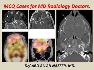
  • 1. MCQ Cases for MD Radiology Doctors. Dr/ ABD ALLAH NAZEER. MD.
  • 2.
  • 3.
  • 4.
  • 5. X-Ray left leg: In the medial part of the calf, there is a heterogeneous soft tissue lesion which contains numerous small spherical calcifications characteristic of phleboliths. The bones appear normal. MRI left leg: In the medial gastrocnemius muscle all sequences show a heterogeneous mass. T1-weighted images show an increase in fat around the circumference of the lesion and multiple interlacing vascular channels in the lesion, giving a “bag of worms” appearance. STIR axial images through the hemangioma at the same level show the vascular component of the lesion to be markedly hyperintense to muscle. On T2-weighted images, the central portion of the mass shows high signal intensity. (High-signal intralesional fat is abundant in many hemangiomas.)Axial T1 post-contrast images show enhancement of the lesion, which is composed of a cluster of tubular structures that represent discrete vessels. Soft tissue hemangioma: intramuscular, leg:
  • 6.
  • 7.
  • 8.
  • 9. X-Ray: There is a well-defined deep soft tissue swelling of the anterior aspect of the lower thigh with no calcification or cortical erosion of the femoral bone. MRI Images: There is a relatively well-circumscribed lobulated lesion centered on the vastus medialis muscle, isointense to the muscles on T1WI, hyperintense on T2WI, and STIR with areas of high signal on T1WI, low signal on T2W, and STIR (hemorrhagic areas) and peripheral rim of fatty signal well-visualized on T1WI sequences. The postcontrast sequences show a prominent enhancement. Diagnosis: Intramuscular hemangioma:
  • 10.
  • 11. A 33-year-old man with history of swelling of his forearm for 6 years. Anteroposterior radiograph of the radius and ulna demonstrates multiple phleboliths in soft tissue, suggesting intramuscular hemangioma. A 33-year-old woman with a history of swelling of her right lower extremity. Coronal T2 weighted MR image (TR/TE 3500/80) of the right lower extremity shows a soft tissue mass, which is homogeneous and very bright in appearance with slightly lobulated margins.
  • 12.
  • 13. Hemangioma, soft tissue. There is a soft tissue mass (white arrows) which contains numerous spherical calcifications characteristic of phleboliths (red oval). The combination is characteristic of a hemangioma of the soft tissue, most likely of the cavernous variety.
  • 14.
  • 15. Hemangioma, soft tissue. There is a huge soft tissue mass containing innumerable phleboliths in the soft tissue of the thigh (white oval) that is seen to contain numerous enlarged and disordered arterial and venous channels (red arrows) on the angiogram done on the right.
  • 16.
  • 17.
  • 18.
  • 19. Conventional radiograph of the bilateral lower limbs shows disparity in limb length. The right limb appears relatively longer and shows soft tissue hypertrophy, periosteal and endosteal thickening (involving middle 1/3rd shaft of the tibia), scalloping of planter surface of right calcaneum and erosion of articular surfaces of the right talocalcaneal joint. Plain computed tomography (bone window) demonstrates a large periosteal defect giving way to the infiltrating mass lesion, periosteal/ endosteal thickening (axial and coronal image-arrows) and cortical erosions at talocalcaneal joint (sagittal image- arrows). T1W sagittal and coronal sequences show marked atrophy and poor differentiation of individual muscles and variegated appearance of masses, ranging from nodular to thick irregular cords, few of them showing a branching pattern. The lesion shows signal intensity similar to as well as slightly higher than that of muscle. On T2W/STIR (sagittal and coronal) sequences, the lesion demonstrates a characteristic “bag of worms” appearance of plexiform neurofibromatosis. Postcontrast T1W fat saturation image reveals marked but slight inhomogeneous enhancement of the soft tissue (asterisk) and subperiosteal component (arrow) of the mass. The subperiosteal component also shows a central area of cystic necrosis. Dynamic 3D postcontrast computed tomography angiography (arterial phase) reveals hypertrophy of the right lower limb main arteries, which otherwise appear to be patent. Multiple tortuous collateral branches infiltrating soft tissues of the calf are also seen.
  • 20.
  • 21. There is resorption of a portion of the ulnar shaft at the junction of the middle and distal thirds. There is a narrow-sharpened pencil-like appearance of the ulnar distal to the absent section with cupping of the ulnar proximal to this, mimicking the appearance of a joint. This abnormality is rare and has been termed ulnar pseudoarthrosis and is usually only seen in patients with neurofibromatosis type 1 (NF1) which was true in this case. Involvement of the ulna is uncommon with tibial pseudoarthrosis being many times more prevalent in patients with NF1.
  • 22.
  • 23. Right sphenoid wing hypoplasia causing deformity of the right orbit which is flattened posteriorly. Slightly asymmetry dilated CSF spaces at the right middle cranial fossa. Right upper eyelid subcutaneous soft tissue lesion, that elicits intermediate signal at T1 & T2 WI showing post contrast enhancement. Right parasellar/cavernous intensely enhancing soft tissue mass lesion is also noted. The brain parenchyma demonstrates no abnormality. Sphenoid wing dysplasia, right orbital plexiform neurofibroma, & cavernous sinus mass that could be; plexiform neurofibroma or schwannoma or less likely meningioma (No biopsy was obtained) Features impressive of Neurofibromatosis type 1,
  • 24.
  • 25.
  • 26.
  • 27. Large confluent masses of low attenuation are seen within the superior, middle and posterior mediastinum. Diffuse abdominal and retroperitoneal plexiform neurofibromatosis with large confluent hypodense masses almost totally occupying the abdominal and pelvic cavities. They are seen within the celiac region, extending to the periportal region, para-aortic region, small bowel mesentery, greater omentum, and retrocrural space. They also encase the patent splanchnic branches without narrowing, displacing and compressing the adjacent organs and bowel loops without invasion. Multiple diffuse and scattered subcutaneous and muscular nodules and masses (plexiform neurofibromas). Multiple low-density well-defined hypodense neurofibromas are seen along lumbosacral spinal canal, in the neural foramina and paravertebral regions bilaterally, along the sacral plexus in the pelvis on both sides and along the course of the nerves in both thighs with large right anterior thigh mass. The patient is known to have neurofibromatosis 1 and presented complaining of increased abdominal girth and recurrent attacks of intestinal obstruction. This is a case of huge abdominal and retroperitoneal plexiform neurofibromas in a known case of neurofibromatosis type 1 (NF1). He underwent abdominal surgical intervention and biopsy proved neurofibromas.
  • 28.
  • 29.
  • 30.
  • 31. NF1, also known as von Recklinghausen disease or peripheral neurofibromatosis, is characterized by a progressive disease that affects the growth of neural tissues and has varied manifestations, such as numerous tumors of peripheral nerves, skin manifestations, ocular disorders, gliomas, and dysplastic skeletal lesions. Skin tumors can take on sessile or pedunculated forms. The second form can reach great dimensions, as in this case.
  • 32.
  • 33. Axial (Fig. 2A) and sagittal (Fig. 2B) T1-weighted MR images of the brain showed multiple masses with cystic degeneration in bilateral CP-angles as low signal intensity. Axial FLAIR image showed high signal intensity of the lesions (Fig. 2C). Axial (Fig. 2D), coronal (Fig. 2E) and sagittal T1-weighted with contrast MR images (Fig. 2F) showed heterogeneously contrast-enhancement of the lesions. The masses were proved to be multiple acoustic neurinomas. One of them extended into the right internal acoustic canal as is shown on axial FLAIR image (Fig. 2C). Bilateral acoustic neurinomas are diagnostic for neurofibromatosis type 2
  • 34.
  • 35. Axial post contrast CT scan image of the brain showed multiple extra-axial coarse calcifications which were proved to be meningiomas including intraventricular meningiomas (Figs. 6A & B). Coronal post-contrast T1-weighted MR image showed the same large calcified mass on CT which is contrast enhanced and well defined with a mass effect in this image. Coronal post Gd injection MR image showed bilateral acoustic schwannomas as enhancement lesions in CP angles. Bilateral acoustic neurinomas and multiple meningiomas are diagnostic for NF2.
  • 36.
  • 37. CT scan of the abdominopelvis with IV and oral contrast revealed a large retroperitoneal lobulated nonenhanced and homogeneous mass with encasement of the aorta and extension into the neural foramina. There is also neural foraminal widening of the lumbar vertebra. The para-spinal muscles contained multiple small non- enhanced, homogeneous masses. The masses were proved to be neurofibromas from the neural foramina to the retroperitoneal space (plexiform neurofibromatosis- type one) and from the para-spinal muscles (Fig. 7A). On the lower slice of the same CT scan, multiple subcutaneous neurofibromas and the extension of the retroperitoneal neurofibroma to the pelvic cavity were demonstrated (Fig. 7B).
  • 38.
  • 39. Axial contrast-enhanced computed tomography scan (a) showing plexiform neurofibroma with calcifications (arrow) in left parapharyngeal and carotid space (b, c) axial magnetic resonance T1- and T2- weighted images showing heterogeneous signal intensity lesion (arrows) with flow voids within the lesion.
  • 40.
  • 41.
  • 42. X-ray of the right wrist: (a) lateral, (b) oblique, and (c) AP—soft tissue swelling overlying ulnar aspect of wrist with remodeling of triquetrum (white arrow), hamate (black arrow), and 4th proximal metacarpal (arrowhead). *posterior subluxation of ulna. Axial T2-weighted wrist MRI with fat suppression— multilobulated right wrist mass (anterior/ulnar aspect). Individual lesions (arrows) show target signs: rim of high-signal intensity surrounding central low-signal intensity. *usual location of ulnar nerve. DR: distal radius; DU: distal ulna; FD: flexor digitorum longus; FCU: flexor carpi ulnaris. Coronal T2-weighted wrist MRI with fat suppression—(3a) anterior section: multilobulated mass abutting flexor digitorum longus (FD). Target signs (white arrows). (3b) Posterior section: remodeling of triquetrum (black arrow), hamate (white arrow), and proximal metacarpal (arrowhead). *distal radioulnar joint widening.
  • 43.
  • 44. Noninfectious spondylodiskitis: Andersson lesion diffuse syndesmophytic ankylosis can give a vertebral body squaring "bamboo spine" appearance syndesmophytes are classically described as paravertebral ossification running parallel to the spine linear ossification along the central spine; representing interspinous ligament ossification can give a "dagger spine" appearance on frontal radiographs; ossification of spinal ligaments, joints and discs apophyseal and costovertebral arthritis and ankylosis enthesophyte formation from enthesopathy pseudoarthroses may form at fracture sites dural ectasia. Diagnosis: Ankylosing spondylitis.
  • 45.
  • 46. Subperiosteal hemorrhage in neurofibromatosis type 1 in addition to a neuroectodermal disorder, is accompanied by mesodermal dysplasia that is accompanied by skeletal changes. The typical osseous findings include bowing of the legs, increase in length of long bones, pseudarthrosis, subperiosteal cyst formation, local bony erosions from adjacent lesions, and intramedullary neurofibromas. Except for the last two, which are due to direct involvement by neurofibromas, the remainder are due to dysplastic changes in bones.
  • 47.
  • 48. Dystrophic Spinal Deformities in a Neurofibromatosis Type 1 Murine Model (A) Anterior-posterior radiographs of two NF1 patients with dystrophic scoliosis involving the thoracic (top) and lumbar spine (bottom). (B) Anterior-posterior radiographs demonstrate representative cases of short-segmented scoliotic deformities in Nf1flox/−;Col2.3Cre mice involving the thoracic (second panel) and lumbar spine (fourth panel). Significant vertebral rotation is further evident in the representative Nf1flox/−;Col2.3Cre mouse with thoracic scoliosis as shown (second panel). (C) Vertebral wedging, another hallmark feature of NF1 dystrophic scoliosis, is illustrated in representative lateral radiographs of the lumbar spine. The wedging angle was measured by the intersection of lines drawn parallel to the rostral and caudal vertebral endplates as shown.
  • 49.
  • 50. Extensive plexiform neurofibromas infiltrating the right leg, ankle and foot in a girl with NF1. Axial T2 weighted image (a), sagittal T2 weighted image (b) showing rounded shaped and tubular lesions located on the nerve route, T2 hyperintense with central hypointense areas. Some of them realize the target image.
  • 51.
  • 52. Neurofibromatosis type 11 with cervical and brain MRI showing scattered spinal neurofibromas and bilateral schwannomas of the acoustic nerve.
  • 53.
  • 54.
  • 55.
  • 56. Neurofibromatosis type 11 with multiple brain and spinal meningioma.
  • 57.
  • 58.
  • 59. 12 -year-old boy with Tuberous sclerosis with cortical tubers. (A) Sagittal T1-weighted MR image. (B) Coronal T2-weighted MR image (C,D) Axial fluid attenuated inversion recovery (FLAIR) MR. Shows high signal in the regions corresponding abnormally large and malformed gyri due to an abnormal or absent myelination within cortical tubers (arrow A,B,C,D). (A,B) Axial fluid attenuated inversion recovery (FLAIR) MR. Showing multiple bilateral linear bright white matter striations along the lines of neuronal migration (arrows A,B). (A) T1-weighted image (B) axial T1 MR images with gadolinium (C) Sagittal sonographic images of the kidney. There are multiple non- enhancing subependymal nodules ( arrow A,B).
  • 60.
  • 61.
  • 62. 22 -year-old boy with Tuberous sclerosis and surgical resection of subependymal giant cell astrocytoma and bypass valve. (A,B) Non-contrast-enhanced CT (C,D) Contrast enhanced CT. Demonstrate a calcified mass occupying the lateral ventricles and enhanced after contrast administration due to subependymal giant cell astrocytoma remaining (arrow A,B,C,D). 22 -year-old boy with Tuberous sclerosis and surgical resection of subependymal giant cell astrocytoma and bypass valve(discussed in the previous case). (A) Sagittal T1- weighted image (B) Coronal T1-weighted image with gadolinium (C) Axial fluid attenuated inversion recovery (FLAIR) and (D) coronal T2-weighted image. Demonstrate a subependymal giant cell astrocytoma remaining (arrow A,B,C,D).
  • 63.
  • 64. 18-year-old boy von Hippel-Lindau syndrome and intracranial hemangioblastoma. (A) Sagittal T1-weighted image (B) Coronal T2-weighted image (C,D) Sagittal and coronal T1-weighted image with gadolinium. It is showing a fronto-temporal cystic lesion with heterogeneous areas( arrows A,B) there is an enhancing in the multiple cystic areas and in the peripheral( Arrow C). Small hemangioblastoma are also shown in the cerebellum as a focal hyperintense lesions.
  • 65.
  • 66. 35-year-old man with progressive headaches. MRI features suggestive of an epidermoid cyst.
  • 67.
  • 68. 60-year-old man. An oval mass is seen on the left CPA, hypointense on 3D-CISS sequences, that enhances after contrast administration. Volume rendering reconstruction of TOF images demonstrate an aneurysm of the left superior cerebellar artery.
  • 69.
  • 70. Skull radiographies, lateral (a) and anteroposterior view (b) showing an important thickening of the diploe associated with the characteristic “hair on end” appearance. Note also the loss of the maxillary sinus aeration. Diagnosis: Thalassemia major.
  • 71.
  • 72. Gross thickening of diploic space of the skull vault with no discrete masses. No intracranial masses, hemorrhage or vascular thrombosis. The 'hair on end' sign is always appreciated on lateral skull radiograph and CT. In this case, it is evident on MRI. As in the above case, it is related to Thalassemia major.
  • 73.
  • 74.
  • 75. 30-year-old male presents with shortness of breath and pleuritic chest pain. His past medical history is significant for Beta thalassemia and multiple venous thromboses. Diagnosis: Extramedullary hematopoiesis in thalassemia.
  • 76.
  • 77. Chest radiograph (a); chest CT scan in coronal plane, mediastinal window (b); axial slice in bone window (c) and mediastinal window (d): Diffuse ribs widening marked on the posterior arches, surrounded by extramedullary hematopoiesis. Note the cortical erosions (arrow) and thickening of residual trabeculae. CT scan, unenhanced and enhanced axial slices (a,b); coronal slice (c) and MRI, coronal slice (d): bilateral paravertebral masses, lobulated, well circumscribed, non osteolytic, slightly enhanced after contrast agent administration, related to Extramedullary hematopoiesis.
  • 78.
  • 79. Computed tomography scan of the thorax showing extramedullary hematopoiesis (EMH) tissue in the transverse process of the vertebrae (A), EMH tissue around the posterior end of the ribs with lytic expansile lesions (B), masses of EMH tissue around the aorta enhanced slightly with contrast (C), expansile lesion of the sternum (D) and EMH at the anterior end of the ribs (E). Diagnosis: Thalassemia with extra-medullary hematopoiesis.
  • 80.
  • 81. Reversal of normal marrow signal seen as predominantly bright PD fat sat and dark T1 signal, indicating conversion of yellow to red marrow. Exaggerated trabeculae and slight bone expansion. Haemopoietic expansion results in abnormal high signal intensity marrow on the PD FS and T2 FS sequences. Normal signal in the patella, indicating it contains normal yellow marrow and normal trabeculae. Beta thalassemia major - Cooley anemia
  • 82.
  • 83. Calvarial (a) and orbital wall (b) brown tumour- It depicted as multiple lytic expansile lesions(Arrows) in the calvaria and right lateral orbital wall. Calvarial brown tumour- Axial T1-weighted FSE(a) and Axial T2-weighted FSE (b) images showing the same lesions in the X-Ray. Because brown tumours usually contain hemosidrin, their classic MRI appearance is hypointense in both T2 and T1WI with blooming artifact on gradient echo sequences.
  • 84.
  • 85. Radiograph of the distal femur in a patient with primary hyperparathyroidism. This images shows scalloped defects along the inner margin of the cortex which denote endosteal resorption. Radiograph of the humerus ad mid-femur diaphysis in a patient with primary hyperparathyroidism. This image depicts a brown tumour. Note the osseous expansion and lucency of the proximal humerus. Brown tumour have varied appearance.
  • 86.
  • 87.
  • 88. 29-years male patient with CRD. The X-ray of the lower pelvis and proximal femur shows Brown tumors in the posterior aspect of the acetabulum bilaterally (arrows). Additionally there is protrusio acetabuli and diffuse osteomalacia. Note the post-operative clips on the left following renal transplantation.
  • 89.
  • 90. -59 female with ROD. AP X-ray of the pelvis with diffuse sclerosis affecting the iliac bones, the sacrum, pubis, ischium and the proximal femura. -X-ray Lateral view of the lumbar spine of a 47 y/o male with ROD. There is sclerosis of the endplates in all the lumbar vertebrae.
  • 91.
  • 92. Thick Skull Generalized cranial thickening with preservation of anatomy in "layers" (Int / ext. tables, diploe) in chronic treatment with phenytoins “Salt and-pepper” secondary to renal osteodystrophy, diffuse sclerosis with multiple lytic lesions (brown tumors). DD. Hyperostosis interna Idiopathic Paget disease Fibrous dysplasia Anemia Metastases/meningioma
  • 93.
  • 94. Scurvy in a 4-year-old boy with speech delay, a history of difficulty walking for 1 month, and a serum vitamin C level of 0.08 mg/dL (normal range, 0.2–2.0 mg/dL). (a) Anteroposterior radiograph of both knees shows diffuse osteopenia, Frankel lines (arrowheads), Trummerfeld zones or scurvy lines (dashed arrows), widening of the growth plate (solid arrows), and subepiphyseal corner fracture (circle). Because of the metaphyseal lucent bands, the patient was initially thought to have leukemia or metastases, but the findings from histopathologic examination of the specimen from bone marrow biopsy were normal. (b) Coronal T2-weighted fat-suppressed MR image of both distal femoral metadiaphysis shows heterogeneously increased T2 signal intensity in the marrow (*) and around the bone (arrows).
  • 95.
  • 96. This sagittal (from the side) CT reconstruction of the spine (low back) in a patient with sickle cell anemia demonstrates typical findings involving the vertebral bodies and marrow texture seen in patients with chronic disease. There is fish mouth vertebrae involving all visualized vertebral bodies secondary to bony endplate infarctions secondary to sludging of blood with occlusion of the vascular supply to the endplates. The marrow texture demonstrates irregular sclerosis of trabeculations secondary to bone marrow infarcts. Sickle Cell Osteopathy.
  • 97.
  • 98. Hypoxic- Ischemic Encephalopathy (HIE) Pathophysiology of HIE Causes in adults: Drowning Asphyxiation Cardiac arrest Cerebrovascular accident Selectively vulnerable areas of the brain:
  • 99.
  • 100.
  • 101. Patient is a 12-year-old male with dysphagia and wheezing. Diagnosis. Double aortic arch.
  • 102.
  • 103.
  • 104. A 42-year-old female, with a longstanding history of anxiety, recently had some problems with subtle chest pain as well as a sensation of mild hanging up of food within her chest upon swallowing. Frontal and lateral plain radiographs of the chest demonstrate a large subcarinal mass. The azygoesophageal line is displaced to the right. Post- contrast chest CT demonstrates a sizeable mass in the posterior mediastinum measuring up to 11 cm in diameter. The mass appears to be solid and well-circumscribed, deviating the esophagus towards the left and abutting the posterior carina, as well as the left and right main-stem bronchi. There is no obvious involvement of the lung parenchyma. There are considerable feeding vessels. Diagnosis: Castleman's Disease.
  • 105.
  • 106.
  • 107. Patient is a 47-year-old-female status post-patent foramen ovale (PFO) closure. Frontal chest radiograph demonstrates a scimitar vein (arrows) extending to the medial right hemidiaphragm with hypoplasia of the right lung. Coronal and 3D reconstruction images demonstrates anomalous pulmonary venous drainage (arrow) of the entire right lung to the IVC. Diagnosis: Scimitar syndrome
  • 108.
  • 109.
  • 110. Patient is a 39-year-old male on highly active anti- retroviral therapy (HAART) who presented with chest pain and shortness of breath to the Emergency Department. An initial chest radiograph was ordered, which revealed no acute infiltrate. Subsequently, the patient underwent computed tomography of the chest. Diagnosis: Poland syndrome
  • 111.
  • 112. A single frontal chest radiograph demonstrates almost complete absence of the clavicles with a small lateral clavicle present on the left. There are bilateral winged scapula. Mild scoliosis is present. Diagnosis: Cleidocranial dysostosis.
  • 113.
  • 114.
  • 115. Patient is a 36-year-old female with history of prior hysterectomy presenting with a 2-month history of cough and dyspnea. X-Ray images show bilateral well-defined rounded opacity, more on the right upper lobe. CT Scan show with homogeneous density lesions with well-defined margins. No calcification seen inside. No density enhancement after intravenous administration of contrast medium. Diagnosis: Benign metastasizing leiomyomas to the lung.
  • 116.
  • 117. Patient is a 51-year-old woman presenting with pleuritic chest pain, shortness of breath and hypoxia. A selected axial post-contrast CT view demonstrates a large loculated right pleural fluid collection containing an air-fluid level. The surrounding pleura are thickened and enhancing. A bronchus originating from the right lower lobe communicates with the pleural space. The right lower lobe is collapsed. Pleural fluid culture grew S. Aureus. Diagnosis: Empyema with bronchopleural fistula.
  • 118.
  • 119. Patient is a 27-year-old female who presented to the emergency department with sharp chest pain radiating to the back. CT was performed for clinical suspicion of pulmonary embolism. The right subclavian artery arises as the last branch of the aortic arch, and passes posterior to the esophagus and trachea. No pulmonary embolism was seen. Diagnosis Aberrant right subclavian artery with left sided arch.
  • 120.
  • 121. Patient is an 82-year-old male with a chronic cough. Axial and coronal high-resolution CT images of the upper chest shows enlarged tracheal diameter measuring 3 cm with apical emphysematous changes (Fig. 1), scalloping of the tracheal wall (arrow on Fig. 2), and extensive basilar bronchiectasis (arrows and arrowheads on Figs. 3 & 4). Diagnosis: Mounier Kuhn syndrome.
  • 122.
  • 123. Patient is a 60-year-old woman noted to have mass effect on the right side of the heart described in cardiology angiogram report. Evaluation requested to rule out cardiac mass. Patient has stable chronic heart disease symptoms; no acute symptoms at time of exam. PA and lateral views of the chest were obtained; frontal view demonstrated deviation of the cardiac silhouette to the left with obliteration of the right heart border. Lateral view demonstrated prominent appearance of sternum projecting posteriorly in the chest, a classic example of severe pectus excavatum deformity of the sternum. Exam was unchanged from comparison exams dating back several years. Diagnosis: Pectus excavatum deformity.
  • 124.
  • 125. Patient is an 83-year-old woman referred from outside institution for evaluation of a hilar/lung mass incidentally seen on chest radiograph. PA and lateral chest radiographs were obtained prior to evaluation by the thoracic surgery service. A mass measuring at least 7 cm in diameter was seen projecting in the anterior right hemithorax. Due to its location, differential diagnoses must be considered arising from the right hilum, right medial lung, or anterior mediastinum. Diagnosis: Thymoma.
  • 126.
  • 127.
  • 128. Patient is a 17-year-old woman presenting with left lower back and left shoulder pain at rest. Not exacerbated by motion. No other significant medical history. Initial chest x-ray demonstrated a large soft tissue mass in the posterior aspect of the left lung base. Subsequent CT images demonstrated the mass projecting into the left lower lobe with small atelectasis; mass abutted the ribs posteriorly, the spine and posterior mediastinum medially, and the diaphragm inferiorly. There was minimal involvement of surrounding structures. Aggressive fibromatosis/Desmoid tumour.
  • 129.
  • 130.
  • 131. A 57-year-old female presents with acute onset abdominal pain. A barium enema (Fig. 1) demonstrates obstruction with focal narrowing of the ascending colon near the hepatic flexure. Overhead images (Fig. 2) from the barium enema demonstrate contrast within a large dilated midline structure, corresponding to the herniated cecum. The ascending colon traverses abnormally toward the midline. A NG tube is identified anterolaterally within the collapsed stomach. CT scan (Figs. 3 and 4) shows a large air-filled structure along the midline, representing the herniated cecum within the lesser sac. The lesser sac is posterior to the liver, anterior to the inferior vena cava and between the stomach and pancreas. There is a tapered narrowing through the foramen of Winslow. The stomach is displaced laterally. Diagnosis: Internal hernia through the foramen of Winslow.
  • 132.
  • 133.
  • 134. A 49-year-old male presented with a history of gastrointestinal bleeding. Multiple sac-like outpouchings arising from the small bowel. Diagnosis: Small bowel diverticulosis.
  • 135.
  • 136.
  • 137. A 67-year-old man presented to a local hospital with sepsis. At that time, he was slightly jaundiced and had slightly elevated transaminases and was diagnosed with primary biliary cirrhosis. He had three further hospitalizations within three months for recurrent infections. He was subsequently referred to our hospital where a CA19-9 level was drawn and was markedly elevated. An MRI/MRCP was performed. Pathology from brushings performed on an ERCP on the day admission confirmed the MRI suspicions of malignant cells derived from adenocarcinoma. A 0.8 cm x 0.9 cm enhancing lesion/focus at the confluence of the right and left hepatic ducts that may represent tumor (Klatskin tumor) vs. scar. There is extensive intrahepatic ductal dilatation proximal to this lesion. The common bile duct is normal in caliber. (Klatskin's Tumor).
  • 138.
  • 139. A 60-year-old female had sudden onset severe abdominal pain. CT exam showed a large U-shaped distended bowel segment in the mid to right abdomen. No associated small bowel obstruction or signs of ischemia, such as mural thickening, infiltration of the mesenteric fat, or pneumatosis, were identified. A possible whirl sign was present with twisting at the base of the cecum and mesentery. Diagnosis: Cecal volvulus.
  • 140.
  • 141. Patient is a 62-year-old female with elevated alkaline phosphatase and clinical suspicion for choledocholithiasis. MR projection image demonstrates non-fusion of the main pancreatic duct (white arrow) with the smaller accessory pancreatic duct (red arrow). The main pancreatic duct drains into the duodenum via the minor papilla and the smaller accessory pancreatic duct drains via the major papilla with the common bile duct. Diagnosis: Pancreatic Divisum.
  • 142.
  • 143.
  • 144. Axial contrast-enhanced CT slice through the level of the adrenals demonstrates a cystic lesion in the upper pole of the right kidney with solid enhancing portion laterally which is suspicious for renal cell carcinoma. Also, there is an intensely enhancing circular lesion in the left adrenal gland suspect for pheochromocytoma in this patient. Concurrent axial contrast-enhanced T1-weighted MR image of the head through the cerebellum demonstrates a cystic lesion in the left cerebellum with an enhancing mural nodule typical for hemangioblastoma. Diagnosis: Van Hippel Lindau disease.
  • 145.
  • 146.
  • 147. Axial CT image with contrast demonstrates a spiculated mesenteric metastatic carcinoid mass. A metastatic soft tissue nodule is seen in the subcutaneous fat along the anterior abdominal wall. Axial CT image with contrast demonstrates 2 small submucosal small bowel wall masses adjacent to the spiculated mesenteric mass compatible with primary small bowel carcinoid tumor. Axial CT image with contrast of the liver demonstrates a hypervascular, arterial-phase enhancing mass in the medial right hepatic lobe compatible with metastasis. Diagnosis: Primary carcinoid tumor of the small bowel
  • 148.
  • 149. Patient is a 72-year-old female presenting to the Emergency Department with abdominal pain and rectal bleeding. CT scan revealed an encapsulated air- fluid collection adjacent to the sigmoid colon. A subsequent single contrast barium enema confirmed the diagnosis of a giant diverticulum of the sigmoid colon. Diagnosis: Giant colonic diverticulum
  • 150.
  • 151.
  • 152. A 50-year-old female with smoking history presents with shortness of breath and decreasing functional ability. Evaluate for emphysema. Plain chest radiographs often demonstrate increased interstitial markings in a reticular pattern. It is difficult to appreciate the cysts due to their nearly imperceptible thin walls. CT demonstrates extensive replacement of the lung parenchyma with thin walled cysts. Diagnosis: Lymphangiomyomatosis
  • 153.
  • 154.
  • 155. A 36-year-old female presented with chest pain. PA and lateral chest radiographs demonstrate a wedge-shaped opacity of the middle lobe that extends to the lung periphery. A selected axial CT image at the level of the right main pulmonary artery demonstrates filling defects of the distal right main pulmonary artery. There is a corresponding area of consolidative lung parenchymal process of the right middle lobe. Diagnosis: Thrombotic Pulmonary Embolism.
  • 156.
  • 157.
  • 158. Patient is a 60-year-old male with history of multiple sclerosis with increased abdominal distention and difficulty urinating. Findings: Dilated, stool-filled colon from the cecum to the sigmoid with a collapsed rectum. Diagnosis: Acute colonic pseudo- obstruction (Ogilvie's syndrome).
  • 159.
  • 160.
  • 161. A 67-year-old female presented with splenomegaly, early satiety, night sweats, fever, weakness, and a 10-pound weight loss. Contrast enhanced coronal CT reconstruction (Fig. 1) demonstrates massive splenomegaly and multiple hypodense splenic mass lesions of varying sizes. The liver appears fatty with cirrhotic morphology however no focal hepatic lesions are appreciated (Fig. 2). The osseous structures of the pelvis (Fig. 3) and spine (not shown) demonstrate diffuse patchy increased bone density Diagnosis: Myelofibrotic extramedullary hematopoiesis.
  • 162.
  • 163.
  • 164. Patient is a 53-year-old female with end stage liver disease presenting for pre-transplant work-up. Findings: Ultrasound showed multiple small hyperechoic foci throughout the enlarged spleen. CT showed multiple hyperdense lesions throughout the enlarged spleen. MRI showed multiple hypointense T1 and T2 lesions throughout the enlarged spleen. The CT and MRI images also showed incidental small gallstones. Diagnosis: Gamna-Gandy bodies.
  • 165.
  • 166. Patient is a 54-year-old male with cirrhosis. Findings: Numerous tiny echogenic foci scattered in the spleen with characteristic low MR signal. Diagnosis: Gamna-Gandy bodies
  • 167.
  • 168. Patient is a 44-year-old female with Caroli disease seen for pre-transplant evaluation Contrast-enhanced CT images through the liver demonstrate several areas of cystic dilatation of the intrahepatic biliary ducts (red arrows). A gallstone is seen (blue arrow) within a dilated intrahepatic biliary duct of the left hepatic lobe. Diagnosis: Caroli disease.
  • 169.
  • 170. Patient is a 55-year-old man complaining of left lower quadrant abdominal pain for several days, intermittent in nature. Non-inflamed appendix herniates within the inguinal canal. Diagnosis: Amyand hernia.
  • 171.
  • 172. De Garengeot hernias are femoral hernias that contain the appendix. It is a rare phenomenon, with only 1% of all femoral hernias containing the appendix (and usually found incidentally at surgery), and only 0.08-0.13% containing an incarcerated acute appendicitis (sometimes detected on pre-operative CT)
  • 173.
  • 174. Patient is a 30-year-old female with right lower quadrant pain with suspicion for appendicitis. Inflammatory changes surrounding diverticula in the right lower quadrant. The appendix is normal. inflamed diverticula with or without colon wall thickening or adjacent abscess. The diagnosis is difficult to make as diverticula may be obscured by adjacent inflammatory changes and the entity may be indistinguishable from colon cancer in an older population. Also, since appendicitis more commonly causes right lower quadrant inflammatory changes visualization of a normal appendix is essential for CT diagnosis. Other than appendicitis and colon cancer the differential diagnosis includes Crohn’s disease and epiploic appendigitis. Diagnosis: Cecal diverticulitis.
  • 175.
  • 176.
  • 177. Findings: Multiple MR and CT images demonstrate cerebellar and spinal hemangioblastomas, pancreatic cyst and enhancing left renal lesion concerning for renal cell carcinoma. Diagnosis: Von Hippel-Lindau Disease.
  • 178.
  • 179. A 64-year-old woman with lupus anticoagulant, status post recent hip replacement, presented with progressive weakness, nausea and hyponatremia. Cosyntropin stimulation test was preformed and was suggestive of adrenal insufficiency. CT and MRI findings may vary. Typical acute CT findings include a non-enhancing hyperdense mass (50-90 HU) that distorts the normal shape of the adrenal gland [3]. There may also be thickening of the adjacent diaphragmatic crura and streakiness of periadrenal fat. Areas of enhancement raise the possibility for underlying tumor, such as adrenal carcinoma or metastatic lung cancer. Chronic adrenal hematomas become low density, may become cystic and/or may calcify. Post-hemorrhage adrenal cysts are termed adrenal pseudocysts. MRI is useful to confirm the presence and chronicity of adrenal hemorrhage. Because of intracellular deoxyhemoglobin, in the acute stages (<7 days), adrenal hemorrhage may show iso-intense to hypo-intense on T1WI and markedly low signal on T2WI [4]. Diagnosis: Bilateral Adrenal Hemorrhage.
  • 180.
  • 181. Chronic hemolytic anemia. Classical “hair-on-end effect. A, At age 15, long, thin vertical spicules. B, At age 31 , they become wide, more sclerotic and distinct. Areas of sclerosis in frontal and anterior parietal areas have distinct lucent central component resembling doughnut’ lesions. Occipital bone is not expanded.
  • 182.
  • 183. Chronic renal disease On peritoneal dialysis in a 25- year old male. Sagittal (a) and axial (b,c) CT images demonstrate unequivocal diffuse bone sclerosis on axial skeleton. Note the peritoneal catheter (arrow).
  • 184.
  • 185. Sickle cell disease in a 54-year old female. Plain films of the abdomen (a) and both knees (b) show heterogeneous diffuse osteosclerosis in a patient with previous history of multiple bone infarctions.
  • 186.
  • 187. Sickle cell disease in a 41-year old male. Coronal (a) and axial (b) CT images illustrate the uncommon pattern of diffuse osteosclerosis in Sickle cell disease.
  • 188.
  • 189. Osteoblastic metastases in a 74-year old male with prostatic carcinoma. Lateral view plain film of lumbar spine (a) demonstrate diffuse osteosclerosis of lumbar vertebrae. Axial CT (b) confirmed coarse areas of hyperdensity across the axial skeleton, some associated to cortical destruction, consistent with diffuse metastatic disease.
  • 190.
  • 191. Systemic mastocytosis in a 49-year old male. Plain films of the hands (a) and pelvis (b) show a generalized increased of bone density. Axial CT image (c) in the same patient reveals the sclerotic process involving the spine and also splenomegaly (*) as an abdominal manifestation.
  • 192.
  • 193. A 42-year-old man with biopsy-proven mastocytosis. a Sagittal, non-enhanced, low dose CT image of the lumbar spine shows diffuse marrow sclerosis (arrow) secondary to mast cell infiltration. b Sagittal, non-enhanced, low-dose CT image of the right humerus shows diffuse sclerosis of the marrow space (arrow) secondary to mast cell infiltration. c Axial, non- enhanced, low-dose CT image of the pelvis shows diffuse sclerosis of the marrow space (arrow) secondary to mast cell infiltration Less
  • 194.
  • 195. Systemic mastocytosis (SM) refers to mast cell infiltration in extra-cutaneous tissues. The symptoms of systemic mastocytosis are due to degranulation of mast cells and/or accumulation of mast cells in target organs. findings refer organ involvement with organ dysfunction. The example above shows hepatic involvement with cirrhosis (white arrow) and ascites (yellow arrow) and nodal involvement with bulky adenopathy (red arrow). We also have involvement with diffuse sclerosis. Interestingly, the non-radiology literature stresses the more common osteoporosis, with scarce mention of the sclerosis that tends to dominate the radiology literature.
  • 196.
  • 197. A 69-year old male with colon adenocarcinoma coexisting with incidental Paget's disease. Post operative anteroposterior plain film of the abdomen (a), axial (b) and coronal (c) CT images demonstrate cortical thickening, diffuse increased bone density and coarsening of trabeculae of the pelvis and proximal left femur characteristic of Paget disease.
  • 198.
  • 199. Osteopathia striata in an asymptomatic 43-year old male. CT images on axial (a), sagittal (b) and coronal (c) views show dense linear striations oriented parallel to the long axis of left femur (arrow).
  • 200.
  • 201. Osteopoikilosis in a 35-year old male. The coronal MIP (a) and axial (b) CT images show multiple bone islands in the pelvis and clustered around the epiphyses and metaphysis of both femurs. The lesions vary in size, ranging between 2-10 mm.
  • 202.
  • 203. Marrow expansion of the skull in a 12-year-old boy with SCA. (a) Collimated Water view of the skull shows striated appearance of the skull caused by diploic space widening and trabecular prominence. (b) Axial CT image (bone window) of the upper skull helps confirm diploic space widening and trabecular prominence. (c) Sagittal T1-weighted MR image of the brain shows diploic space widening with intermediate-signal- intensity material, representing red marrow (∗). Note the sparing of the occipital bone (arrows), which contains no marrow elements.
  • 204.
  • 205. Medullary bone infarcts in SCA. (a) Frontal radiograph of the right shoulder in a 22-year-old patient shows an area of patchy sclerosis and radiolucency. (b) Sagittal fat-suppressed T1- weighted magnetic resonance (MR) image of the right hip in a 19-year-old patient, obtained after intravenous injection of contrast material, shows multiple irregular areas of high signal intensity forming a rim around central areas of lower signal intensity, representing enhancement in areas of maturing medullary infarct. Note the high-signal- intensity regions in the medial acetabular wall, also representing infarct (arrowheads).
  • 206.
  • 207. Acute infarct with extraosseous soft-tissue abnormality in a 24-year-old man with SCA who presented with acute thigh pain. (a) Coronal T1- weighted MR image of the left femur shows heterogeneous abnormal signal intensity throughout the lower diaphysis extending to the articular surface, as well as surrounding soft-tissue edema. There are also areas of low signal intensity in the tibial plateau indicating infarct. (b) Coronal inversion recovery MR image of the same area shows heterogeneous high signal intensity of the lower shaft (including condyles), with considerable high signal intensity of the edematous soft tissue surrounding the femur.
  • 208.
  • 209. Moya moya disease in a 7-year-old girl with SCA. (a) Lateral view from a left common carotid angiogram reveals occlusion of the supraclinoid internal carotid artery and multiple small collateral vessels arising from the distal internal carotid artery and extending into the regions of the basal ganglia and thalamus, giving the classic “puff of smoke” appearance. (b) Sagittal T1-weighted MR image of the left hemisphere helps confirm multiple tiny serpentine areas of signal void (arrows) in the region of the basal ganglia and thalamus, findings that represent the collateral vessels.
  • 210.
  • 211. Brown tumour: Plain radiograph well-defined, purely lytic lesions that provoke little reactive bone. The cortex may be thinned and expanded but will not be penetrated. The zone of transition is narrow with no evidence of cortical destruction or para-osseous soft tissue component.
  • 212.
  • 213.
  • 214. Lead Poisoning. There are multiple metallic foreign bodies in the bowel (black arrows) including a nail in the region of the cecum. This is pica. Anytime one sees evidence of pica, look for lead lines. In this case, there are dense metaphyseal lines forming the articular cortices of the acetabuli (red lines). There are dense metaphyseal bands at the distal femurs and proximal tibias (white arrows). Note also there are similar dense bands at the heads of both fibulas (red arrows). There are dense metaphyseal bands at the distal radius and ulna and in all of the metacarpals (red arrows).
  • 215.
  • 216. Radiographs demonstrate large intra- articular irregular ossified masses in suprapatellar and infrapatellar locations associated with moderate joint effusion. The superior lesion demonstrates incomplete lucent cleavage plane between the lesion and the periosteum. A subtle band of dense tissue is seen extending into anterior-lateral distal femoral diaphysis. Diagnosis: Parosteal Osteogenic Sarcoma.
  • 217.
  • 218.
  • 219.
  • 220. MRI images demonstrate enhancing low T1 weighted- image and intermediate to high T2 weighted-image signal mass anterior to distal femoral metaphysis with evidence of cortical breakthrough. A second mass with similar characteristics is visualized within the Hoffa’s fat pad without attachment to the osseous structures. A large joint effusion is also noted. A low T1-weighted signal elongated abnormality is seen under the endosteum extending into the distal femoral diaphysis corresponding to the area of sclerosis seen on the plain film. Diagnosis: Parosteal Osteogenic Sarcoma.
  • 221.
  • 222. Figure 1A-C: AP (A), oblique (B) and lateral (C) views of the left foot demonstrate severe osteolysis of the forefoot, distal to the mid-tarsal bones. Note diffuse osteopenia, osteophytosis and subchondral cysts of the phalanges, and relative sparing of the mid and hindfoot. Diagnosis: Neuropathic Osteoarthropathy.
  • 223.
  • 224. There is diffuse osteopenia of the visualized bones of the hand and wrist. There are significant destructive changes in the wrist with extensive bony fusion. There is significant destruction of all visualized metacarpophalangeal joints which demonstrate postsurgical changes of previous joint replacement. No evidence of acute fracture. Diagnosis: Rheumatoid Arthritis.
  • 225.
  • 226. Plain radiographs of the foot demonstrate bony deformity with flattening of the second metatarsal head and collapse of the articular surface (Figs. 1 & 2). There is also change in bone density with sclerosis and cystic change of the distal metatarsal. Compare to the normal appearing third metatarsal (Fig. 3). Diagnosis: Freiberg Infraction (AVN of the 2nd metatarsal head)
  • 227.
  • 228.
  • 229. Plain film and CT show a solitary, permeative, metadiaphyseal lytic lesion with extensive layered periosteal reaction, findings highly suggestive for malignant tumor. Radiographically the differential diagnosis for this age group includes osteosarcoma, metastatic disease and including lymphoma. MR shows a soft-tissue mass involving the marrow and extending through the cortex into adjacent soft tissues. Bone marrow replacement on MR narrows the differential to small, round, blue cell tumors, such as lymphoma and Ewing sarcoma. Between these two tumors the patient's age determines their relative likelihood, with Ewing sarcoma being more likely in patients younger than 30 years old. Bone scan and PET imaging are used to evaluate for metastatic or multifocal involvement. In this case, bone scan reveals increased perfusion, blood pool and uptake in the region of the tumor. These features reflect the increased vascular demand of the tumor and increased bone remodeling secondary to bone destruction by the tumor. These findings could also be seen in Paget's disease and osteomyelitis. Diagnosis: Large B Cell Lymphoma
  • 230.
  • 231. Plain radiographs show mixed sclerotic and lytic areas within the body of the calcaneus, navicular, cuboid, cuneiforms, and proximal second metatarsal. Diagnosis: Mycetoma (Madura Foot)
  • 232.
  • 233. Large subchondral cyst formation, widened trochlear and radial notch, and overgrowth with flattening of the radial head (best seen on the lateral view). There is also loss of the joint space. Diagnosis: Hemophilia Arthropathy
  • 234.
  • 235. Soft tissue mass located in the right lower extremity predominantly in the subcutaneous fat demonstrates T1 low- signal, T2 high-signal and enhancement on post-contrast imaging. Diagnosis: Malignant fibrous histiocytoma Differential Diagnosis: Liposarcoma, leiomyosarcoma, rhabdomyosarcoma, fibrosarcoma.
  • 236.
  • 237. Magnetic resonance imaging is helpful for surgical planning: defines the extent of the tear, reveals the quality of the tendon edges and demonstrates degree of retraction. Findings include a fluid-filled tendon sheath, absence of the tendon distally, an antecubital fossa mass and muscle edema and/or atrophy. Typical treatment for biceps rupture is surgical repair with insertion of the tendon at the apex of the radial tuberosity or tenodesis. Diagnosis: Biceps tendon rupture
  • 238.
  • 239. Plain films in corresponding areas to those of increased radiotracer uptake demonstrate enchondromas with deformity of the right hemipelvis, femur, tibia and fibula. Diagnosis: Ollier's disease.
  • 240.
  • 241. There is a large knee joint effusion. Frond-like synovial projections are evident. These follow fat signal on all sequences. Diagnosis: Lipoma arborescens
  • 242.
  • 243. Frontal and lateral views of spine demonstrate bambooing of the spine and fusion of the bilateral sacroiliac joints. There is calcification of the intervertebral discs and interspinous ligament. Diagnosis: Ankylosing spondylitis.
  • 244.
  • 245. AP view of left hand, wrist, and forearm demonstrating ulnar angulation of the distal radius and decreased carpal angle. Oblique and lateral views demonstrating dorsal subluxation of the ulna. Radiographic images of the bilateral forearms and wrists demonstrated features of Madelung deformity.
  • 246.
  • 247.
  • 248. Radiographs: Nonspecific soft tissue mass. Ossification within the tumor may occur about 10% of the time. MR: Since the fat content is often less than 10-25% of the tumor volume, MRI may not show the typical features of lipomatous tumor. A myxoid liposarcoma that contains abundant water can mimic a cystic lesion. Contrast- enhanced MRI is useful in distinguishing cystic or necrotic lesions from solid, cellular lesions. Myxoid liposarcoma exhibits low signal intensity on non-enhanced T1WI and high SI on T2WI. In contrast, lipomas and well- differentiated liposarcomas typically show high SI secondary to the relatively high fat content. Myxoid liposarcomas has three enhancement patterns: homogeneous, heterogeneous, and no enhancement.
  • 249.
  • 250. MRI of the thoracic spine demonstrates a non-enhancing hyperintense intramedullary lesion on STIR sequence (Fig. 1) from the level of T8 through the conus which is mildly expanded. Axial T2 weighted image (Fig. 2) shows the lesion to be centrally located. There are no significant degenerative changes, central spinal canal stenosis or evidence of spinal dural fistula. MRI of the cervical spine demonstrates no additional abnormality. A subsequent MRI of the brain (Fig. 3) demonstrates T2 and FLAIR hyperintensities in the left thalamus and scattered throughout predominantly subcortical areas without periventricular involvement; features that are consistent with acute disseminated encephalomyelitis (ADEM). Diagnosis: Acute Transverse Myelitis
  • 251.
  • 252. Axial CT of the cervical spine demonstrates an expansile cystic mass arising from the C2 vertebral body. No evidence of cortical breakthrough or paravertebral soft tissue involvement. Sagittal MRI T1 and T2 weighted images demonstrate the expansile cystic mass at C2 which extends into the bilateral pedicles and odontoid process. Diagnosis: Plasmacytoma
  • 253.
  • 254. A solid/cystic 3.2 x 3.6 cm mass lesion is seen in the left parafalcine frontal region. This lesion appears to be extra- axial in location with enhancing components seen along the falx. A small dural tail is noted. The lesion also has a cystic component. The ventricles and basal cisterns are preserved. Findings are most consistent with a cystic meningioma.
  • 255.
  • 256. (A, B) WHO grade I meningioma with diffuse infiltration of bone and occlusion of (C) anterior superior sagittal sinus on angiography and (D) MRV with venous rerouting.
  • 257.
  • 258. On CT there is a large mixed cystic and solid lesion within the right frontal lobe. On MRI the lesion is mostly cystic and contains an enhancing solid component. Diagnosis: Cystic meningioma
  • 259.
  • 260. Ovoid cystic dilatation of the distal central spinal cord between the conus medullaris tip and filum terminale origin. Diagnosis: Ventriculus terminalis.
  • 261.
  • 262. Axial and coronal post contrast SPGR images demonstrate cutaneous vessels at the forehead with intracranial extension into the superior sagittal sinus with thickened, enhancing lesion at the anterior falx. Sagittal T1 post-contrast image demonstrates the intracranial component of the lesion in the sagittal view. Non-contrast CT of the head shows calcification in the anterior interhemispheric fissure within the known sinus pericranii.
  • 263.
  • 264. Sagittal T2 MR of the lumbar spine demonstrates posterior vertebral scalloping secondary to pulsations of ectatic dura matter. Axial T2 MR of the lumbar spine again demonstrates vertebral scalloping secondary to dural pulsations with widening of the interpediculate distance and scalloping of the pedicles. Diagnosis: Dural ectasia.
  • 265.
  • 266. A single lateral fluoroscopic image of the neck, during a pharyngiogram, demonstrates enlargement of the prevertebral soft tissue with areas of focal lucency representing air. CT show fluid collection with air bubble inside. Diagnosis: Retropharyngeal abscess.
  • 267.
  • 268.
  • 269. On CT, there was an 1.9 x 1.3 cm high attenuation lesion in the right frontal lobe, with areas of calcifications. The surrounding parenchyma was normal, without signs of edema or mass effect. On MR, there was heterogeneously high signal intensity on T1 and T2 images in the right frontal lobe. On T2WI images there was a ring of low attenuation surrounding the lesion. Diagnosis: Cavernous angioma.
  • 270.
  • 271. Clinical presentation is most often asymptomatic, usually an incidental finding on imaging. Rarely this anomaly may cause trigeminal neuralgia or pituitary dysfunction. A large artery arising from the cavernous internal carotid artery (ICA), coursing posterior to the sella, supplying the basilar artery. Diagnosis: Persistent Trigeminal Artery.
  • 272.
  • 273. Intertrochanteric region well defined lytic lesion in the proximal femur with sclerotic rim and internal matrix calcifications. Diagnosis: Liposclerosing myxofibrous tumor (LSMFT).
  • 274.
  • 275.
  • 276. Abdominal radiograph showed a large mass in the lower abdomen and pelvis with displacement of the adjacent structures. CT and MRI images showed a predominately cystic lesion with some areas containing fat and some enhancing septa. Diagnosis: Lipoblastoma
  • 277.
  • 278. Bilateral parotid hemangiomas: Contrast enhanced axial CT images demonstrate bilateral, lobulated, enhancing masses in the expected region of the parotid glands. No cystic areas are seen within the masses. No normal parotid tissue is seen. Prominent enhancing vessels can be seen adjacent to these masses. Bilateral large hyperintense parotid masses with flow voids are consistent with bilateral parotid hemangiomas on this T2 coronal fat-saturated MRI of the face and neck.
  • 279.
  • 280. Images of the right humerus, forearm and femur demonstrated multiple expansile lytic lesion with septation involving the proximal radius. Diagnosis: multiple non- ossifying fibromas.
  • 281.
  • 282. Left Hand X-Ray: There is marked expansion of cortices with squared metaphysis and decreased tubulation. By comparison with the Radiographic Atlas of Development of the Hand by Greulich and Pyle, the patient's skeletal age is at the lower limits of normal for the patient's chronologic age. AP and Lateral Chest X-ray: There is borderline cardiomegaly, prominently involving the right side of the heart. Hila, pulmonary vasculature, lungs and pleural spaces are normal. There is generalized osteopenia, best seen in the humerus and scapula. Diagnosis: Thalassemia.
  • 283.
  • 284. Frontal view of the chest demonstrate cardiomegaly, particularly of the right heart border, with normal pulmonary vasculature. In some cases, decreased pulmonary vasculature would be seen. Although not present with this patient, lateral views of the chest sometimes demonstrate posterior bulging of the heart, representing the enlarged right atrium or displacement of the left heart by the enlarged right heart. Differential diagnosis for enlarged right heart with normal or decreased pulmonary vascularity includes tricuspid atresia, tricuspid stenosis, tricuspid regurgitation, pulmonic valve stenosis, pulmonic valve regurgitation, Tetralogy of Fallot, Ebstein's anomaly, and Uhl's anomaly.
  • 285.
  • 286. Chest radiograph demonstrated an enlarged heart and shunt vascularity. There was a small right apical pneumothorax. Pneumomediastinum is manifest by two findings: a lucent stripe along the border of the right atrium, and adjacent to the pulmonary artery, a thin lucent line elevating a soft tissue structure that is likely the thymus. Diagnosis: Spinnaker sign in pneumomediastinum.
  • 287.
  • 288. Arnold Chiari III Malformation: A 2 day neonate came with visible swelling at back and enlarged head.. MRI brain demonstrates small posterior fossa with wrapping of cerebellum around brain stem and low-lying tentorium. Tectal beaking also appreciated. Associated CC agenesis, small cephalocele and hydrocephalus also seen.
  • 289.
  • 290. Semilobar Holoprosencephaly: Multisequential MRI brain in a neonate show failure of division of midline structures, fused thalami, monoventricle with partially developed occipital and temporal horns. Incomplete falx and absent interhemispheric fissure anteriorly and absent corpus callosum.
  • 291.
  • 292. Optic Pathway Glioma in a Patient with Neurofibromatosis I: 6 years old patient, known case of Neurofibromatosis 1 presented with loss of vision, MRI brain with contrast reveals abnormal enhancing lesions in suprasellar location involving optic chiasma, right optic nerve, proximal part of left optic nerve with extension posteriorly into bilateral optic tracts measuring 3.2x2.5mm. Cystic component abutting left medial temporal lobe measuring 16x14.6mm. It is causing mass effect over intra conal structure resulting in mild proptosis, widening of optic canal and distended subarachnoid space around right optic nerve.
  • 293.
  • 294. Pericallosal Lipoma with partial agenesis of corpus callosum: Unenhanced CT Brain axial slice shows low attenuation area (-45HU) with marginal curvilinear calcification representing bracket sign. It is causing splaying of frontal horns &body of lateral ventricles. Associated genu and proximal part of body of corpus callosum are absent.
  • 295.
  • 296. TRIGONOCEPHALY: Multisequential MRI brain reveals abnormal triangular configuration of frontal region.
  • 297.
  • 298. Pachygyria with bilateral Schizencephaly: Multisequential MR imaging in a 10 year old boy reveals bilateral grey matter lined clefts in frontal region down to the lateral ventricle. Grey matter overlying cleft appear thickened. Small abnormal nodular grey matter also seen along subependymal lining of lateral ventricle. Dysgenesis of corpus callosum seen with interdigitation of falx.
  • 299.
  • 300.
  • 301. ARNOLD CHIARI MALFORMATION TYPE II. T2 AXIAL AND SAGITTAL MR IMAGES of a 9-month old female patient showing herniation of brainstem in spinal can. ARNOLD CHIARI MALFORMATION TYPE 2. T2 AXIAL AND SAGITTAL MR IMAGES OF LUMBAR SPINE of a 8 month old male patient showing meningomyelocele at lower lumbar level.
  • 302.
  • 303.
  • 304. HYDRANENCEPHALY. CT SCAN AXIAL IMAGES BRAIN of a 3-month old female patient showing complete replacement of cerebral hemisphere with CSF with no evidence of any cortical tissue. HYDRANENCEPHALY. CT SCAN SAGITTAL IMAGES BRAIN of a 3-month old female patient showing complete replacement of cerebral hemisphere with CSF with no evidence of any cortical tissue.
  • 305.
  • 306. FOCAL CORTICAL DYSPLASIA. T2 AXIAL MR IMAGES of 5-month old male patient showing fronto- parietal cortical region is large, the conical gyral pattern is abnormal, with broad gyri and large, irregular sulci, more on the left side.
  • 307.
  • 308. Occipital Meningoencephalocele Sagittal T1-weighted (a) and axial T2-weighted (b) MR images and MR venogram (c) show an occipital meningoencephalocele with no evidence of involvement of venous structures. The lesion consists predominantly of herniated meninges, but a small amount of brain tissue was found at surgery.
  • 309.
  • 310. Naso-orbital Frontoethmoidal Encephalocele: Naso-orbital frontoethmoidal encephalocele in a 2-year-old patient. (a) Three-dimensional shaded-surface-display image from CT data shows a large, left-sided fronto-orbital mass. (b) Axial CT scan shows a left frontal lobe encephalocele that extends through the ethmoid bone into the orbit. (c) Axial T2-weighted MR image obtained at the same level as b helps confirm the presence of a frontal lobe encephalocele. The contents of the orbital vault were formed normally.
  • 311.
  • 312. Vertically Positioned Straight Sinus with Persistent Fetal Anatomy: Vertically positioned straight sinus with persistent fetal anatomy. Sagittal T2-weighted MR image (a) and sagittal (b) and coronal (c) MR venograms demonstrate a vertically positioned straight sinus (black arrow in a, arrow in b) and a fenestrated superior sagittal sinus (arrowheads in c) resulting from deflection around the tract of a histologically proved atretic parietal encephalocele (white arrows in a).
  • 313.
  • 314. Lacunar skull (luckenschadel) Deep scalloping between the bony septations that characterize the lacunar skull (luckenschadel) (arrows) are best appreciated on an axial computed tomography section, as in this patient with a Chiari II malformation.
  • 315.
  • 316. Chiari III malformation Large herniated sac (open arrows) with enlarged portion of fourth ventricle (arrowheads), cerebellum (C), and cervical cord (black arrow) are seen. Osseous defects in the infraoccipital area, posterior rim of foramen magnum, and posterior arch of C1and C2 .
  • 317.
  • 318. Chiari IV Sagittal T1-weighted MR image showing Chiari IV malformation with thin brainstem. The only part of the cerebellum is a small portion of the superior vermis (arrow).With type IV Chiari malformation, cerebellar hypoplasia is present, without inferior displacement .
  • 319.
  • 320. Dandy-Walker cyst Axial T1-weighted MR image shows a large posterior fossa cyst (c) compressing the cerebellar hemispheres (arrows).b) Sagittal T1-weighted MR image showing Dandy-Walker cyst (large arrows) with absence of the inferior vermis and a small remnant of the superior vermis (small arrow) present. c) Dandy-Walker cyst. Axial noncontrast head CT scan shows Dandy- Walker cyst with associated agenesis of the corpus callosum
  • 321.
  • 322. Joubert's syndrome Axial noncontrast T1-weighted MR image shows a narrow isthmus and large superior cerebellar peduncles (molar tooth appearance of the midbrain). The “molar tooth” appearance of the midbrain on axial MR images is characteristic. The diagnosis of Joubert syndrome is based on the presence of characteristic clinical features and the "molar tooth sign" on cranial magnetic resonance imaging (MRI), resulting from hypoplasia of the cerebellar vermis and accompanying brain stem abnormalities on axial imaging through the junction of the midbrain and pons (isthmus region).
  • 323.
  • 324. Lissencephaly, Total agyria MRI is the modality of choice to show characteristic features. The smooth brain with absent or a few broad gyri is well shown, especially in the sagittal plane. Fig38. a) Axial T1-weighted MR images showing lissencephaly, Total agyria. b) Sagittal T1-weighted MR image shows total agyria with marked increased gray matter.
  • 325.
  • 326. Pachygyria refers to gyri that are focally or diffusely thick, broad and flat, and associated with a thick cortex . Dysmorphic facies occur in children with pachygyria unrelated to any syndrome. Coronal T1-weighted MR image showing multiple areas of pachygyria (arrows).
  • 327.
  • 328. Polymicrogyria: It may be unilateral (40%) or bilateral (60%). The cortex surrounding the sylvian fissure is involved in approximately 80% of cases. The frontal lobe is most commonly involved. Sagittal T1-weighted MR image showing polymicrogyria that involves the frontal lobe with small irregular gyri (arrows) and increased gray matter.
  • 329.
  • 330. 5a, b. A 42-year-old woman with unilateral polymicrogyria in the right Sylvian fissure. a Axial first echo of fast SE (4,500/18,108 [TR/TE]) image shows thick cortex (arrowheads) in the right Sylvian fissure. b Coronal inversion recovery (1,800/20/630 [TR/TE/ TI]) image depicts the cortical junction irregularities clearly (arrowheads). The cortical abnormality is confined to the right Sylvian fissure
  • 331.
  • 332. Hemimegalencephaly: The involved area is enlarged to a varying extent on CT and MRI. The gray-white matter is poorly differentiated. Gray matter heterotopia is common. The ipsilateral lateral ventricle is enlarged. Axial T1-weighted MR image showing hemimegalencephaly of the left calvaria and cerebral hemisphere with abnormal pachygyric-appearing cortex (arrows).
  • 333.
  • 334. Series of axial CT images of an adolescent female who presented with sudden left sided hemiplegia. A) NCCT showed an intraparenchymal bleed in right basal ganglia extending into right frontal region (white arrow) with perilesional edema and is seen to cause mass effect in the form of effacement of frontal horn of ipsilateral lateral ventricle. B) CECT showed serpentine enhancement (white arrow) adjacent to the hemorrhage. C) CTA shows enlarged right posterior communicating artery (white arrow) along with several other feeding arteries from right ICA and right ACA. D) CTA shows early enlarged and tortuous draining vein draining into right internal cerebral vein (white arrow) further into vein of Galen. This right basal ganglia AVM is Spetzler-Martin Grade 4.
  • 335.
  • 336. Series of axial CT images illustrating classic findings of AVM in a child who presented with sudden loss of consciousness. A) On NCCT, there is intracranial hemorrhage in the body of left ventricle (white arrow). B) On CECT, there is hyperdense serpentine enhancement in the medial part of trigone of left lateral ventricle (white arrow). C)CTA shows AVM with intranidal aneurysm (white arrow). The lesion is seen to be supplied by lateral branch of left posterior choroidal artery (black arrow). No obvious dilatation of supplying artery is seen. D)3D rendering shows tangle of vessels (red arrow) along with its feeding artery and draining vein draining into the vein of Galen (white arrow). This left peri-trigonal AVM is Spetzler-Martin Grade 3.
  • 337.
  • 338. 7-year-old male child presented with complaints of headache. A) NCCT scan showed left fronto-temporal extra-axial tangle of vessels (white arrow) with adjacent brain atrophy and dystrophic calcification (red arrow). B) CECT shows intense enhancement of all components. C)T1WI,D)T2WI,E)FLAIR,F)SWAN shows multiple honey comb of flow voids with adjacent gliotic and hyperintense brain parenchyma & blooming on GRE.G),H)CTA, I)T1C,J)MRA shows enlarged supplying artery(left MCA), tightly packed nidus & early draining tortuous, prominent cortical veins into enlarged superior sagittal sinus. A direct A-V communication is seen from a branch from left M2 segment (black arrow) is seen in H).
  • 339.
  • 340. 16-year -old male presented with complains of seizures with loss of consciousness. A) NCCT-AXIAL showed hyperdense intraparenchymal bleed (white arrow) density in left frontal and left gangliocapsular region with adjacent white matter edema. Multiple hyperdense serpentine vascular channels are seen adjacent to this bleed. There is mass effect in the form of effacement of ipsilateral sulcus spaces and compression of ipsilateral lateral ventricle with a midline shift to the right side. B) CECT-AXIAL shows intense enhancement of all the components with a diffuse type of nidus (red arrow) with intervening brain parenchyma with in. C), D), E), F) CTA shows arterial supply from left MCA (black arrow) and ACA (yellow arrow) with diffuse type of nidus with brain parenchyma within(red arrow in B) and venous drainage in left internal cerebral (blue arrow) and superior sagittal sinus (green arrow).
  • 341.
  • 342. 3-year old male child presenting with seizures. MR scan A) B) AXIAL T2WI, C)CORONAL T2WI, D)SAGITTAL T2WI, E)AXIAL T1WI, F)TOF MIP, G)H) TOF : shows multiple dilated, tortuous arterial feeders(white arrow) from posterior/mesencephalic group draining into an aneurysmal sac(red arrow in B & C), behind third ventricle in the midline, draining further into prominent sinus confluence. Cavernous sinus and Jugular veins were normal.
  • 343.
  • 344. In this figure we can see the vein of Galen malformation with MRI and US.
  • 345.
  • 346. Muscle-eye-brain disease and POMGnT1 mutation in a 5- month-old infant who presented with muscular hypotonia, weakness, intractable seizures, and poor visual function. (a) Sagittal T2-weighted MR image shows hypoplasia of the vermis, flattening of the ventral pons, a dysmorphic tectum and midbrain (arrow), an abnormal concave posterior border of the brainstem (*), an enlarged fourth ventricle, and supratentorial ventriculomegaly (arrowhead). (b) Coronal T2-weighted MR image shows cerebellar hypoplasia, multiple bilateral subcortical cysts in the cerebellar hemispheres (arrow), cerebellar dysplasia, generalized polymicrogyria (arrowheads), abnormal signal intensity of the supratentorial white matter, absence of the septum pellucidum, and marked ventriculomegaly.
  • 347.
  • 348. Pontine tegmental cap dysplasia in a 3-year-old girl who presented with sensorineural hearing loss and global developmental delay. (a) Sagittal T1-weighted MR image shows a flattened ventral pons and a cap covering the dorsal pons (arrow) and protruding into the fourth ventricle. (b) Axial T2-weighted MR image reveals bilateral hypoplastic middle cerebellar peduncles (arrows). (c) Axial color-coded fractional anisotropic map obtained at the level of the pons shows the absence of transverse pontine fibers in the ventral and middle pons (horizontal red band), an ectopic band of fibers (horizontal orientation) dorsal to the pons (*), and small middle cerebellar peduncles (anteroposterior orientation) (arrowheads).
  • 349.
  • 350. Cerebellar agenesis in a 15-year-old girl who presented with severe cognitive impairment and tetraspasticity. (a) Sagittal T2-weighted MR image shows near-complete absence of cerebellar structures except for a rudimentary remnant of the anterior vermis (arrow), an enlarged posterior fossa, and marked hypoplasia of the pons. (b) Axial T2- weighted MR image shows rudimentary cerebellar tissue (arrows) projecting lateral to the brainstem.
  • 351.
  • 352. Global cerebellar hypoplasia in a 2-year-old child with a history of congenital CMV infection. The patient presented with impaired motor and cognitive functions, seizures, and sensorineural hearing loss. (a) Axial T2-weighted MR image shows global loss of cerebellar volume (more pronounced in the left cerebellar hemisphere) and reduced prominence of the subarachnoid spaces. (b) Axial susceptibility-weighted image of the supratentorial brain shows scattered punctate foci of hypointense signal (arrows), suggestive of calcification in the setting of congenital CMV infection.
  • 353.
  • 354. Vanishing cerebellum in a 5-month-old male infant after surgical closure of an open myelomeningocele at birth. (a) Sagittal T2-weighted MR image shows a small posterior fossa, flattening of the ventral pons (white arrow), and small remnants of cerebellar tissue herniating into the spinal canal (white arrowhead). Typical findings of a Chiari 2 malformation, including an abnormal tectum (black arrowhead), a prominent massa intermedia (*), and dysgenesis of the corpus callosum (black arrows), are also seen. (b) Coronal T2-weighted MR image shows marked reduction in the size of the cerebellar hemispheres, with only small remnants being visualized (arrows).
  • 355.
  • 356. Cerebellar disruption in a 3-year-old boy who had been born at 25 weeks gestation and had undergone shunt placement for post hemorrhagic hydrocephalus. (a) Sagittal T1-weighted MR image shows a small posterior fossa, a small vermis (arrowhead), and pontine hypoplasia (white arrow). Note also the thin corpus callosum (black arrow), a sequela of prematurity. (b) Coronal T2-weighted MR image shows marked reduction in the size of the cerebellar hemispheres, which have a skeletonized appearance; T2 hypointensity of the left folia (arrow), suggestive of remote hemosiderin deposition; and a relatively well-preserved cerebellar vermis compared with the hemispheres. Note also the encephalomalaciac changes in the supratentorial brain, a sequela of prematurity.
  • 357.
  • 358. DAI in a 16-year-old boy with altered consciousness after a high-speed motor vehicle collision. (a) Sagittal T2-weighted MR image shows edema of the splenium of the corpus callosum (black arrow) and, to a lesser extent, of the isthmus (black arrowhead) and genu (white arrowhead). Note the scalp hematoma at the vertex (white arrow). (b) Sagittal diffusion-weighted MR image shows restricted diffusion in the genu (white arrowhead), isthmus (black arrowhead), and splenium (arrow) of the corpus callosum, consistent with findings of DAI.
  • 359.
  • 360. Transient splenial lesion in a 5-year-old boy with prolonged seizure activity. (a) Sagittal T2-weighted MR image demonstrates an expansile splenium (black arrow) of the corpus callosum with central T2 prolongation. Note the minimal to non- expansile T2 prolongation in the region of the genu (white arrow) of the corpus callosum. (b) Sagittal diffusion-weighted MR image shows reduced water diffusion, corresponding with the abnormalities in the splenium (black arrow) and genu (white arrow) of the corpus callosum. (c) Sagittal T2-weighted MR image shows a normal corpus callosum, without residual signal intensity abnormality and resultant volume loss, 3 months after a and b were obtained. (d) Sagittal diffusion-weighted MR image shows no residual diffusion abnormality within the corpus callosum. This represents a transient splenial lesion, which also involves the genu of the corpus callosum.
  • 361.
  • 362. Craniopharyngioma in an 8-year-old boy with visual disturbances. (a) Sagittal contrast- enhanced T1-weighted image shows a cystic suprasellar mass (arrow), with a thin peripheral rim of enhancement (arrowhead). The enhancement is slightly thicker along the inferior margin of the cyst wall. (b) Sagittal CT image shows areas of calcification (arrow) along the wall, in particular along the inferior aspect of the cyst wall. A cystic suprasellar mass with areas of calcification is pathognomonic for craniopharyngioma.
  • 363.
  • 364. Pineoblastoma in a 3-year-old girl with nausea and vomiting. (a) Sagittal CT image shows a high- attenuation mass (arrow) in the pineal region, with an internal fluid level (arrowhead), suggestive of hemorrhagic necrosis. (b) Sagittal contrast-enhanced T1-weighted MR image shows the layering hematocrit level (black arrow) within the large heterogeneously enhancing lesion. The lesion results in obstructive hydrocephalus, with splaying of the chiasmatic (arrowhead) and infundibular (white arrow) recesses of the third ventricle. (c) Sagittal apparent diffusion coefficient (ADC) map shows restricted diffusion in the solid nonhemorrhagic portions of the lesion (arrow). This helped to confirm a Pineoblastoma.
  • 365.
  • 366. Pineal teratoma in a 4-year-old boy with headache. (a) Sagittal CT image of the head shows a heterogeneous mass in the pineal region with areas of internal fat (arrows) and calcification (arrowheads). (b) Sagittal contrast-enhanced T1-weighted contrast- enhanced MR image shows heterogeneous solid areas of enhancement (arrow) and internal cystic areas (arrowhead). This helped confirm a pineal teratoma.
  • 367.
  • 368. Vein of Galen aneurysmal malformation in a 3-month-old infant with high-output cardiac failure. Sagittal T2-weighted MR image shows a dilated venous pouch (*) in the quadrigeminal cistern, representing an aneurysmally dilated vein of Galen. The straight sinus (arrowhead) is atretic, and the outflow is through a dilated persistent falcine sinus (black arrow). The torcula is also dilated (white arrow).
  • 369.
  • 370. Glio-ependymal cyst (a) Axial section of noncontrast T1 weighted MRI scan showing cystic lesion within right frontal lobe of case 1. (b) Sagittal section showing similar cyst in case 1. (c) Axial section of contrast-enhanced MRI scan showing no contrast enhancement of cystic lesion and absence of any mural nodule of case 1. (d) Photomicrograph showing a cyst lined by a single layer of columnar epithelium and the wall composed of glial tissue (hematoxylin and eosin, ×40) in case 1
  • 371.
  • 372. Congenital Glioependymal Cyst Presenting with Severe Proptosis Noncontrast MR imaging of axial T2 (A) and sagittal T1 (B) images show the cystic structure after CSF signal intensity on all sequences. Note proptotic left globe.
  • 373.
  • 374. Ring-shaped lateral ventricular nodules(RSLVNs): MRI, axial balanced, a three-dimensional steady-state free precession T2-weighted imaging (A), FLAIR (B) and T1-post gadolinium (C), obtained at the level of the roof of the lateral ventricles show ring-shaped left lateral ventricular nodules (arrows) with no contrast enhancement. The RSLVNs are an incidental finding on MRI in patients who undergo neuroimaging mainly for headache.1 The nature of the RSLVNs is controversial and includes neuroglial/glioependymal cyst, inflammatory or reactive formation of ependyma, astrocytic gliosis reaction near subependymal veins, redetachment of the fused portion of coarctation of the frontal horn or a variant of subependymoma1,2. In the differential diagnosis, the most common pathological entities are subependymal heterotopias, usually found in patients with epilepsy and subependymal nodules in patients with tuberous sclerosis.
  • 375.
  • 376. Neuroenteric cysts of the brain. (A-L): Plain CT (A) shows a hyperdense lesion in the posterior fossa extending to the left cerebellopontine angle. T2W (B) and FLAIR (C,D) axial images show an extra-axial cyst with hypointense fluid and resultant obstructive hydrocephalus (D). A smaller hypointense focus is also seen (white arrow). T1W sagittal (E) and fat-saturated axial images (F) show that the cyst content is hyperintense without loss of signal intensity on fat suppression. Post- contrast axial images (G) do not show contrast enhancement. Histopathology-cyst wall is composed of pseudostratified ciliated columnar epithelium (black arrow) with extensive areas of squamous metaplasia-neuroenteric cyst (H) Diffusion Trace image (I) and ADC map (J) shows facilitated diffusion. SWI image (K) shows a focus of blooming corresponding to the hypointense focus seen on conventional imaging. MRS TE 135 msec (L) shows a large NAA-like peak at 2.02 ppm
  • 377.
  • 378. Retroclival epidural hematoma in a 7-year-old boy after a high-speed motor vehicle collision. (a) Sagittal CT image demonstrates a high-attenuation retroclival collection (arrow). (b) Sagittal T1-weighted MR image shows the retroclival collection (arrow), which helped confirm that the uplifted retroclival dura is contiguous with the tectorial membrane (arrowhead).
  • 379.
  • 380. Chiari malformation I: Sagittal section T2-weighted shows elongated pointed "peg like" cerebellar tonsils herniating through foramen magnum into upper cervical spinal canal.
  • 381.
  • 382. Optic Nerve Glioma: MRI appearance consists of variable thickening and enhancement, acquiring a beading appearance (folding on itself).
  • 383.
  • 384. Low grade glioma of superior vermis.
  • 385.
  • 387.
  • 389.
  • 391.
  • 393.
  • 395.
  • 396. Plexiform neurofibroma in the first branch of the trigeminal nerve control of the same neurofibroma after 8 years, having increased in size with a redundant aspect
  • 397.
  • 398. Plexiform neurofibroma that affects the three trigeminal nerve branches in a patient with neurofibromatosis type I
  • 399.
  • 400. Bilateral globus pallidus hamartomas with unilateral involvement of cerebellar white matter.
  • 401.
  • 402. Gadolinium enhancement of a hamartomatous lesion in right superior cerebellar peduncle in a patient with a chiasmatic glioma
  • 403.
  • 404. Innumerable deep and superficial roots neurofibromas in pelvis and thigh with neurofibromatosis type 1
  • 405.
  • 406.
  • 407. Multiples schwannomas of posterior roots on lumbar spine Axial view of the same patient than image 43 showing the relationship of the schwannomas with the spinal cord and the subarachnoid space
  • 408.
  • 409. Tuberous sclerosis: Examples of the four patterns of benign white matter lesions in tuberous sclerosis Subependymal nodules (with enhancement after gadolinium) and benign lesions of white matter in the same patient. Todd et al. Netter´s Pediatric Chapter: Neurocutaneous Disorders (left).
  • 410.
  • 411. Tumors of the pancreatic islets; early arterial phase enhancement (left) becoming isointense on venous phase (right) in Von Hippel-Lindau disease.
  • 412.
  • 414.
  • 415. Superior sagittal sinus thrombosis in a 15-year old patient affected by acute lymphoblastic leukemia (ALL) treated with L-asparaginase with sudden onset of headache, papilledema and vomiting. Sagittal T1 WI demonstrates a hyperintense thrombus within superior sagittal sinus (a, arrow). FLAIR (b), DWI (c) and ADC maps (d) show associated parenchymal infarction with cytotoxic edema in the left frontal lobe with evidence of dilated, thrombosed, cortical veins as demonstrated by axial T2* GE images (e). Post-contrast reformatted sagittal and coronal reformatted MPRAGE sequence demonstrates the filling defect within the venous sinus (f, g), best depicted with MR venogram (h).
  • 416.
  • 417. Metronidazole toxicity in a 17-year old patient with chronic liver failure and sudden onset of cognitive impairment and somnolence after 3 days from metronidazole therapy initiation for bacterial colitis. Axial FLAIR images show presence of diffuse symmetric and bilateral hyperintense signal involving subcortical white matter and U-fibers (a, e, arrows), internal capsule (b), corpus callosum (especially splenium, b, i; star), mesencephalic red nuclei (with the so-called “giant panda face with bright eyes” appearance (f, box, see ref.13)), pontine grey matter and cerebellar dentate nuclei (j; arrow head). DWI (c, g, k) and ADC maps (d, h, l) demonstrate reduced diffusivity within the same regions, suggesting cytotoxic edema. The patient recovered well after therapy discontinuation.
  • 418.
  • 419. Posterior reversible encephalopathy syndrome (PRES) in a 10-year old patient affected by Hemophagocytic lymphohistiocytosis (HLH) with cyclosporine therapy. Axial FLAIR and coronal T2 WI (a, d) demonstrate diffuse hyperintensities involving occipito-parietal cortical and subcortical regions with increased diffusion on DWI and ADC maps (b,c) and mild contrast enhancement on axial T1WI after gadolinium administration (f) compared to non contrast T1WI (e).
  • 420.
  • 421. Atypical posterior reversible encephalopathy syndrome (PRES) in a 3-year old patient with chronic renal insufficiency, secondary to severe renal malformations, and systemic hypertension. After sudden onset of somnolence and altered mental status urgent MR examination demonstrates diffuse T2 hyperintense signal alterations involving bilateral cerebellar hemispheres (a) with significant cerebellar swelling on sagittal and axial T1 WI(d,e). The lesions show foci of restricted diffusion on DWI (b) and ADC maps (c) and contrast enhancement on post-contrast isotropic MPRAGE sequence (f). There was coexistence of supratentorial T2 hyperintense lesions (d) without contrast enhancement (g). MR examination 3 months later demonstrated diffuse cerebellar atrophy with foci of gliosis in both cerebellar hemispheres as shown on T2 coronal images (h) with resolution of diffusion restriction on DWI and ADC maps (i, j). Supratentorial lesions resolved spontaneously (k).
  • 422.
  • 423. Vigrabatrin-related neurotoxicity in a 9-month- old patient suffering from epileptic seizures. Coronal and axial T2 WI demonstrate diffuse symmetric and bilateral hyperintense signal involving globus pallidi, thalami, mesencephalon, bulb and dentate nuclei (a, e, i). Axial DWI and ADC maps show restricted diffusion in the same regions consistent with cytotoxic edema. Follow-up MR scan 2 months after therapy discontinuation demonstrates complete resolution of the neurotoxic pattern on coronal T2WI (d), axial DWI (h) and ADC maps (l).
  • 424.
  • 425. Learn the diagnostic criteria for overgrowth syndromes namely, Klippel-Trenaunay (KT) Syndrome, Parkes Weber (PW) syndrome and Congenital Lipomatous Overgrowth, Vascular malformations, Epidermal nevi Skeletal, spinal anomalies (CLOVES) syndrome and Neurofibromatosis Type 1.
  • 426.
  • 427. Klippel–trenaunay syndrome X-ray pelvis with both hip joints and thigh showed increased lengthening of the right femur as compared to the left causing a pelvic tilt. Bones reveal normal density. There is increased girth of the soft tissue on right side as compared to left (a) T1-weighted coronal image, (b) T2-weighted coronal image, (c) short-tau inversion recovery coronal image diffuse irregular tortuous channels in the subcutaneous plane involving the entire thigh appearing hypointense on T1, hyperintense on T2-weighted and short-tau inversion recovery images with flow voids suggestive of venous collaterals. Furthermore, hypertrophy of muscle of both compartments noted
  • 428.
  • 429. Sagittal coronal T2-weighted- images with Fat Saturation show lipomatous hypertrophy of both lower and upper extremities. Note the prominent subcutaneous fat (between arrows).
  • 430.
  • 431. PARKES WEBER SYNDROME- VR CT images show prominent arterial branches in left lower limb (a) and multiple small collateral veins, but no a large marginal vein (of Servelle). Note the hypertrophy of the affected limb PARKES WEBER SYNDROME- MIP Gd-enhanced MRA (a) shows left limb hypertrophy and multiple dysplastic arterial branches from superficial femoral artery with precocious vein drainage, confirmed with the arteriogram (b)
  • 432.
  • 433. Maximum intensity projection (MIP) images from a time resolved MRA of the upper and lower extremities does not demonstrate any connection of the cystic spaces with the vessels. This is consistent with a lymphatic malformation. Note the large caliber venous structures in right upper and lower extremities
  • 434.
  • 435. 17-Month old female baby with Metachromatic Leukodystrophy. Axial T2 and FLAIR hyperintensities are seen in bifrontal, temporal and parietal peri-ventricular white matter with sparing of subcortical U fibers. The hyperintensities is more marked in bifrontal lobes. Oval hyperintense foci with central cystic area are seen involving bilateral thalami.
  • 436.
  • 437. 6-Year old boy with Adrenoleukodystrophy. Bilateral symmetrical T2 and FLAIR hyperintensities are seen involving the parieto-temporo-occipital peri-ventricular white matter with sparing of subcortical U fibers, splenium of corpus callosum, posterior internal and external capsules, anterolateral mid brain. Sagittal post- contrast T1 weighted images shows peripheral edge enhancement of the bilateral parietal white matter.
  • 438.
  • 439. 2-Years old male baby with CANAVAN Disease. Axial T2WI shows high signal in white matter typically a diffuse bilateral involvement of the subcortical U fibers. MR spectroscopy- Markedly elevated NNA creatine ratio.
  • 440.
  • 441. A 5 -Month old male baby with Krabbe's Disease. Axial T2WI show bilateral hyperintensities are seen in parietal, occipital, deep gray matter and cerebellar white matter with sparing of subcortical white matter.
  • 442.
  • 443. Alexander's disease: T2-weighted axial MRI sections of the patient's brain (Magnetom 1.0 Tesla, TR 2.8 ms, TE 120 ms). Symmetric confluent lesions and proliferation in the white matter of the temporal, parietal, and, most markedly, of the frontal lobes. Cerebellar white matter is less affected. Basal ganglia and brain stem are spared. Ventricles are narrow Less
  • 444.
  • 445. A 1-Year old male baby with Leighs Disease. Axial T2 and FLAIR images show hyperintense lesions in midbrain and pons posteriorly.
  • 446.
  • 447. 6 months old with altered sensorium: A) Axial T2-weighted image reveals symmetrical hyperintense lesions involving the substantia nigra. B-C) Axial T2 and Diffusion-weighted images reveal swollen basal ganglia with hyperintense signal and restricted diffusion. Leigh disease.
  • 448.
  • 449. A 5 –Years old female baby with Non-specific Leukodystrophy. Axial T2 and FLAIR show bilateral subcortical cerebral white matter hyperintensities.
  • 450.
  • 451. A 3-Year old male baby with Megalencephalic Leukodystrophy. Axial T2 and FLAIR cortical hyperintensities are seen in bilateral high frontal lobe and also in posterior peri-ventricular white matter.
  • 452.
  • 453. A 3-Year old female baby with cavitating Leukoencephalopathy. Axial T2 and FLAIR show diffuse white matter abnormality is noted in both cerebral hemispheres with involvement of the periventricular and subcortical white matter (up to U fibres). Central cavitation are also noted within involving both basal ganglia. Sagittal T1-weighted images show involvement of the corpus callosum with atrophy and central cavitations. Ex-vacuo dilatation of frontal horns of both ventricles.
  • 454.
  • 455. A 4-Year old male baby with Vanishing white matter disease. Axial T2 show diffuse white matter hyperintensity similar to CSF intensity extending from periventricular white matter to the subcortical arcuate fibres. Axial FLAIR images shows white matter vanished and replaced by near-CSF intensity fluid i.e., it attenuated. Axial T1WI show diffuse white matter hypointensity similar to CSF intensity.
  • 456.
  • 457. 4 months old child with encephalopathy (Case 1): Axial T2- weighted images reveal symmetrical hyperintense lesions involving the substantia nigra(yellow arrow), central tegmental tracts in the pons (red arrow) as well as the cervical cord (Dotted arrow).
  • 458.
  • 459. Leigh Disease: 6 months old child with altered sensorium (Case2): A) Coronal T2-weighted image reveals symmetrical hyperintense lesions involving the thalamus(dotted arrow), subthalamic nuclei(black long arrows) and substantia nigra(arrow).B and C) Diffusion-weighted images reveal restricted diffusion in substantia nigra(black small arrow), tegmentum and periaqueductal location(red arrow), basal ganglia (Dotted yellow arrow) and thalamus (Dotted white arrow).
  • 460.
  • 461. 18 months old with encephalopathy (Case 5): (A) Axial T2-weighted image reveals symmetrical lesions in the substantia nigra (arrows). B) Axial T2-weighted image reveals diffuse hyperintense in splenium of corpus callosum (yellow arrows) with symmetrical hyperintense signal involving the white matter(Black dotted arrows).C-D) Corresponding DWI and ADC images reveal areas of restricted diffusion in white matter (red arrows) and corpus callosum (yellow arrows)
  • 462.
  • 463. 1 and 2:Lissencephaly with band heterotopia . Axial T2WI shows Agyria/pachygyria complex, band of GM isointense area within b/l cerebral white matter hyperintensity-representing band heterotopia.
  • 464.
  • 465. 3 and 4 Axial T1/T2WI shows periventricular subependymal nodules in GM representing Subependymal GM heterotopia.
  • 466.
  • 467. Microcephaly, cortical malformation, and brain calcification; Axial CT image (A) shows many small dystrophic calcifications in the junction between cortical and subcortical white matter (white arrows) and noticeable reduction of the brain parenchyma thickness. Sagittal T2 weighted image (B) shows hypogenesis of the corpus callosum (black arrow), enlarged cisterna magna (long white arrow), and pons hypoplasia (white arrow). Axial T2 weighted image (C) shows simplified gyral pattern (white arrows), ventriculomegaly (long black arrow) wildly open Sylvius fissure as well as enlargement of subarachnoid space (black arrow). Coronal T2 weighted image (D) shows pachygyria in frontal lobes (black arrows). Note the bilateral cortical thickness in the pachygyric frontal lobe (black arrows), shown on axial and sagittal T2 weighted images(E and F).
  • 468.
  • 469. Classic septo-optic dysplasia(SOD) in a 7 y/o boy with low vision and growth retardation. A: Coronal T2WI shows absence of septum pellucidum (black arrow) and hypoplastic optic chiasm (white arrow). B: Optic nerves look hypoplastic as well on coronal T2WI through orbits (white arrowheads).
  • 470.
  • 471. SOD plus in an 11 y/o boy with low vision and seizures. A: Coronal T2WI demonstrates bilateral opened-lip Schizencephaly (white arrows) and optic chiasm hypoplasia (black arrow). B: No septum pellucidum is identified (black arrow). Note flattening of the roof of lateral ventricles (black arrowhead) and pachygyria covering the lips of Schizencephaly (short white arrows). C: Sagittal T1WI also shows optic chiasm thinning (long white arrow) and a small pituitary gland (short white arrow). There´s continuity between fornices and corpus callosum (black arrowheads) due to septum pellucidum agenesis. D: Axial T2WI through orbits depicts bilateral optic nerve hypoplasia (white arrowheads).
  • 472.
  • 473. SOD-like in a 2 m/o girl with congenital nystagmus and history of neonatal hypoglycemia and jaundice. A, B, C: FIESTA T2WI show bilateral optic nerve hypoplasia on MPR images (white arrows). D: Coronal T2WI demonstrates the presence of a cavum septum pellucidum (black arrow) and hypoplasia of optic chiasm (white arrow). E: On sagittal BRAVO T1WI there’s an ectopic posterohypophysis at the median eminence (white circle). Anterohypophysis looks small (white arrow) and the stalk seems thinned (black arrowhead). Corpus callosum is normal (black arrow).
  • 474.
  • 475. Classic SOD associated with olfactory bulbs absence in a 2 m/o boy with history of neonatal hypoglycemia and hypocortisolism. A: Coronal T2WI shows absence of septum pellucidum (black arrowhead) and optic chiasm thinning (black arrow). B: Axial fat-sat T2WI depicts optic nerves hypoplasia (white arrow). C: On sagittal T1WI there’s a small anterohypophysis (white arrow) and optic chiasm look hypoplastic (black arrow). No posterohypophysis is evident within the sella. D: Coronal fat-sat T2WI shows absence of olfactory bulbs (white arrowheads).
  • 476.
  • 477. Unilateral optic nerve hypoplasia in a 3 y/o girl with SOD-like. A: Coronal T2WI shows right optic chiasm hypoplasia (white arrow), with normal septum pellucidum (black arrow). B: There’s also right optic nerve hypoplasia (white arrow) and absence of olfactory bulbs (white circle), indicating a possible Kallmann syndrome association. C: No olfactory sulci are evident (white arrows). D: A third ventricle lipoma (black arrow) and a small pituitary gland (white arrow) are shown on sagittal T1WI. Corpus callosum looks normal (black arrowheads).
  • 478.
  • 479. A and B: 9-year old children with Joubert syndrome. A: Axial T2-weighted MRI of the brain shows the molar tooth sign (circle) formed by thickened and horizontalized cerebellar peduncles with a prominent interpeduncular fossa, corresponding to the roots of the tooth. B. Sagittal T2- weighted MRI depicts an elongated 4th ventricle (*) with a hypoplastic cerebellar vermis (arrow). C and D: comparison with a normal individual.
  • 480.
  • 481. T1-weighted MRI of an 8 years old patient. A. Axial view shows upper cerebellar peduncles horizontalized and the fourth ventricle with enlarged anteroposterior diameter (arrow heads). B. Coronal view pointing a median cleft in rostral cerebellar vermis (arrow). C. Axial view depicts disorganization of the regional cortical architecture in the upper vermian portion (*).(Joubert syndrome).
  • 482.
  • 483. Polymicrogyria. Focal area of closely packed numerous gyri and shallow sulci(blue arrow) associated with focal sub-cortical white matter atrophy and enlarged sub-arachnoid space, absent septum pellucidum (black arrow). Corrugated gray white matter junction on 3D-MPRAGE (white box). Prominent anomalous cortical veins in sub-arachnoid space on T1 weighted IR. (red arrow)
  • 484.
  • 485. Bilateral fronto-parietal pachygyria (blue arrow)with parieto-occipital Polymicrogyria(white arrow), absent septum pellucidum, thin corpus callosum and fronto- parietal leukodystrophy. Hypoplastic pons (red arrow), cerebellar vermis with enlarged retrocerebellar space communicating with fourth ventricle. Blooming on GRE (black arrow) suggestive of peri-ventricular calcifications. Better appreciation of grey white matter junction on IR coronal (white circle).
  • 486.
  • 487. Incomplete Lissencephaly. Smooth contour of brain with few broad thick gyri (blue arrow)with antero-posterior gradient and bilateral frontal leukodystrophy. Wide, posteriorly directed bilateral sylvian fissures(black arrow), smooth underlying grey white matter junction better appreciated on 3D-MPRAGE(red arrow) and coronal IR (white circle).
  • 488.
  • 489. On T2WI, dysplastic frontal cortex (blue arrow)appears blurred with indistinct grey- white matter junction due to marked white matter volume loss. Better appreciation of grey white matter junction on IR reveals bilateral frontal polymicrogyria (white circle)and posterior pachygyria. There is associated marked enlargement of sub arachnoid space (white arrow) and ventriculomegaly. Poor contrast discrimination of grey-white matter on FLAIR (white circle) for comparison.