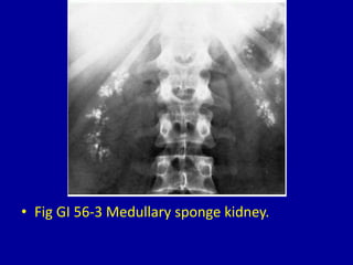
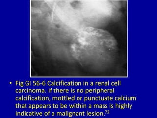
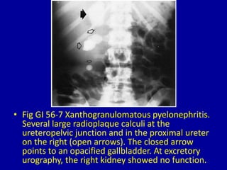
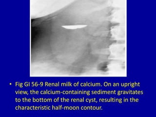
6. • Fig GI 56-4 Lipoid pneumonia. Multifocal areas
of consolidation in the right lung containing
areas of fat attenuation. (Courtesy of Michael
B. Gotway, MD, Scottsdale, A Z.)
7. • Fig GI 56-5 Simple renal cyst. Curvilinear,
peripheral calcification outlines part of the
cyst wall (arrows). Smooth splaying of upper
pole calyces is demonstrated on this film from
an excretory urogram.71
8. • Fig GI 56-6 Calcification in a renal cell
carcinoma. If there is no peripheral
calcification, mottled or punctuate calcium
that appears to be within a mass is highly
indicative of a malignant lesion.72
9. • Fig GI 56-7 Xanthogranulomatous pyelonephritis.
Several large radioplaque calculi at the
ureteropelvic junction and in the proximal ureter
on the right (open arrows). The closed arrow
points to an opacified gallbladder. At excretory
urography, the right kidney showed no function.
10. • Fig GI 56-8 Calcification in a renal artery
aneurysm. (A) Plain abdominal radiograph
demonstrates the circular calcification with a
cracked-eggshell appearance at the renal hilum.
(B) Selective right arteriogram shows contrast
material filling the saccular aneurysm (arrow).
11. • Fig GI 56-9 Renal milk of calcium. On an upright
view, the calcium-containing sediment gravitates
to the bottom of the renal cyst, resulting in the
characteristic half-moon contour.
12. • Fig GI 56-10 Congenital unilateral multicystic
kidney. There are three peripherally calcified
masses, with no excretion of contrast material
on excretory urography.72