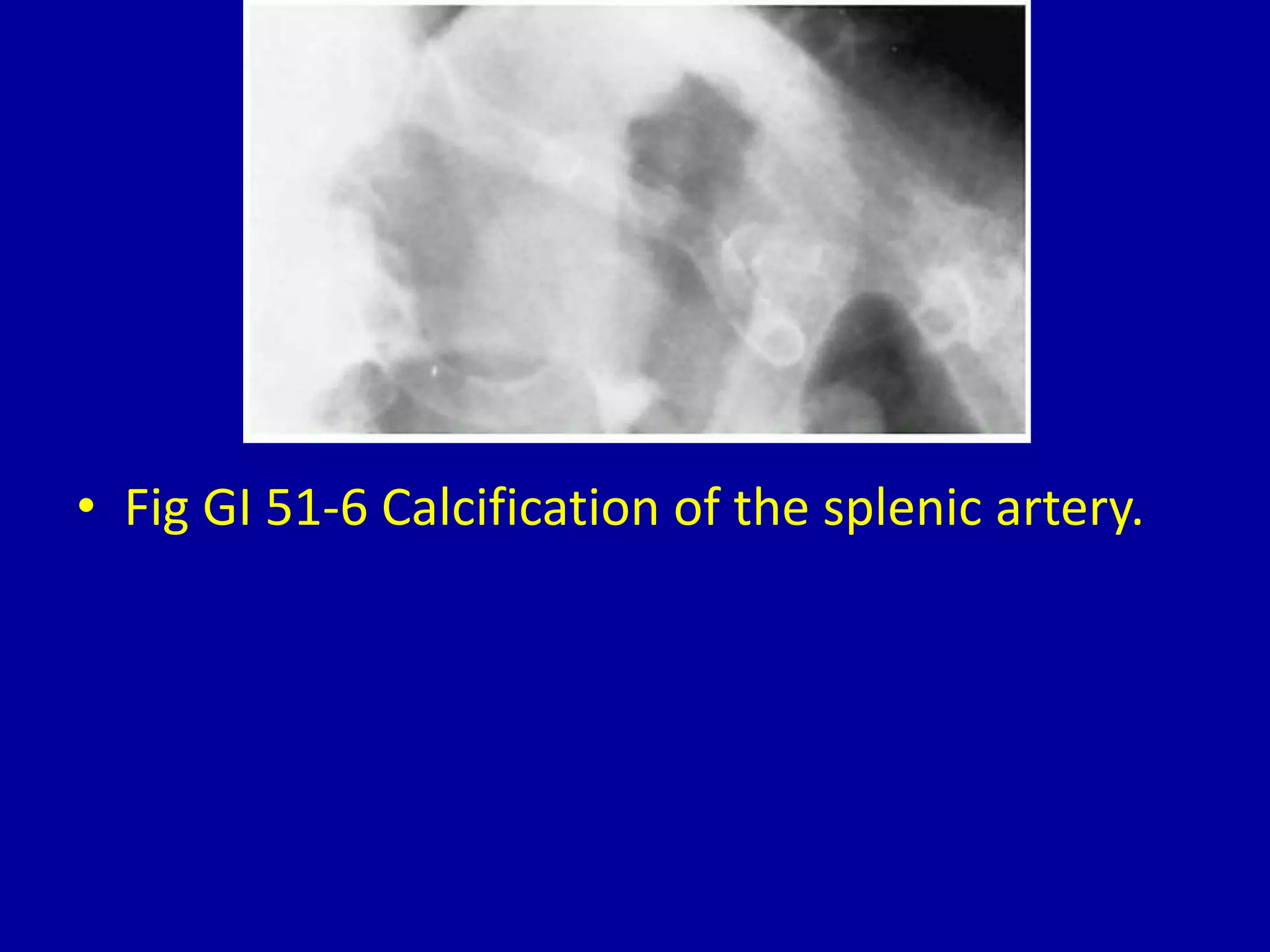
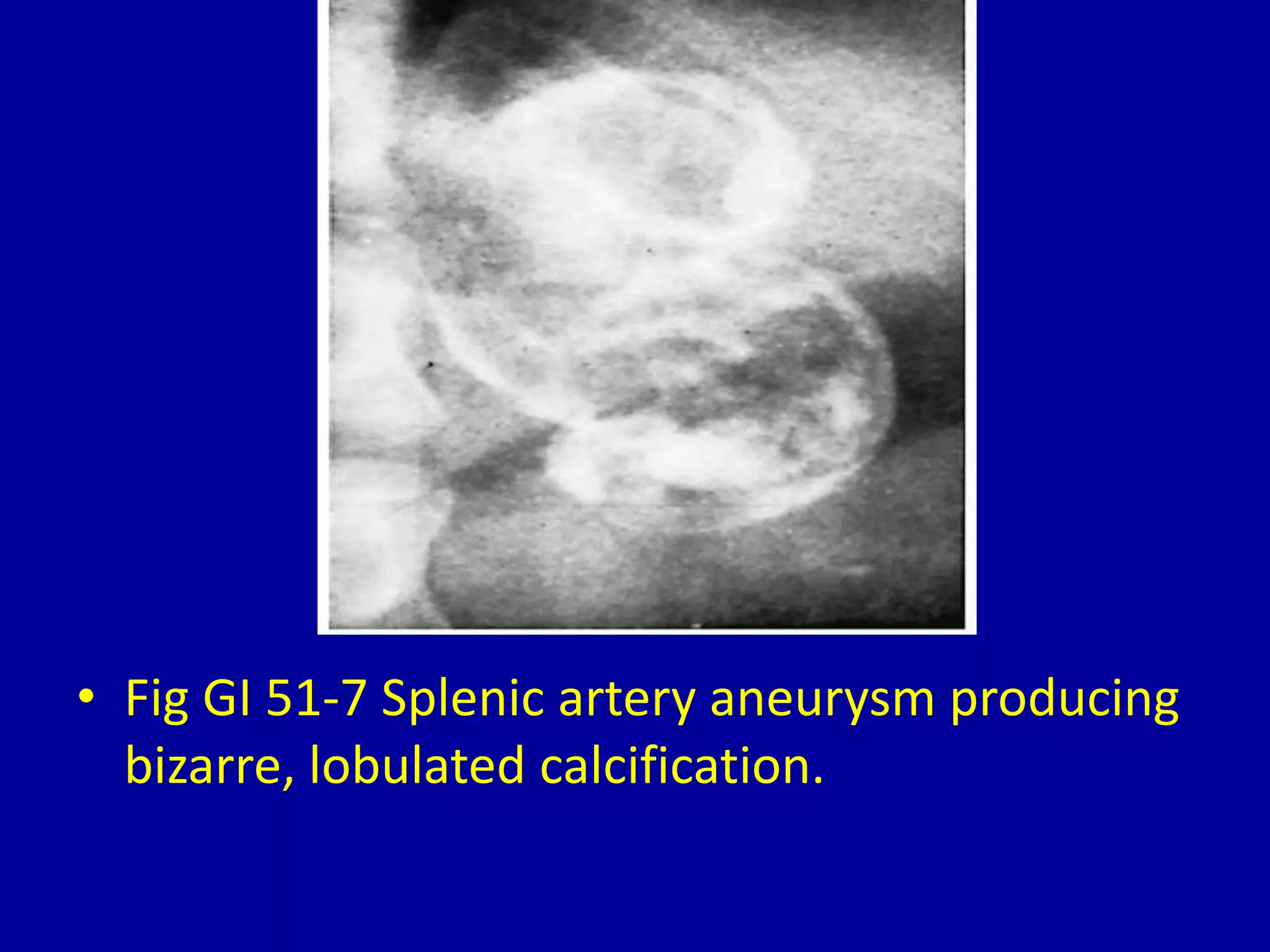
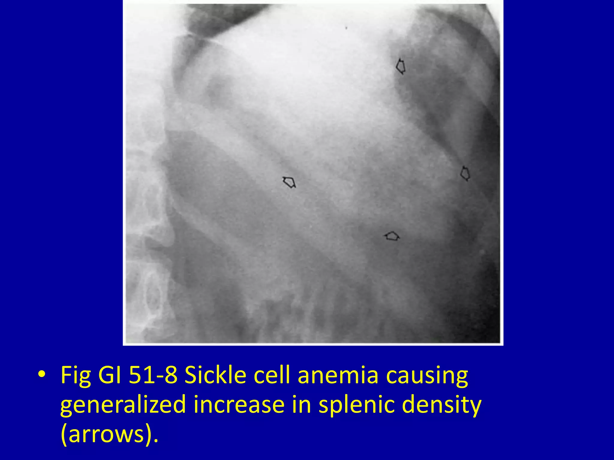
51Spleen Calcification
CLINICAL IMAGAGING
AN ATLAS OF DIFFERENTIAL DAIGNOSIS
EISENBERG
DR. Muhammad Bin Zulfiqar
PGR-FCPS III SIMS/SHL
• Fig GI 51-1 Histoplasmosis. CT scan shows
multiple small calcifications in the spleen.
• Fig GI 51-2 Brucellosis. Large calcified splenic
granulomas.
• Fig GI 51-3 Huge calcified nonparasitic splenic
cyst.
• Fig GI 51-4 Calcified hydatid cyst of the spleen
(echinococcal disease).
• Fig GI 51-5 Calcified splenic hematoma.
• Fig GI 51-6 Calcification of the splenic artery.
• Fig GI 51-7 Splenic artery aneurysm producing
bizarre, lobulated calcification.
• Fig GI 51-8 Sickle cell anemia causing
generalized increase in splenic density
(arrows).
51 spleen calcification

51 spleen calcification

  • 1.
  • 2.
    CLINICAL IMAGAGING AN ATLASOF DIFFERENTIAL DAIGNOSIS EISENBERG DR. Muhammad Bin Zulfiqar PGR-FCPS III SIMS/SHL
  • 3.
    • Fig GI51-1 Histoplasmosis. CT scan shows multiple small calcifications in the spleen.
  • 4.
    • Fig GI51-2 Brucellosis. Large calcified splenic granulomas.
  • 5.
    • Fig GI51-3 Huge calcified nonparasitic splenic cyst.
  • 6.
    • Fig GI51-4 Calcified hydatid cyst of the spleen (echinococcal disease).
  • 7.
    • Fig GI51-5 Calcified splenic hematoma.
  • 8.
    • Fig GI51-6 Calcification of the splenic artery.
  • 9.
    • Fig GI51-7 Splenic artery aneurysm producing bizarre, lobulated calcification.
  • 10.
    • Fig GI51-8 Sickle cell anemia causing generalized increase in splenic density (arrows).