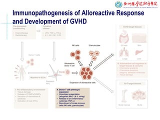
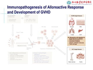
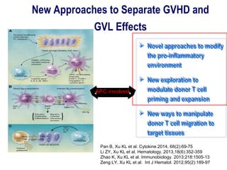
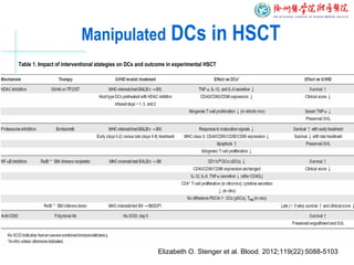
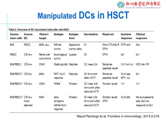
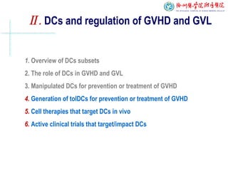
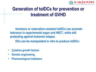
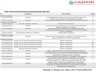
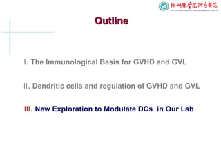
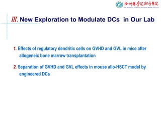
The Advances of regulatory dendritic cells inThe Advances of regulatory dendritic cells in
GVHD and GVL after allogeneic bone marrowGVHD and GVL after allogeneic bone marrow
transplantationtransplantation
Yihong Huang, Kailin Xu ( 黄一虹,徐 林开 )
Institute of Hematology
The Affiliated Hospital of Xuzhou Medical College
(徐州医学院血液病研究所)
OutlineOutline
Ⅱ. Dendritic cells and regulation of GVHD and GVL
Ⅰ. The Immunological Basis for GVHD and GVL
Ⅲ. New Exploration to Modulate DCs in Our Lab
The classical Model of the Immunopathogenesis of
GVHD Described by Three Interlinked Phases
 Pro-inflammatory environment
 Donor T cell proliferation after encountering activated
recipient APCs bearing alloantigen
 Alloreactive cell migration to target tissues and resulting in
tissue damage in the form of GVHD
Ferrara JT, et al. Stem Cells. 1996,14:473-489.
Ferrara JT, et al. Semin Hematol.2006,43:3-10
Immunopathogenesis of Alloreactive Response
and Development of GVHD
Immunopathogenesis of Alloreactive Response
and Development of GVHD
Immunopathogenesis of Alloreactive Response
and Development of GVHD
New Approaches to Separate GVHD and
GVL Effects
 Novel approaches to modify
the pro-inflammatory
environment
 New exploration to
modulate donor T cell
priming and expansion
 New ways to manipulate
donor T cell migration to
target tissues
 Novel approaches to modify
the pro-inflammatory
environment
 New exploration to
modulate donor T cell
priming and expansion
 New ways to manipulate
donor T cell migration to
target tissues
APC involved
Pan B, Xu KL et al. Cytokine.2014, 68(2):69-75
Li ZY, Xu KL et al. Hematology. 2013,18(6):352-359
Zhao K, Xu KL et al. Immunobiology. 2013;218:1505-13
Zeng LY, Xu KL et al. Int J Hematol. 2012,95(2):189-97
OutlineOutline
Ⅱ. Dendritic cells and regulation of GVHD and GVL
Ⅰ. The Immunological Basis for GVHD and GVL
Ⅲ. New Exploration to Modulate DCs in Our Lab
Ⅱ. DCs and regulation of GVHD and GVL
2. The role of DCs in GVHD and GVL
3. Manipulated DCs for prevention or treatment of GVHD
4. Generation of tolDCs for prevention or treatment of GVHD
5. Cell therapies that target DCs in vivo
6. Active clinical trials that target/impact DCs
1. Overview of DCs subsets
Overview of DC subsets
Based on the source of DC Based on the function of DC
Mature DCs(mDCs): enhance T cell
immune response;
ImmatureDCs (iDCs): involved in the
induction of peripheral T cell tolerance under
steady state conditions ,but likely change
into mature under inflammatory conditions;
Regulatory DCs (rDCs)/Tolerogenic
DCs(tolDCs): established DCs with potent
immunoregulatory property, to impair the
functions of alloreactive/xeno reactive T cells.
Elizabeth O. Stenger et al. Blood. 2012;119(22):5088-5103
Ⅱ. DCs and regulation of GVHD and GVL
1. Overview of DCs subsets
2. The role of DCs in GVHD and GVL
3. Manipulated DCs for prevention or treatment of GVHD
4. Generation of tolDCs for prevention or treatment of GVHD
5. Cell therapies that target DCs in vivo
6. Active clinical trials that target/impact DCs
The Important role of Dendritic Cells in GVHD and GVL
 Acute GVHD can be induced by either host or donor APCs, whereas
host APCs are required for the initiation of aGVHD and donor APCs
amplify the process.
 Dendritic cells(DCs) as professional APCs, have been well recognized
for their role in the induction of GVHD and GVL responses .
 Traditional therapies have targeted T cells, yet immunostimulatory
DCs are critical in the pathogenesis of GVHD.
Zhao K, Xu KL et al. J Interferon Cytokine Res. 2014,34(9):707-15 .
Pan B, Xu KL et al. Clinical Immunology (2014) 150, 101–8.
Sang W, Xu KL et al. Immunol Lett . 2011,136(2):194-202 .
The role of DCs in GVHD
 Initial tissue injury may trigger DCs within
tissues and induce DCs to migrate to
draining lymph nodes;
 Migrating DCs “trap” autoreactive T
cells , where they induce T cell activation
and may “imprint” a homing phenotype
that permits selective trafficking of T cells
to GVHD target organs;
 DCs within inflamed tissues may act to
amplify the developing GVHD by providing
further priming signals to T cells.
DCs can influence development of GVHD at multiple levels:
Ronjon Chakraverty et al. Blood. 2007;110:9-17
The role of DCs in GVL
Host DCs are required for full GVL effects, although donor DCs
can initiate GVL activity when low burden of tumor are present..
 Depletion of donor BM CD11b myeloid cells in an experimental
model enhanced survival of recipients with tumor;
 Addition of precursor pDCs, augmented GVL without increase in
GVHD;
 Increased graft pDC is associated with relapse and decreased
patient overall survival (OS).
Li JM et al .Biol Blood Marrow Transplant. 2004;10(8):540-551.
Li JM et al. Immunol. 2009;183(12):7799-7809.
Rajasekar R et al.Biol Blood MarrowTransplant. 2010;16(6):854-860.
Ⅱ. DCs and regulation of GVHD and GVL
1. Overview of DCs subsets
2. The role of DCs in GVHD and GVL
3. Manipulated DCs for prevention or treatment of GVHD
4. Generation of tolDCs for prevention or treatment of GVHD
5. Cell therapies that target DCs in vivo
6. Active clinical trials that target/impact DCs
Manipulated DCs for prevention or
treatment of GVHD
DCs may be targeted using antibodies to cell surface
molecules or can be manipulated in vitro to skew their function
to suppress GVHD.
 Pharmacologic interventions
 Biologic interventions
 Vaccination strategies
Manipulated DCs in HSCT
Table 1. Impact of interventional stategies on DCs and outcome in experimental HSCT
Elizabeth O. Stenger et al. Blood. 2012;119(22):5088-5103
Manipulated DCs in HSCT
Table 2. Overview of DC vaccination trials after allo-HSCt
Maud Plantinga et al. Frontiers in immunology. 2014,5:218
Ⅱ. DCs and regulation of GVHD and GVL
1. Overview of DCs subsets
2. The role of DCs in GVHD and GVL
3. Manipulated DCs for prevention or treatment of GVHD
4. Generation of tolDCs for prevention or treatment of GVHD
5. Cell therapies that target DCs in vivo
6. Active clinical trials that target/impact DCs
Generation of tolDCs for prevention or
treatment of GVHD
Immature or maturation-resistant tolDCs can promote
tolerance in experimental organ and HSCT, while still
protecting against leukemia relapse.
DCs can be manipulated in vitro to produce tolDCs:
• Cytokine growth factors
• Genetic engineering
• Phamacological mediators
Generation of tolerogenic DC in vitro
Usage of tolDC therapy to inbibit GVHD in mice
Table 3. Summary of tolDC used in allo-HSCT
Elizabeth O. Stenger et al. Blood. 2012;119(22):5088-5103
Ⅱ. DCs and regulation of GVHD and GVL
1. Overview of DCs subsets
2. The role of DCs in GVHD and GVL
3. Manipulated DCs for prevention or treatment of GVHD
4. Generation of tolDCs for prevention or treatment of GVHD
5. Cell therapies that target DCs in vivo
6. Active clinical trials that target/impact DCs
Mesenchymal stem cells (MSCs): have shown promise in the
prevention and treatment of GVHD.
• Human MSCs impair DC maturation and induce T-cell
hyporesponsiveness.
• TolDCs generated by coculture of DCs with human MSCs (MSC-
DCs) induce Ag specific Treg via activation of the Notch pathway.
Cell therapies that target DCs in vivo
Myeloid-derived suppressor cells (MDSCs) : modulate both innate
and adaptive immunity with immunosuppressive properties.
•MDSCs inhibit the differentiation and maturation of DCs.
•MDSCs were more potent inhibitors of MHC-mismatched GVHD.
Perla Filippini et al. Expert Rev. Clin. Immunol. 2014,10(10), 1357–1374
Chen W, Xu KL et al. Immunol Lett. 2012,143(2):161-9.
Chen W, Xu KL et al. Cell Biochem Biophys. 2013, 67:1181–1191 .
Cell therapies that target DCs in vivo
Regulatory T cells(Treg):
• Treg therapy supports tolerance through effects on DCs, while DCs
also control the number and function of Treg.
• Human Treg can abrogate GVHD via induction of PD-L1 expression
on conditioned DCs and on effector T cells.
• Ag-specific Treg can be induced and expanded by monDCs in an
IDO-dependent manner.
Cao J, Xu KL et al. Leuk Res. 2010,34(10): 1374-82
Chen C, Xu KL et al. Transplant Proc. 2011,43(5): 2041-8 .
Perla Filippini et al. Expert Rev. Clin. Immunol. 2014,10(10), 1357–1374
DC based methods of transplant tolerence
Adrian E. Morelli et al. Nature review immunology. 2007,7:610-321
Ⅱ. DCs and regulation of GVHD and GVL
1. Overview of DCs subsets
2. The role of DCs in GVHD and GVL
3. Manipulated DCs for prevention or treatment of GVHD
4. Generation of tolDCs for prevention or treatment of GVHD
5. Cell therapies that target DCs in vivo
6. Active clinical trials that target/impact DCs
There are numerous open clinical trials for the prevention
or treatment of GVHD currently studying pharmacologic or
biologic interventions and cellular therapies that target or
impact DCs.
Active clinical trials that target/impact DCs
Table 4. Active clinical trials using interventions that target/ impact DCs
Elizabeth O. Stenger et al. Blood. 2012;119(22):5088-5103
OutlineOutline
Ⅱ. Dendritic cells and regulation of GVHD and GVL
Ⅰ. The Immunological Basis for GVHD and GVL
Ⅲ. New Exploration to Modulate DCs in Our Lab
1. Effects of regulatory dendritic cells on GVHD and GVL in mice after
allogeneic bone marrow transplantation
2. Separation of GVHD and GVL effects in mouse allo-HSCT model by
engineered DCs
Part 1:
Effects of regulatory dendritic cells on GVHD and GVL in mice after
allogeneic bone marrow transplantation
Generation of inducible DCreg
A, imDC ; B, mDC ; C, DCreg ; D, LPS stimulated regDC
Figure 1. The cell morphological observation of DC subsets
 Compared with mDCs, shorter synapsis was shown in DCreg;
and rare wrinkles were observed in DCreg.
Surface molecules expression on DCs
mDCimDC DCreg
 Co-stimulatory molecules were down regulated in DCreg
Figure 2. FACS was used to detected the molecules of expressed on DCs
Mice survival after transplantation
 The first death in the allo-
BMT group was found on 10
d, and all mice died within
25 d;
 Most of the mice in the
imDC group died within 21
d, while 20% of the mice
were still alive at 30 d;
 The first death in the DCreg
group was found on 19 d,
while 46.7% of the mice
were still alive at 30 d.
Figure 3. The survival rate of mice with allo-BMT
Chen C, Xu KL. Leukemia Research, 2014 , 38(12):1460-8
Effects of DCreg on pathological changes in
GVHD tissues after transplantation
 Liver cell edema, focal necrosis, lymphocyte infiltration at the portal area,
intestinal epithelial cells necrosis, significant inflammatory cell infiltration
in the pulmonary vessels in the allo-BMT and imDC groups;
 Mice in the DCreg group, we found only mild or even no pathological
damage in target organs.
Normal allo-BMT imDC DCreg
Liver
Gut
Lung
Figure 4. Target organs were detected after HE staining
Pathological GVHD score was low in DCreg group
Groups Liver Intestine Lung
allo-BMT 6.33±0.58 7.00±1.00 6.67±0.58
imDC 4.67±0.58* 4.67±0.58* 2.33±1.16*
DCreg 3.00±1.00*#
2.67±0.58*#
2.00±1.00*#
Table 1. GVHD pathologic scoring of liver, small intestine, and lung in mice after transplantation (n = 3, )sx ±
*P < 0.05 vs allo-BMT group; #P < 0.05 vs imDC group
Cytokine levels in plasma
 IFN-γ levels increased significantly in the allo-BMT group, whereas low
level of that was detected in DCreg group.
 IL-10 as immune surppressor cytokine increased obviously in Dcreg
mice.
Figure 5. cytokines from allo-BMT mice were detected by ELISA
Part 2:
Suppression of graft-vs-host disease and retention of graft-vs-
leukemia reaction by murine genetic engineering dendritic cells
after bone marrow transplantation
Establishment of engineered DCs

Lentivirus-mediated expression of soluble tumor necrosis factor
receptor 1 (sTNFR1) converted immature DC were established in
vitro, named sTNFR-DC;

PXZ9-DC were used as negetive vector control;
Mice survival following transplantation
 Mice from the leukemia model
group began to die after 13 days,
and all died due to leukemia
within 18 days;
 allo-BMT group began to die
after 8 days, and all died by 18
days
 Most of the pXZ9-imDC group
mice died within 21 days
 sTNFR1-imDC group mice died
starting at 15 day, and 40% of
mice survived up to 30 days
Figure 6. The survival rate of mice after allo-BMT
Pan B, Xu KL. Immunol Lett . 2012,142(1-2):48-54
Evaluation of GVHD
 High GVHD clinical score were
evaluated in allo-BMT mice ,
whereas injected sTNFR-DC
mice showed slight GVHD score.
 More severe pathological
damage were observed in allo-
BMT group, but mild changes
were shown in sTNFR-DC mice.
Skin
Liver
Gut
Allo-BMT sTNFR1-DC PXZ9-DC
Figure 6. The clinical score and pathological changes in mice
Cytokine levels in plasma
 IFN-γ levels increased
significantly in the allo-BMT
group and PXZ9-DC group,
whereas low level of IFN-γ was
detected in sTNFR-DC group.
 IL-4 which was considered as
immune surppressor cytokine
increased obviously in sTNFR-
DC group.
Figure 7. Cytokine expression in mice after allo-BMT
Prospective

Synergistic therapies or those that
target both T and DC cells may be
more effective.

The injection site, dose and frequency
of the tolDCs induced in vitro should be
carefully considered.
Potential DC-based therapies for GVHD :
Thank You for Your Attention

5 huang yihong

  • 1.
    The Advances ofregulatory dendritic cells inThe Advances of regulatory dendritic cells in GVHD and GVL after allogeneic bone marrowGVHD and GVL after allogeneic bone marrow transplantationtransplantation Yihong Huang, Kailin Xu ( 黄一虹,徐 林开 ) Institute of Hematology The Affiliated Hospital of Xuzhou Medical College (徐州医学院血液病研究所)
  • 2.
    OutlineOutline Ⅱ. Dendritic cellsand regulation of GVHD and GVL Ⅰ. The Immunological Basis for GVHD and GVL Ⅲ. New Exploration to Modulate DCs in Our Lab
  • 3.
    The classical Modelof the Immunopathogenesis of GVHD Described by Three Interlinked Phases  Pro-inflammatory environment  Donor T cell proliferation after encountering activated recipient APCs bearing alloantigen  Alloreactive cell migration to target tissues and resulting in tissue damage in the form of GVHD Ferrara JT, et al. Stem Cells. 1996,14:473-489.
  • 4.
    Ferrara JT, etal. Semin Hematol.2006,43:3-10 Immunopathogenesis of Alloreactive Response and Development of GVHD
  • 5.
    Immunopathogenesis of AlloreactiveResponse and Development of GVHD
  • 6.
    Immunopathogenesis of AlloreactiveResponse and Development of GVHD
  • 7.
    New Approaches toSeparate GVHD and GVL Effects  Novel approaches to modify the pro-inflammatory environment  New exploration to modulate donor T cell priming and expansion  New ways to manipulate donor T cell migration to target tissues  Novel approaches to modify the pro-inflammatory environment  New exploration to modulate donor T cell priming and expansion  New ways to manipulate donor T cell migration to target tissues APC involved Pan B, Xu KL et al. Cytokine.2014, 68(2):69-75 Li ZY, Xu KL et al. Hematology. 2013,18(6):352-359 Zhao K, Xu KL et al. Immunobiology. 2013;218:1505-13 Zeng LY, Xu KL et al. Int J Hematol. 2012,95(2):189-97
  • 8.
    OutlineOutline Ⅱ. Dendritic cellsand regulation of GVHD and GVL Ⅰ. The Immunological Basis for GVHD and GVL Ⅲ. New Exploration to Modulate DCs in Our Lab
  • 9.
    Ⅱ. DCs andregulation of GVHD and GVL 2. The role of DCs in GVHD and GVL 3. Manipulated DCs for prevention or treatment of GVHD 4. Generation of tolDCs for prevention or treatment of GVHD 5. Cell therapies that target DCs in vivo 6. Active clinical trials that target/impact DCs 1. Overview of DCs subsets
  • 10.
    Overview of DCsubsets Based on the source of DC Based on the function of DC Mature DCs(mDCs): enhance T cell immune response; ImmatureDCs (iDCs): involved in the induction of peripheral T cell tolerance under steady state conditions ,but likely change into mature under inflammatory conditions; Regulatory DCs (rDCs)/Tolerogenic DCs(tolDCs): established DCs with potent immunoregulatory property, to impair the functions of alloreactive/xeno reactive T cells. Elizabeth O. Stenger et al. Blood. 2012;119(22):5088-5103
  • 11.
    Ⅱ. DCs andregulation of GVHD and GVL 1. Overview of DCs subsets 2. The role of DCs in GVHD and GVL 3. Manipulated DCs for prevention or treatment of GVHD 4. Generation of tolDCs for prevention or treatment of GVHD 5. Cell therapies that target DCs in vivo 6. Active clinical trials that target/impact DCs
  • 12.
    The Important roleof Dendritic Cells in GVHD and GVL  Acute GVHD can be induced by either host or donor APCs, whereas host APCs are required for the initiation of aGVHD and donor APCs amplify the process.  Dendritic cells(DCs) as professional APCs, have been well recognized for their role in the induction of GVHD and GVL responses .  Traditional therapies have targeted T cells, yet immunostimulatory DCs are critical in the pathogenesis of GVHD. Zhao K, Xu KL et al. J Interferon Cytokine Res. 2014,34(9):707-15 . Pan B, Xu KL et al. Clinical Immunology (2014) 150, 101–8. Sang W, Xu KL et al. Immunol Lett . 2011,136(2):194-202 .
  • 13.
    The role ofDCs in GVHD  Initial tissue injury may trigger DCs within tissues and induce DCs to migrate to draining lymph nodes;  Migrating DCs “trap” autoreactive T cells , where they induce T cell activation and may “imprint” a homing phenotype that permits selective trafficking of T cells to GVHD target organs;  DCs within inflamed tissues may act to amplify the developing GVHD by providing further priming signals to T cells. DCs can influence development of GVHD at multiple levels: Ronjon Chakraverty et al. Blood. 2007;110:9-17
  • 14.
    The role ofDCs in GVL Host DCs are required for full GVL effects, although donor DCs can initiate GVL activity when low burden of tumor are present..  Depletion of donor BM CD11b myeloid cells in an experimental model enhanced survival of recipients with tumor;  Addition of precursor pDCs, augmented GVL without increase in GVHD;  Increased graft pDC is associated with relapse and decreased patient overall survival (OS). Li JM et al .Biol Blood Marrow Transplant. 2004;10(8):540-551. Li JM et al. Immunol. 2009;183(12):7799-7809. Rajasekar R et al.Biol Blood MarrowTransplant. 2010;16(6):854-860.
  • 15.
    Ⅱ. DCs andregulation of GVHD and GVL 1. Overview of DCs subsets 2. The role of DCs in GVHD and GVL 3. Manipulated DCs for prevention or treatment of GVHD 4. Generation of tolDCs for prevention or treatment of GVHD 5. Cell therapies that target DCs in vivo 6. Active clinical trials that target/impact DCs
  • 16.
    Manipulated DCs forprevention or treatment of GVHD DCs may be targeted using antibodies to cell surface molecules or can be manipulated in vitro to skew their function to suppress GVHD.  Pharmacologic interventions  Biologic interventions  Vaccination strategies
  • 17.
    Manipulated DCs inHSCT Table 1. Impact of interventional stategies on DCs and outcome in experimental HSCT Elizabeth O. Stenger et al. Blood. 2012;119(22):5088-5103
  • 18.
    Manipulated DCs inHSCT Table 2. Overview of DC vaccination trials after allo-HSCt Maud Plantinga et al. Frontiers in immunology. 2014,5:218
  • 19.
    Ⅱ. DCs andregulation of GVHD and GVL 1. Overview of DCs subsets 2. The role of DCs in GVHD and GVL 3. Manipulated DCs for prevention or treatment of GVHD 4. Generation of tolDCs for prevention or treatment of GVHD 5. Cell therapies that target DCs in vivo 6. Active clinical trials that target/impact DCs
  • 20.
    Generation of tolDCsfor prevention or treatment of GVHD Immature or maturation-resistant tolDCs can promote tolerance in experimental organ and HSCT, while still protecting against leukemia relapse. DCs can be manipulated in vitro to produce tolDCs: • Cytokine growth factors • Genetic engineering • Phamacological mediators
  • 21.
  • 22.
    Usage of tolDCtherapy to inbibit GVHD in mice Table 3. Summary of tolDC used in allo-HSCT Elizabeth O. Stenger et al. Blood. 2012;119(22):5088-5103
  • 23.
    Ⅱ. DCs andregulation of GVHD and GVL 1. Overview of DCs subsets 2. The role of DCs in GVHD and GVL 3. Manipulated DCs for prevention or treatment of GVHD 4. Generation of tolDCs for prevention or treatment of GVHD 5. Cell therapies that target DCs in vivo 6. Active clinical trials that target/impact DCs
  • 24.
    Mesenchymal stem cells(MSCs): have shown promise in the prevention and treatment of GVHD. • Human MSCs impair DC maturation and induce T-cell hyporesponsiveness. • TolDCs generated by coculture of DCs with human MSCs (MSC- DCs) induce Ag specific Treg via activation of the Notch pathway. Cell therapies that target DCs in vivo Myeloid-derived suppressor cells (MDSCs) : modulate both innate and adaptive immunity with immunosuppressive properties. •MDSCs inhibit the differentiation and maturation of DCs. •MDSCs were more potent inhibitors of MHC-mismatched GVHD. Perla Filippini et al. Expert Rev. Clin. Immunol. 2014,10(10), 1357–1374 Chen W, Xu KL et al. Immunol Lett. 2012,143(2):161-9. Chen W, Xu KL et al. Cell Biochem Biophys. 2013, 67:1181–1191 .
  • 25.
    Cell therapies thattarget DCs in vivo Regulatory T cells(Treg): • Treg therapy supports tolerance through effects on DCs, while DCs also control the number and function of Treg. • Human Treg can abrogate GVHD via induction of PD-L1 expression on conditioned DCs and on effector T cells. • Ag-specific Treg can be induced and expanded by monDCs in an IDO-dependent manner. Cao J, Xu KL et al. Leuk Res. 2010,34(10): 1374-82 Chen C, Xu KL et al. Transplant Proc. 2011,43(5): 2041-8 . Perla Filippini et al. Expert Rev. Clin. Immunol. 2014,10(10), 1357–1374
  • 26.
    DC based methodsof transplant tolerence Adrian E. Morelli et al. Nature review immunology. 2007,7:610-321
  • 27.
    Ⅱ. DCs andregulation of GVHD and GVL 1. Overview of DCs subsets 2. The role of DCs in GVHD and GVL 3. Manipulated DCs for prevention or treatment of GVHD 4. Generation of tolDCs for prevention or treatment of GVHD 5. Cell therapies that target DCs in vivo 6. Active clinical trials that target/impact DCs
  • 28.
    There are numerousopen clinical trials for the prevention or treatment of GVHD currently studying pharmacologic or biologic interventions and cellular therapies that target or impact DCs. Active clinical trials that target/impact DCs
  • 29.
    Table 4. Activeclinical trials using interventions that target/ impact DCs Elizabeth O. Stenger et al. Blood. 2012;119(22):5088-5103
  • 30.
    OutlineOutline Ⅱ. Dendritic cellsand regulation of GVHD and GVL Ⅰ. The Immunological Basis for GVHD and GVL Ⅲ. New Exploration to Modulate DCs in Our Lab
  • 31.
    Ⅲ. New Explorationto Modulate DCs in Our Lab 1. Effects of regulatory dendritic cells on GVHD and GVL in mice after allogeneic bone marrow transplantation 2. Separation of GVHD and GVL effects in mouse allo-HSCT model by engineered DCs
  • 32.
    Part 1: Effects ofregulatory dendritic cells on GVHD and GVL in mice after allogeneic bone marrow transplantation
  • 33.
    Generation of inducibleDCreg A, imDC ; B, mDC ; C, DCreg ; D, LPS stimulated regDC Figure 1. The cell morphological observation of DC subsets  Compared with mDCs, shorter synapsis was shown in DCreg; and rare wrinkles were observed in DCreg.
  • 34.
    Surface molecules expressionon DCs mDCimDC DCreg  Co-stimulatory molecules were down regulated in DCreg Figure 2. FACS was used to detected the molecules of expressed on DCs
  • 35.
    Mice survival aftertransplantation  The first death in the allo- BMT group was found on 10 d, and all mice died within 25 d;  Most of the mice in the imDC group died within 21 d, while 20% of the mice were still alive at 30 d;  The first death in the DCreg group was found on 19 d, while 46.7% of the mice were still alive at 30 d. Figure 3. The survival rate of mice with allo-BMT Chen C, Xu KL. Leukemia Research, 2014 , 38(12):1460-8
  • 36.
    Effects of DCregon pathological changes in GVHD tissues after transplantation  Liver cell edema, focal necrosis, lymphocyte infiltration at the portal area, intestinal epithelial cells necrosis, significant inflammatory cell infiltration in the pulmonary vessels in the allo-BMT and imDC groups;  Mice in the DCreg group, we found only mild or even no pathological damage in target organs. Normal allo-BMT imDC DCreg Liver Gut Lung Figure 4. Target organs were detected after HE staining
  • 37.
    Pathological GVHD scorewas low in DCreg group Groups Liver Intestine Lung allo-BMT 6.33±0.58 7.00±1.00 6.67±0.58 imDC 4.67±0.58* 4.67±0.58* 2.33±1.16* DCreg 3.00±1.00*# 2.67±0.58*# 2.00±1.00*# Table 1. GVHD pathologic scoring of liver, small intestine, and lung in mice after transplantation (n = 3, )sx ± *P < 0.05 vs allo-BMT group; #P < 0.05 vs imDC group
  • 38.
    Cytokine levels inplasma  IFN-γ levels increased significantly in the allo-BMT group, whereas low level of that was detected in DCreg group.  IL-10 as immune surppressor cytokine increased obviously in Dcreg mice. Figure 5. cytokines from allo-BMT mice were detected by ELISA
  • 39.
    Part 2: Suppression ofgraft-vs-host disease and retention of graft-vs- leukemia reaction by murine genetic engineering dendritic cells after bone marrow transplantation
  • 40.
    Establishment of engineeredDCs  Lentivirus-mediated expression of soluble tumor necrosis factor receptor 1 (sTNFR1) converted immature DC were established in vitro, named sTNFR-DC;  PXZ9-DC were used as negetive vector control;
  • 41.
    Mice survival followingtransplantation  Mice from the leukemia model group began to die after 13 days, and all died due to leukemia within 18 days;  allo-BMT group began to die after 8 days, and all died by 18 days  Most of the pXZ9-imDC group mice died within 21 days  sTNFR1-imDC group mice died starting at 15 day, and 40% of mice survived up to 30 days Figure 6. The survival rate of mice after allo-BMT Pan B, Xu KL. Immunol Lett . 2012,142(1-2):48-54
  • 42.
    Evaluation of GVHD High GVHD clinical score were evaluated in allo-BMT mice , whereas injected sTNFR-DC mice showed slight GVHD score.  More severe pathological damage were observed in allo- BMT group, but mild changes were shown in sTNFR-DC mice. Skin Liver Gut Allo-BMT sTNFR1-DC PXZ9-DC Figure 6. The clinical score and pathological changes in mice
  • 43.
    Cytokine levels inplasma  IFN-γ levels increased significantly in the allo-BMT group and PXZ9-DC group, whereas low level of IFN-γ was detected in sTNFR-DC group.  IL-4 which was considered as immune surppressor cytokine increased obviously in sTNFR- DC group. Figure 7. Cytokine expression in mice after allo-BMT
  • 44.
    Prospective  Synergistic therapies orthose that target both T and DC cells may be more effective.  The injection site, dose and frequency of the tolDCs induced in vitro should be carefully considered. Potential DC-based therapies for GVHD :
  • 45.
    Thank You forYour Attention

Editor's Notes

  • #5 移植前化疗或照射等预处理,使APC细胞活化,进而促使CD4+T细胞活化,产生大量的效应性细胞因子,导致靶器官的损伤,其中CD4+T细胞活化为中心环节
  • #6 移植前化疗或照射等预处理,使APC细胞活化,进而促使CD4+T细胞活化,产生大量的效应性细胞因子,导致靶器官的损伤,其中CD4+T细胞活化为中心环节
  • #7 移植前化疗或照射等预处理,使APC细胞活化,进而促使CD4+T细胞活化,产生大量的效应性细胞因子,导致靶器官的损伤,其中CD4+T细胞活化为中心环节