Download to read offline

![Dash et al.: Immunophenotypic enumeration of CD4+ T-lymphocytes in HIV negative adult
Int J Med Biomed Res 2012;1(3):242-249
243
INTRODUCTION
T-lymphocytes are defined by the expression of CD3+ T cell subpopulations by the co-expression of CD4+ or CD8+ or HLA-DR.[1] CD4+ T-helper lymphocytes play a central role in regulation of immune response.[2] These cells have capacity to help B cells for generating antibodies, to recruit and activate macrophages, to recruit neutrophils, eosinophils, and basophils to sites of infection and inflammation.[3] As the CD4+ T- lymphocytes are main targets of human immunodeficiency virus (HIV), CD4+-lymphocyte counts (LCs) are recognized as the most important measurement of overall HIV-induced immune impairment.[4] The enumeration of CD4+ T-lymphocytes in HIV infected individuals is an essential tool for staging HIV disease, to make decisions for initiation of anti-retroviral therapy (ART), for monitoring response to ART and to initiate chemoprophylaxis against opportunistic infections.[5,6,7] Besides HIV disease, the clinical applications of CD4+ LCs include diagnosis of primary and secondary immunodeficiency disorders, evaluation of immune-mediated diseases and the assessment of immune reconstitution following stem cell transplantation.[8,9,10]
Variability in CD4+ LCs among healthy HIV- seronegative adults has been widely reported and has been attributed to biological, ethnic group influences as well as differences in the methodologies used for T-cell enumeration. Therefore, it is important to know the level of immunocompetence of a particular geographical region by enumerating the baseline CD4+ LCs in HIV-seronegative healthy adults.
There have been studies of CD4+ LCs among HIV-seronegative healthy adults reported from northeast, north, west, northwest, south region of India including a multi-centric study.[11-23] Also studies have been reported from different parts of the world.[24-36] To the best of our knowledge, no study has been conducted from eastern region of India. Hence, we undertook this present study to determine the baseline CD4+ LCs among HIV-seronegative healthy adults who attended integrated counselling and testing centre 1 (ICTC-1) for HIV information in this part of the country to access feasibility of extending it to a larger study group with the purpose of establishing a normal reference range among Indian population.
MATERIALS AND METHODS
Study area and population
The present study was carried out in the Department of Microbiology, integrated counselling and testing centre 1 (ICTC-1), which is a tertiary referral hospital of eastern India. Hundred HIV-seronegative healthy adult volunteers who attended ICTC-1 for HIV information aged between 18 to 55 years were included in the study. These are sexually active and thus more vulnerable to HIV infection and other sexually transmitted diseases.. The exclusion criteria were (a) Any minor illness during the past one month (b) Any major illness, including surgery, trauma and accident during the past six months (c) Any chronic illness (d) Vaccination within past six months (e) Pregnant women (f) Active drug administration (g) HIV seropositive volunteers. Subjects willing to participate were included in this study after obtaining informed verbal consent. All the tests were done in accordance with the Medical College ethical committee guidelines. The findings were analyzed over a period of one year from July 2011 to June 2012.
Sample collection and processing
Five milliliters (ml) of unlysed whole-blood sample was collected at fixed time interval between 10 am and noon. Two ml of blood was transferred to a sterile vial for HIV serology and 3 ml of blood transferred to K2 EDTA containing vacutainer tube for CD4+ LCs.
HIV serology for screening
Samples were subjected to a rapid screening HIV 1 and 2 Immunodot Test (COMBAIDS® - RS Advantage- ST kit, Span Diagnostics Ltd., Surat, India). The test was done according to manufacturer’s instructions. All hundred volunteers were HIV negative.
Immunophenotypic enumeration of CD4+ T-lymphocytes by using flow cytometer
Immunophenotyping of lymphocytes was carried out by BD FACS™ Calibour system (Becton Dickinson, Fluorescent antibody cell sorter,](https://image.slidesharecdn.com/cd4lymphocytes-140913201552-phpapp01/85/Immunophenotypic-enumeration-of-CD4-T-lymphocyte-values-in-human-immunodeficiency-virus-seronegative-adults-in-Eastern-India-2-320.jpg)
![Dash et al.: Immunophenotypic enumeration of CD4+ T-lymphocytes in HIV negative adult
Int J Med Biomed Res 2012;1(3):242-249
244
Singapore) by using 50 μl of well mixed whole- blood collected in K2 EDTA containing vacutainer tube. Three antibody panels were used i.e., BD Tri TEST™ CD3 fluorescein isothiocyanate (FITC)/CD4 phycoerythrin (PE)/CD45 peridinin chlorophyll protein (PerCP), a three-color direct immunofluorescence reagent to identify and determine the percentages and absolute counts of mature T-lymphocytes (CD3+) and helper T-lymphocyte (CD3+CD4+) subsets in erythrocyte-lysed whole-blood, by using Tru Count™ tubes. The absolute CD4+ LCs in the present study were measured with the FACS Calibour system, using single platform technology which is regarded as a reliable and robust method for the enumeration of CD4+ lymphocytes.[37,38]
Quality control
All tests were done according to manufacturer’s recommendations. Manufacturer’s guidelines were strictly adhered to with regard to biosafety practices, trouble-shooting, and maintenance of equipments. The laboratory also participates in the external quality assurance scheme (EQAS) conducted by National AIDS Research Institute (NARI), Pune, India.
Statistical analysis
The values of mean, median and standard deviation of CD4+ T-lymphocytes were calculated using GraphPad® InStat statistical software. Statistical significance was defined when P-value < 0.05.
RESULTS
One hundred HIV-seronegative healthy adults
who attended ICTC I for HIV information were included in the study: 67(67%) were male and 33(33%) were female. The age of the subjects included in the present study were ranged from 18 to 55 years, with a mean age of 32.6 ±11.3 years and median of 30.5 years. The mean age of male was 33.6 ±11.6 years (range, 18 to 55 years) and female was 30.7 ±10.7 years (range, 18 to 55 years).
The overall mean absolute CD4+ LCs in the present study population was 823.9±243.4 cells/μl, median 847 cells/μl and reference range of 338 to 1321 cells/μl. The male showed mean
absolute CD4+ LCs of 793.4 ±243.5 (median=820), and female revealed mean CD4+
LCs of 885.9 ±234.8 (median=896 cells/μl). The reference range of CD4+ LCs was 338 to 1292 in male and 402 to 1321 cells/μl in female. The P value of mean absolute CD4+ LCs equals to 0.001 (Table 1). In 11 subjects (one female and ten male), the absolute CD4+ LCs were less than 500 cells/μl. The overall mean percentage of CD4+ LCs in this present study was 40.5±8.9. The percentage of CD4+ LCs in male and female was 40.1 (±9.2) and 41.3 (±8.8) respectively. The reference range in percentage was 22.6 to 61.2 and 26.1 to 60.4 in males and females respectively.
The subjects were grouped by age; 18-27, 28- 37, 38-47 and 48-55 years. The distribution of mean CD4+ LCs declined with age. Individuals between 18 to 27 years of age group had 893.3±43.4 cells/μl, followed by 832.4±43.6 in 28 to 37 years, 803.1±67 in 38 to 47 years and least 758.4±95.2 cells/μl in the age group of 48 to 55 years (Table 2).
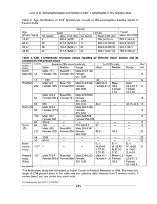
Table 1: CD4+ lymphocyte counts in HIV-seronegative healthy adults in Eastern India
P-value of mean absolute CD4 cell count equals to 0.0001, considered to be extremely statistically significant.
Study
group
No. of subjects
Absolute CD4 count (cells/μl)
% of CD4 cells
Mean (SD)
Median
Reference range
Mean (SD)
Median
Reference range
Male
67
793.4(±243.5)
820
338-1292
40.1(±9.2)
40.9
22.6-61.2
Female
33
885.9(±234.8)
896
402-1321
41.3(±8.8)
39.5
26.1-60.4
Total
100
823.9(±243.4)
847
338-1321
40.5(±8.9)
39.7
22.6-61.2](https://image.slidesharecdn.com/cd4lymphocytes-140913201552-phpapp01/85/Immunophenotypic-enumeration-of-CD4-T-lymphocyte-values-in-human-immunodeficiency-virus-seronegative-adults-in-Eastern-India-3-320.jpg)
![Dash et al.: Immunophenotypic enumeration of CD4+ T-lymphocytes in HIV negative adult
Int J Med Biomed Res 2012;1(3):242-249
246
DISCUSSION
The primary objective of this present study was to characterize CD4+ LCs among representative populations of HIV-seronegative healthy adults in eastern India, the first estimates of CD4+ LCs in this part of the country. The CD4+ LCs has been shown to be influenced by sex, age, race, time of specimen collection (diurnal rhythms), physical and psychological stress, pregnancy, drug administration (zidovudine, cephalosporin,
cancer chemotherapy, nicotine and steroids), tuberculosis, viral infections, presence of anti- lymphocyte auto antibodies and procedures like spleenectomy.[39,40] Other factors that cause variations in the CD4+ LCs were type of instrument used, processing and analyzing the whole-blood samples, integrity of the blood samples, staining reagents and fluorochromes, equipment calibration, preference and gating strategies used for the analysis of the results.[41,42]
Table 4: CD4 T-lymphocyte reference values reported by different countries worldwide and its comparison with present study
Geographical location
No. of subjects
Mean (SD) of absolute CD4 count (cells/μl)
Mean (SD) of percentage CD4 count
Reference No.
Shanghai, China
614
727 (±255)
--
24
Thailand
150
910 (±300)
25
Saudi Arabia
209
869 (±310)
39.4(±7.9)
26
Asian
population
including
China, Malaysia
and India
232
838(±268)
35.6(±6.3)
27
Turkey
220
1095(±341)
47.37(±9.1)
28
Botswana
437
759 (±245)
--
29
Tanzania
147
980(±310)
--
30
Cameroon
203
980
--
31
Uganda
183
1256
--
32
Ethiopia
142
775(±225)
--
33
Central
African Republic
150
933(±320)
--
34
Netherlands
1356
993(±319)
33
United Kingdom
676
830(±290)
43.6(±8.9)
35
Italy
965
940.5
45.1
1
United
States
(Caucasian population)
304
--
44(±7.6)
36
Present study
(East India)
100
823.9(±243.4)
40.5(±8.9)
The mean absolute CD4+ LCs in the present study population was 823.9±243.4 cells/μl, median 847 cells/μl and reference range from 338 to 1321 cells/μl. Similar mean absolute CD4+ LCs of 818.4 cells/μl were noted by Attili et al.[18] in north India, 834.6 cells/μl by Shahapur et al.[22] in south India and 848 cells/μl by Singh et al.[12] in northeast India. A wide variation in](https://image.slidesharecdn.com/cd4lymphocytes-140913201552-phpapp01/85/Immunophenotypic-enumeration-of-CD4-T-lymphocyte-values-in-human-immunodeficiency-virus-seronegative-adults-in-Eastern-India-5-320.jpg)
![Dash et al.: Immunophenotypic enumeration of CD4+ T-lymphocytes in HIV negative adult
Int J Med Biomed Res 2012;1(3):242-249
247
mean absolute CD4+ LCs has been reported from studies conducted in different parts of India. In south India, Kannangai et al.[21] and Murugavel et al.[20] had reported mean CD4+ LCs of 1048 cells/μl and 926 cells/μl respectively. Uppal et al. in west India had revealed a mean of 865 cells/μl.[15] Kannangai et al. reported a mean of 1048 cells/μl in south India, Murugavel et al. 926 cells/μl in south India and Uppal et al. 865 cells/μl in west India. These results were higher than our study.[15,20,21] In comparison, lower mean absolute CD4+ LCs values were reported by Das et al.[13] 771 cells/μl in west India, Ramalingam et al.[19] 799 cells/μl in south India and Ray et al.[17] 703 cells/μl in north India (Table 3). Wide variations in mean absolute CD4+ LCs have also been reported from various parts of the world. Similar mean absolute CD4+ LCs of 838 cells/μl was observed by chng et al. among healthy Asian population comprising of individuals from China, India and Malaysia.[27] Jannossy et al. in Netherlands, Santagostino et al. in Italy, Yaman et al. in Turkey, Vithayasai et al. in Thailand, Al Quozi in Saudi Arabia, Jones et al. in Uganda, Jannossy et al. in Tanzania and Schnizlein-Bick et al. in Cameroon have reported higher absolute CD4+ LCs values, while Jiang et al. in China, Bussmann et al. in Botswana and Jannossy et al. in Ethiopia have reported lower mean absolute CD4+ LCs of 727, 759 and 775 cells/μl respectively (Table-4).[24-36] The mean percentage (%) of CD4+ LCs in our study was 40.5±8.9 (median=39.7 and reference range from 22.6 to 61.2). A multicentric study was carried out by Indian Council of Medical Research (ICMR) in west, north and south parts of the country had revealed mean percentage of CD4+ LCs of 39.46, 37.38, and 32.43% respectively.[23]
Categorization of data based upon the sex of the subjects indicated a significantly higher mean absolute CD4+ LCs in females (885.9±234.4cells/μl) in comparison to males (793.4±243.5). Our findings are consistent with most of the studies conducted in India and other countries.[11,13,16,20,32,33] It is possible that sex hormones sex hormones influence lymphocyte subpopulations and may be responsible for these sex-related differences.
The distribution of mean absolute CD4+ LCs among different age groups in our study showed that the 18 to 27 age group had a significantly higher CD4+ LCs of (893.3±43.4 cells/μl), followed by gradual decrease in CD4+ LCs among subsequent higher age groups, while lowest count of 758.4±95.2 was recorded in the age group of 47 to 55 years. Similar age-related variations were observed by Oladepo et al. among healthy Nigerian adults.[43] They found out that the 18 to 25 year age group had a significantly higher mean CD4+ LC of 861±288 cells/μl, while the lowest count of 774±433 cells/μl was observed among those older than 60 years of age. This might explain why the elderly people fall ill more often than the younger ones, who have more competent immune systems. However, Uppal et al.[15] in west India and Murugavel et al.[20] in South India revealed that none of the parameters differed significantly in any age groups, implying that in adulthood age had no significant influence on various parameters in their studies.
The mean absolute CD4+ LCs in 11% (one female and ten males) of subjects in the present study were < 500 cells/μl. This implies 11% of healthy adult subjects had some amount of immunosupression.[44] Similar value of 10.6% reported by Ramalingam et al. in normal south Indian healthy individuals.[19] Rungta et al. in northwest India observed mean CD4+ LCs in 20% of the controls were < 500 cells/μl.[14]
There were several limitations to this study. The smaller sample size and the confinement to a single geographical area that restricted the performance of other descriptive investigations. In addition, recent seroconversion would not have been detected by rapid screening for HIV 1 and 2 and might have been included in the analysis of samples from HIV-negative individuals.
CONCLUSIONS
Our findings of CD4+ LCs among HIV- seronegative adults in east India adds to the emerging data supporting the presence of significant differences in reference CD4+ LCs between different populations within and outside the country. The pivotal role of CD4+ LCs is in making decisions on the initiation and monitoring of ART in India and other developing countries. The establishment of normal reference ranges](https://image.slidesharecdn.com/cd4lymphocytes-140913201552-phpapp01/85/Immunophenotypic-enumeration-of-CD4-T-lymphocyte-values-in-human-immunodeficiency-virus-seronegative-adults-in-Eastern-India-6-320.jpg)
![Dash et al.: Immunophenotypic enumeration of CD4+ T-lymphocytes in HIV negative adult
Int J Med Biomed Res 2012;1(3):242-249
248
within the local population is a helpful tool to clinicians for the better clinical management of HIV disease in eastern India. Further cohort study with greater sample size may be required to define the staging of HIV disease in relation to the normal CD4+ T-lymphocyte count subsets in the local general population.
REFERENCES
1. Santagostino A, Garbaccio G, Pistorio A, Bolis V, Camisasca G, Pagliaro P, Girotto M. An
Italian national multicenter study for the definition of reference ranges for normal values of peripheral blood lymphocyte subsets in healthy adults. Haematologica 1999;84:499-504.
2. Zhu J, Paul WE. CD4 T cells: fates, functions, and faults. Blood 2008;112:1557-1569.
3. Mosmann TR, Coffman RL. TH1 and TH2 cells: different patterns of lymphokine secretion lead to different functional properties. Annu Rev Immunol 1989;7:145-173.
4. Mellors JW, Muñoz A, Giorgi JV, Margolick JB, Tassoni CJ, Gupta P, Kingsley LA, Todd JA, Saah AJ, Detels R, Phair JP, Rinaldo CR Jr. Plasma viral load and CD4+ lymphocytes as prognostic markers of HIV-1 infection. Ann Intern Med 1997;126:946-954.
5. CDC.gov [Internet]. 1995 Revised Guidelines for Prophylaxis Against Pneumocystis carinii Pneumonia for Children Infected with or Perinatally Exposed to Human Immunodeficiency Virus. c1995 - [cited 2012 Aug 5]. Available from: http://www.cdc.gov/mmwr/preview/mmwrhtml/00037275.html.
6. Friedland GH. Early treatment for HIV-The Time Has Come. N Engl J Med 1990;322:1000-1002.
7. Gebo KA, Gallant JE, Keruly JC, Moore RD. Absolute CD4 vs. CD4 percentage for predicting the risk of Opportunistic illness in HIV infection. J Acquir Immune Defic Syndr 2004;36:1028-1033.
8. Jain A, Atkinson TP, Lipsky PE, Slater JE, Nelson DL, Strober W. Defects of T-cell effector function and post-thymic maturation in X-linked hyper-IgM syndrome. J Clin Invest 1999;103:1151-1158.
9. Bleesing JJ, Straus SE, Fleisher TA. Autoimmune lymphoproliferative syndrome. A human disorder of abnormal lymphocyte survival. Pediatr Clin North Am 2000;47:1291-1310.
10. Storek J, Witherspoon RP. Immunologic reconstitution after haematopoietic stem cell transplantation. In: Atkinson K, editor, Clinical bone marrow and blood stem cell transplantation, 2nd ed. Cambridge, United Kingdom: Cambridge University Press; 2000. p. 111-146.
11. Singh YG, Dar L, Singh NG. Levels of CD4 and CD8 among the inhabitants of Manipur, India. J Commun Dis 2000;32:201-206.
12. Singh HR, Singh NG, Singh TB. Estimation of CD4+ and CD8+ T-lymphocytes in human immunodeficiency virus infection and acquired immunodeficiency syndrome patients in Manipur. Indian J Med Microbiol 2007;25:126-132.
13. Das BR, Bhanushali AA, Khadapkar R, Jeswani KD, Bhavsar M, Dasgupta A. Reference ranges for lymphocyte subsets in adults from western India: Influence of sex, age and method of enumeration. Indian J Med Sci 2008;62:397-406.
14. Rungta A, Hooja S, Vyas N, Rishi S, Rao A, Gupta S. Enumeration of CD4 and CD8 T lymphocytes in healthy HIV seronegative adults of northwest India: a preliminary study. Indian J Pathol Microbiol 2008;51:127-129.
15. Uppal SS, Verma S, Dhot PS. Normal values of CD4 and CD8 lymphocyte subsets in healthy indian adults and the effects of sex, age, ethnicity, and smoking. Cytometry B Clin Cytom 2003;52:32-36.
16. Nag VL, Agarwal P, Venkatesh V, Rastogi P, Tandon R, Agrawal SK. A pilot study on observations on CD4 and CD8 counts in healthy HIV seronegative individuals. Indian J Med Res 2002;116:45-49.
17. Ray K, Gupta SM, Bala M, Muralidhar S, Kumar J. CD4/CD8 lymphocyte counts in healthy, HIV-positive individuals and AIDS patients. Indian J Med Res 2006;124:319-330.
18. Attili VS, Sundar S, Singh VP, Rai M. Validity of existing CD4+ classification in north Indians, in predicting immune status. J Infect 2005;51:41-46.
19. Ramalingam S, Kannangai R, Zachariah A, Mathai D, Abraham C. CD4 counts of normal and HIV-infected south Indian adults: do we need a new staging system? Natl Med J India 2001;14:335-339.
20. Murugavel KG, Balakrishnan P, Mohanakrishnan J, Solomon SS, Shankar EM, Muthu Sundaram SP, Kumarasamy N, Piwowar-Manning E, Livant E, Mayer KH, Thyagarajan SP, Solomon S. Establishment of T- lymphocyte subset reference intervals in a healthy adult population in Chennai, India. Indian J Med Res 2009;129:59-63.
21. Kannangai R, Kandathil AJ, Ebenezer DL, Nithyanandam G, Samuel P, Abraham OC, Sudarsanam TD, Pulimood SA, Sridharan G. Evidence for lower CD4+ T cell and higher viral load in asymptomatic HIV-1 infected individuals of India: implications for therapy initiation. Indian J Med Microbiol 2008;26:217-221.
22. Shahapur PR, Bairy I, Shivananda PG. CD4 and CD8 reference counts in normal healthy south-Indian adults. Indian J Med Microbiol 2008;26:280-281.
23. Saxena RK, Choudhry V, Nath I, Das SN, Paranjape RS, Babu G. Normal ranges of some select lymphocyte subpopulations in peripheral blood of normal healthy Indians. Curr Sci 2004;86:969-975.
24. Jiang W, Kang L, Lu HZ, Pan X, Lin Q, Pan Q, Xue Y, Weng X, Tang YW. Normal values for CD4 and CD8 lymphocyte subsets in healthy Chinese adults from Shanghai. Clin Diagn Lab Immunol 2004;11:811-813.
25. Vithayasai V, Sirisanthana T, Sakonwasun C, Suvanpiyasiri C. Flow cytometric analysis of T-](https://image.slidesharecdn.com/cd4lymphocytes-140913201552-phpapp01/85/Immunophenotypic-enumeration-of-CD4-T-lymphocyte-values-in-human-immunodeficiency-virus-seronegative-adults-in-Eastern-India-7-320.jpg)
![Dash et al.: Immunophenotypic enumeration of CD4+ T-lymphocytes in HIV negative adult
Int J Med Biomed Res 2012;1(3):242-249
249
lymphocytes subsets in adult Thais. Asian Pac J Allergy Immunol 1997;15:141-146.
26. Al Qouzi A, Al Salamah A, Al Rasheed R, Al Musalam A, Al Khairy K, Kheir O, Al Ajaji S, Hajeer AH. Immunophenotyping of peripheral blood lymphocytes in Saudi men. Clin Diagn Lab Immunol 2002;9:279-281.
27. Chng WJ, Tan GB, Kuperan P. Establishment of adult peripheral blood lymphocyte subset reference range for an Asian population by single-platform flow cytometry: influence of age, sex, and race and comparison with other published studies. Clin Diagn Lab Immunol 2004;11:168-173.
28. Yaman A, Cetiner S, Kibar F, Taşova Y, Seydaoğlu G, Dündar IH. Reference ranges of lymphocyte subsets of healthy adults in Turkey. Med Princ Pract 2005;14:189-193.
29.Bussmann H, Wester CW, Masupu KV, Peter T, Gaolekwe SM, Kim S, Reich AM, Ahn S, Wu Y, Thior I, Essex M, Marlink R. Low CD4+ T-Lymphocyte Values in Human Immunodeficiency Virus-Negative Adults in Botswana. Clin Diagn Lab Immunol 2004;11:930-935.
30. Janossy G, Jani IV, Bradley NJ, Bikoue A, Pitfield T, Glencross DK. Affordable CD4(+)-T-cell counting by flow cytometry: CD45 gating for volumetric analysis. Clin Diagn Lab Immunol 2002;9:1085-1094.
31.Schnizlein-Bick CT, Spritzler J, Wilkening CL, Nicholson JK, O'Gorman MR. Evaluation of TruCount absolute-count tubes for determining CD4 and CD8 cell numbers in humanimmunodeficiency virus- positive adults. Site Investigators and The NIAID DAIDS NewTechnologies Evaluation Group. Clin Diagn Lab Immunol 2000;7:336-343.
32. Jones AR, Twedt D, Swaim W, Gottfried E. Diurnal change of blood count analytes in normal subjects. Am J Clin Pathol 1996;106:723-727.
33. Janossy G, Jani I, Göhde W. Affordable CD4 (+)T- cell counts on ‘single-platform’ flow cytometers I. Primary CD4 gating. Br J Haematol 2000;111:1198- 1208.
34. Malone JL, Simms TE, Gray GC, Wagner KF, Burge JR, Burke DS. Sources of variability in repeated T-helper lymphocyte counts from human immunodeficiency virus type 1-infected patients: total lymphocyte count fluctuations and diurnal cycle are important. J Acquir Immune Defic Syndr 1990;3:144- 151.
35. Bofill M, Janossy G, Lee CA, MacDonald-Burns D, Phillips AN, Sabin C, Timms A, Johnson MA, Kernoff PB. Laboratory control values for CD4 and CD8 T lymphocytes: Implications for HIV-1 diagnosis. Clin Exp Immunol 1992;88:243-252.
36. Reichert T, DeBruyère M, Deneys V, Tötterman T, Lydyard P, Yuksel F, Chapel H, Jewell D, Van Hove L, Linden J. Lymphocyte subset reference ranges in adult Caucasians. Clin Immunol Immunopathol 1991;60:190-208.
37. National AIDS Control Organization [Internet]. National Guidelines for Enumeration of CD+ T- Lymphocytes with single platform technology for initiation and monitoring of ART in HIV infected individuals. c2007 – [cited 2012 Aug 25]. Available from: http://www.nacoonline.org/Quick_Links/Publication/Blood_Safety_Lab_Services/.
38. Strauss K, Hannet I, Engels S, Shiba A, Ward DM, Ullery S, Jinguji MG, Valinsky J, Barnett D, Orfao A, Kestens L. Performance evaluation of the FACSCount system: a dedicated system for clinical cellular analysis. Cytometry 1996;26:52-59.
39. Maini MK, Gilson RJ, Chavda N, Gill S, Fakoya A, Ross EJ, Phillips AN, Weller IV. Reference ranges and sources of variability of CD4 counts in HIV- seronegative women and men. Genitourin Med 1996;72:27-31.
40. Miyawaki T, Taga K, Nagaoki T, Seki H, Suzuki Y, Taniguchi N. Circadian changes of T lymphocyte subsets in human peripheral blood. Clin Exp Immunol 1984;55:618-622.
41. Gelman R, Cheng SC, Kidd P, Waxdal M, Kagan J. Assessment of the effects of instrumentation, monoclonal antibody, and fluorochrome on flow cytometric immunophenotyping: a report based on 2 years of the NIAID DAIDS flow cytometry quality assessment program. Clin Immunol Immunopathol 1993;66:150-162.
42. Gratama JW, Kraan J, Van den Beemd R, Hooibrink B, Van Bockstaele DR, Hooijkaas H. Analysis of variation in results of flow cytometric lymphocyte immunophenotyping in a multicenter study. Cytometry 1997;30:166-177.
43. Oladepo DK, Idigbe EO, Audu RA, Inyang US, Imade GE, Philip AO, Okafor GO, Olaleye D, Mohammed SB, Odunukwe NN, Harry TO, Edyong- Ekpa M, Idoko J, Musa AZ, Adedeji A, Nasidi A, Ya’aba Y, Ibrahim K. Establishment of Reference Values of CD4 and CD8 Lymphocyte Subsets in Healthy Nigerian Adults. Clinical and Vaccine Immunology 2009;16:1374-1377.
44. WHO [Internet]. Interim WHO clinical staging of HIV/AIDS and HIV/AIDS case definitions for surveillance. c2005 – [cited 2012 Sept 13]. Available from: http://www.who.int/hiv/pub/guidelines/clinicalstaging.pdf.
How to cite this article: Dash M, Padhi S, Panda P, Parida B, Patra GC. Immunophenotypic enumeration of CD4+ T-lymphocyte values in human immunodeficiency virus-seronegative adults in Eastern India. Int J Med Biomed Res 2012;1(3):242-249 Conflict of Interest: None declared](https://image.slidesharecdn.com/cd4lymphocytes-140913201552-phpapp01/85/Immunophenotypic-enumeration-of-CD4-T-lymphocyte-values-in-human-immunodeficiency-virus-seronegative-adults-in-Eastern-India-8-320.jpg)

This study aimed to establish baseline CD4+ T-lymphocyte counts in 100 HIV-negative adults in eastern India. The mean CD4+ count was 823.9 cells/μl, with women having significantly higher counts than men. Younger age groups between 18-27 years had the highest mean count of 893.3 cells/μl, with counts declining with increasing age. The study provides reference ranges for this population to assess HIV disease progression and treatment responses.


