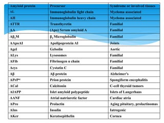
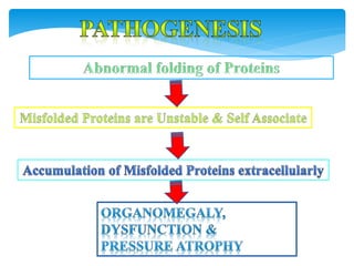
 Amyloid= ‘starch-like’ (Amylum—Latin)
 Virchow in mid 19th century
 Amyloids: Aggregations of un-degraded, pathologic
protein fibrils because of misfolding mechanisms.
 Amyloidosis: a group of diseases.
Background:
Definition: Amyloid
 “ A pathologic protein deposited between
the cells in various tissues and organs of the
body in a wide variety of clinical settings”.
 Non-branching fibres
 Indefinite length
 Diameter of 7.5 –10 nm.
 Cross-Beta-pleated sheet conformation:
X-ray crystallography
Infra-red spectroscopy
Physical nature of Amyloid:
 95%--consists of fibril proteins.
 5%--other glycoproteins
 Three most important forms:
1. AL (amyloid light chain): from plasma cells,
2. AA (amyloid associated): from liver,
3. A-Beta amyloid (seen in cerebral lesions of Alzheimer's
disease)
Chemical nature:
SYSTEMIC:
 Multiple myeloma—AL
 Reactive systemic am—AA
 Hemodialysis associated am—Ab2 microglobulins
 Hereditary am—AA
 Familial Mediterranean fever—AA
 Familial amyloidotrophic neuropathy—AATR
 Systemic Senile am—ATTR
Classification:
LOCALIZED AMYLOIDOSIS:
 Alzheimer's disease –A-beta
 Medullary carcinoma –A cal
 Islet of Langerhans: AIAPP
 Isolated atrial amyloidosis—AANF
 Prion disease –PrPsc
Amyloid protein Precursor Syndrome or involved tissues
AL Immunoglobulin light chain Myeloma associated
AH Immunoglobulin heavy chain Myeloma associated
ATTR Transthyretin Familial
AA (Apo) Serum amyloid A Familial
Aβ2M β2 Microglobulin Familial
AApoAI Apolipoprotein AI Joints
Agel Gelsolin Aortic
ALys Lysosomes Familial
AFib Fibrinogen α chain Familial
Acys Cystatin C Familial
Aβ Aβ protein Alzheimer's
APrPsc Prion protein Spongiform encephalitis
ACal Calcitonin C-cell thyroid tumors
AIAPP Islet amyloid polypeptide Islets of Langerhans
AANF Atrial natriuretic factor Cardiac atria
APro Prolactin Aging pituitary, prolactinomas
AIns Insulin Iatrogenic
AKer Keratoepithelin Cornea
 Normal proteins—when produced in excess; have inherent
tendency to fold improperly and form fibrils.
 Mutant proteins—that are structurally unstable, prone for
misfolding and aggregation.
Two categories of proteins
 AL type
 Abnormal B cell lesion
 Commonly associated with Plasma cell Myeloma & Other
monoclonal B cell lesions
 Increased Light chain production.
 Associated with Bence Jones Protein in Urine
 AA type
 Secondary to Chronic Inflammatory lesion:
 Tuberculosis, Bronchiectasis, Osteomyelitis, Rheumatoid
arthritis, Inflammatory bowel disease, Ankylosing
Spondylitis, IV drug abusers.
Affected organ gets enlarged, firm and waxy appearance.
 Painting the cut surface with iodine imparts a yellow color,
and that is transformed to blue-violet after application of
sulphuric acid.
 Diagnosis: Congo-red dye: ordinary light gives pink/red color;
polarized light gives apple-green birefringence.
Morphology: Gross
1. Congo Red—View under Polarizing Microscopy—Apple
Green birefringence: Gold standard test.
2. Thioflavin T
3. Van Giessen (Khaki color)
4. PAS (Periodic Acid Schiff )Stain.
 Amorphous, eosinophilic, hyaline extra-cellular
protein.
 With progressive accumulation, produces pressure
atrophy of adjacent cells.
Microscopically:
 Kidney: Focal to diffuse mesangial deposits, Nodular
Basement membrane deposits, Capillary loops, Interstitial
space, Wall of blood vessels
 Liver: Hepatomegaly, Waxy surface
 Heart: Dew drop like subendocardial nodules, In between
Myocardial fibres
 Tongue: Macroglossia, Gingival swellings
 Carpel tunnel syndrome.
Morphology:
SPLEEN:
 Splenomegaly +
 Splenic follicles, white pulp involved—SAGO spleen:
tapioca like granules on gross examination.
 Splenic sinuses, red pulp involved– LARDACEOUS spleen
(map like-large deposits).
Qn
 Depend on the site involved
 Can be unsuspected finding in Autopsy
 Can lead to serious organ dysfunction
 Renal disease: Nephrotic syndrome
 Cardiac: Conduction defects, Restrictive
cardiomyopathy
 Hepatosplenomegaly
1. Abdominal fat
2. Tongue
3. Rectal mucosa
4. Renal biopsy
5. Bone marrow biopsy
Biopsy site:
5. amyloidosis dr. sinhasan, mdzah

5. amyloidosis dr. sinhasan, mdzah

  • 2.
     Amyloid= ‘starch-like’(Amylum—Latin)  Virchow in mid 19th century  Amyloids: Aggregations of un-degraded, pathologic protein fibrils because of misfolding mechanisms.  Amyloidosis: a group of diseases. Background:
  • 3.
    Definition: Amyloid  “A pathologic protein deposited between the cells in various tissues and organs of the body in a wide variety of clinical settings”.
  • 4.
     Non-branching fibres Indefinite length  Diameter of 7.5 –10 nm.  Cross-Beta-pleated sheet conformation: X-ray crystallography Infra-red spectroscopy Physical nature of Amyloid:
  • 5.
     95%--consists offibril proteins.  5%--other glycoproteins  Three most important forms: 1. AL (amyloid light chain): from plasma cells, 2. AA (amyloid associated): from liver, 3. A-Beta amyloid (seen in cerebral lesions of Alzheimer's disease) Chemical nature:
  • 6.
    SYSTEMIC:  Multiple myeloma—AL Reactive systemic am—AA  Hemodialysis associated am—Ab2 microglobulins  Hereditary am—AA  Familial Mediterranean fever—AA  Familial amyloidotrophic neuropathy—AATR  Systemic Senile am—ATTR Classification:
  • 7.
    LOCALIZED AMYLOIDOSIS:  Alzheimer'sdisease –A-beta  Medullary carcinoma –A cal  Islet of Langerhans: AIAPP  Isolated atrial amyloidosis—AANF  Prion disease –PrPsc
  • 8.
    Amyloid protein PrecursorSyndrome or involved tissues AL Immunoglobulin light chain Myeloma associated AH Immunoglobulin heavy chain Myeloma associated ATTR Transthyretin Familial AA (Apo) Serum amyloid A Familial Aβ2M β2 Microglobulin Familial AApoAI Apolipoprotein AI Joints Agel Gelsolin Aortic ALys Lysosomes Familial AFib Fibrinogen α chain Familial Acys Cystatin C Familial Aβ Aβ protein Alzheimer's APrPsc Prion protein Spongiform encephalitis ACal Calcitonin C-cell thyroid tumors AIAPP Islet amyloid polypeptide Islets of Langerhans AANF Atrial natriuretic factor Cardiac atria APro Prolactin Aging pituitary, prolactinomas AIns Insulin Iatrogenic AKer Keratoepithelin Cornea
  • 10.
     Normal proteins—whenproduced in excess; have inherent tendency to fold improperly and form fibrils.  Mutant proteins—that are structurally unstable, prone for misfolding and aggregation. Two categories of proteins
  • 11.
     AL type Abnormal B cell lesion  Commonly associated with Plasma cell Myeloma & Other monoclonal B cell lesions  Increased Light chain production.  Associated with Bence Jones Protein in Urine
  • 12.
     AA type Secondary to Chronic Inflammatory lesion:  Tuberculosis, Bronchiectasis, Osteomyelitis, Rheumatoid arthritis, Inflammatory bowel disease, Ankylosing Spondylitis, IV drug abusers.
  • 13.
    Affected organ getsenlarged, firm and waxy appearance.  Painting the cut surface with iodine imparts a yellow color, and that is transformed to blue-violet after application of sulphuric acid.  Diagnosis: Congo-red dye: ordinary light gives pink/red color; polarized light gives apple-green birefringence. Morphology: Gross
  • 14.
    1. Congo Red—Viewunder Polarizing Microscopy—Apple Green birefringence: Gold standard test. 2. Thioflavin T 3. Van Giessen (Khaki color) 4. PAS (Periodic Acid Schiff )Stain.
  • 15.
     Amorphous, eosinophilic,hyaline extra-cellular protein.  With progressive accumulation, produces pressure atrophy of adjacent cells. Microscopically:
  • 16.
     Kidney: Focalto diffuse mesangial deposits, Nodular Basement membrane deposits, Capillary loops, Interstitial space, Wall of blood vessels  Liver: Hepatomegaly, Waxy surface  Heart: Dew drop like subendocardial nodules, In between Myocardial fibres  Tongue: Macroglossia, Gingival swellings  Carpel tunnel syndrome. Morphology:
  • 17.
    SPLEEN:  Splenomegaly + Splenic follicles, white pulp involved—SAGO spleen: tapioca like granules on gross examination.  Splenic sinuses, red pulp involved– LARDACEOUS spleen (map like-large deposits). Qn
  • 18.
     Depend onthe site involved  Can be unsuspected finding in Autopsy  Can lead to serious organ dysfunction  Renal disease: Nephrotic syndrome  Cardiac: Conduction defects, Restrictive cardiomyopathy  Hepatosplenomegaly
  • 19.
    1. Abdominal fat 2.Tongue 3. Rectal mucosa 4. Renal biopsy 5. Bone marrow biopsy Biopsy site: