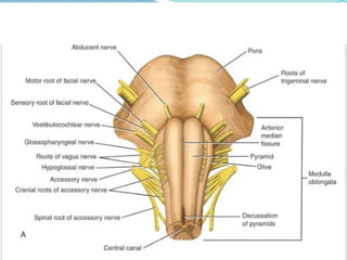
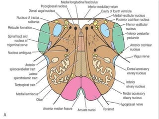
Brain Stem
Learning Objectives
 To review the anatomy of the brainstem
 To develop a three-dimensional picture of the
interior of the brainstem
 To know the positions of several of the cranial nerve
nuclei, the olivary nuclear complex, and the paths
taken by the various ascending and descending
nerve tracts as they ascend to the higher brain
centers or descend to the spinal cord
 To assess the signs and symptoms presented by a
patient and identify the exact location of a structural
lesion
Introduction to the Brainstem
 The brainstem is made up of the medulla
oblongata, the pons, and the midbrain and
occupies the posterior cranial fossa of the skull.
 It is stalk like in shape and connects the narrow
spinal cord with the expanded forebrain
Functions of Brain Stem
 it serves as a conduit for the ascending tracts
and descending tracts connecting the spinal cord
to the different parts of the higher centers in the
forebrain
 it contains important reflex centers associated
with the control of respiration and the
cardiovascular system and with the control of
consciousness; and
 it contains the important nuclei of cranial
nerves III through XII.
Gross Appearance of the Medulla
Oblongata
 connects the pons superiorly with the spinal cord
inferiorly
 The junction of the medulla and spinal cord is at the
origin of the anterior and posterior roots of the first
cervical spinal nerve, which corresponds approximately
to the level of the foramen magnum.
 The medulla oblongata is conical in shape, its broad
extremity being directed superiorly.
 The central canal of the spinal cord continues upward
into the lower half of the medulla; in the upper half of
the medulla, it expands as the cavity of the fourth
ventricle
Anterior surface
 On the anterior surface of the medulla is the anterior
median fissure, which is continuous inferiorly with the
anterior median fissure of the spinal cord. On each
side of the median fissure, there is a swelling called the
pyramid.
 The pyramids are composed of bundles of nerve fibers,
called corticospinal fibers, which originate in large
nerve cells in the precentral gyrus of the cerebral
cortex. The pyramids taper inferiorly, and it is here
that the majority of the descending fibers cross over to
the opposite side, forming the decussation of the
pyramids
Posterolateral to the pyramids are the
olives, which are oval elevations produced
by the underlying inferior olivary nuclei.
In the groove between the pyramid and
the olive emerge the rootlets of the
hypoglossal nerve.
Posterior to the olives are the inferior
cerebellar peduncles, which connect the
medulla to the
cerebellum. In the groove between the
olive and the inferior cerebellar peduncle
emerge the roots of the glossopharyngeal
and vagus nerves and the cranial roots of
Posterior surface
 The posterior surface of the superior half of the medulla
oblongata forms the lower part of the floor of the fourth
ventricle .
 The posterior surface of the inferior half of the medulla
is continuous with the posterior aspect of the spinal cord
and possesses a posterior median sulcus. On each side of
the median sulcus, there is an elongated swelling, the
gracile tubercle, produced by the underlying gracile
nucleus.
 Lateral to the gracile tubercle is a similar swelling, the
cuneate tubercle, produced by the underlying cuneate
nucleus
internal structure of the medulla
 The internal structure of the medulla oblongata
is considered at four levels:
 (1) level of decussation of pyramids,
 (2) level of decussation of lemnisci,
 (3) level of the olives, and
 (4) level just inferior to the pons
Level of Decussation of Pyramids
 A transverse section through the inferior half of the
medulla oblongata passes through the decussation of the
pyramids, the great motor decussation.
 In the superior part of the medulla, the corticospinal
fibers occupy and form the pyramid, but inferiorly, about
three-fourths of the fibers cross the median plane and
continue down the spinal cord in the lateral white
column as the lateral corticospinal tract.
 As these fibers cross the midline, they sever the
continuity between the anterior column of the gray
matter of the spinal cord and the gray matter that
surrounds the central canal.
 The fasciculus gracilis and the fasciculus
cuneatus continue to ascend superiorly
posterior to the central gray matter. The nucleus
gracilis and the nucleus cuneatus appear as
posterior extensions of the central gray matter.
 The substantia gelatinosa in the posterior gray
column of the spinal cord becomes continuous
with the inferior end of the nucleus of the
spinal tract of the trigeminal nerve.
Level of Decussation of Lemnisci
 A transverse section through the inferior half of the medulla
oblongata, a short distance above the level of the decussation of
the pyramids, passes through the decussation of lemnisci, the
great sensory decussation
 The decussation of the lemnisci takes place anterior to the
central gray matter and posterior to the pyramids. It should be
understood that the lemnisci have been formed from the
internal arcuate fibers, which have emerged from the anterior
aspects of the nucleus gracilis and nucleus cuneatus.
 The internal arcuate fibers first travel anteriorly and laterally
around the central gray matter. They then curve medially
toward the midline, where they decussate with the
corresponding fibers of the opposite side
 The nucleus of the spinal tract of the trigeminal
nerve lies lateral to the internal arcuate fibers. The
spinal tract of the trigeminal nerve lies lateral to the
nucleus
 The lateral and anterior spinothalamic tracts and the
spinotectal tracts occupy an area lateral to the
decussation of the lemnisci. They are very close to one
another and collectively are known as the spinal
lemniscus.
 The spinocerebellar, vestibulospinal, and the
rubrospinal tracts are situated in the anterolateral
region of the medulla oblongata.
Level of the Olives
 A transverse section through the olives passes
across the inferior part of the fourth ventricle.
 The amount of gray matter has increased at this
level owing to the presence of the olivary nuclear
complex; the nuclei of the vestibulocochlear,
glossopharyngeal, vagus, accessory, and
hypoglossal nerves; and the arcuate nuclei
Olivary Nuclear Complex
 The largest nucleus of this complex is the inferior olivary
nucleus. The gray matter is shaped like a crumpled bag
with its mouth directed medially; it is responsible for the
elevation on the surface of the medulla called the olive.
 Smaller dorsal and medial accessory olivary nuclei also
are present. The cells of the inferior olivary nucleus send
fibers medially across the midline to enter the cerebellum
through the inferior cerebellar peduncle. Afferent fibers
reach the inferior olivary nuclei from the spinal cord (the
spino-olivary tracts) and from the cerebellum and
cerebral cortex. The function of the olivary nuclei is
associated with voluntary muscle movement
Vestibulocochlear Nuclei
 The vestibular nuclear complex is made up of
the following nuclei:
 (1) medial vestibular nucleus,
 (2) inferior vestibular nucleus,
 (3) lateral vestibular nucleus, and
 (4) superior vestibular nucleus.
 The medial and inferior vestibular nuclei can be
seen on section at this level
cochlear nuclei
 There are two cochlear nuclei.
 The anterior cochlear nucleus is situated on the
anterolateral aspect of the inferior cerebellar
peduncle,
 and the posterior cochlear nucleus is situated
on the posterior aspect of the peduncle lateral to
the floor of the fourth ventricle
The Nucleus Ambiguus
 The nucleus ambiguus consists of large motor
neurons and is situated deep within the reticular
formation.The emerging nerve fibers join the
glossopharyngeal, vagus, and cranial part of the
accessory nerve and are distributed to voluntary
skeletal muscle
Central Gray Matter
 The central gray matter lies beneath the floor of the
fourth ventricle at this level. Passing from medial to
lateral ,the following important structures may be
recognized:
 (1) the hypoglossal nucleus,
 (2) the dorsal nucleus of the vagus,
 (3) the nucleus of the tractus solitarius, and
 (4) the medial and inferior vestibular nuclei.
 The nucleus ambiguus, referred to above, has
become deeply placed within the reticular formation
 The arcuate nuclei are thought to be inferiorly displaced
pontine nuclei and are situated on the anterior surface
of the pyramids . They receive nerve fibers from the
cerebral cortex and send efferent fibers to the cerebellum
through the anterior external arcuate fibers.
 The pyramids containing the corticospinal and some
corticonuclear fibers are situated in the anterior part of
the medulla separated by the anterior median fissure; the
corticospinal fibers descend to the spinal cord, and the
corticonuclear fibers are distributed to the motor nuclei
of the cranial nerves situated within the medulla.
 The medial lemniscus forms a flattened tract on each
side of the midline posterior to the pyramid. These fibers
emerge from the decussation of the lemnisci and convey
sensory information to the thalamus
 The medial longitudinal fasciculus forms a small tract of nerve
fibers situated on each side of the midline posterior to the medial
lemniscus and anterior to the hypoglossal nucleus.It consists of
ascending and descending fibers
 The inferior cerebellar peduncle is situated in the posterolateral
corner of the section on the lateral side of the fourth ventricle.
 The spinal tract of the trigeminal nerve and its nucleus are
situated on the anteromedial aspect of the inferior cerebellar
peduncle.
 The anterior spinocerebellar tract is situated near the surface in
the interval between the inferior olivary nucleus and the nucleus of
the spinal tract of the trigeminal nerve.
 The spinal lemniscus, consisting of the anterior spinothalamic,
the lateral spinothalamic, and spinotectal tracts, is deeply placed
 The reticular formation, consisting of a diffuse mixture of nerve
fibers and small groups of nerve cells, is deeply placed posterior to
the olivary nucleus.
 The reticular formation represents, at this level, only a small part
of this system, which is also present in the pons and midbrain.
 The glossopharyngeal, vagus, and cranial part of the accessory
nerves can be seen running forward and laterally through the
reticular formation .
 The nerve fibers emerge between the olives and the inferior
cerebellar peduncles.
 The hypoglossal nerves also run anteriorly and laterally through
the reticular formation and emerge between the pyramids and the
olives
Level Just Inferior to the Pons
 There are no major changes, in comparison to
the previous level, in the distribution of the gray
and white matter. The lateral vestibular nucleus
has replaced the inferior vestibular nucleus, and
the cochlear nuclei now are visible on the
anterior and posterior surfaces of the inferior
cerebellar peduncle
brainstem,medulla.pptx......................

brainstem,medulla.pptx......................

  • 1.
  • 2.
    Learning Objectives  Toreview the anatomy of the brainstem  To develop a three-dimensional picture of the interior of the brainstem  To know the positions of several of the cranial nerve nuclei, the olivary nuclear complex, and the paths taken by the various ascending and descending nerve tracts as they ascend to the higher brain centers or descend to the spinal cord  To assess the signs and symptoms presented by a patient and identify the exact location of a structural lesion
  • 3.
    Introduction to theBrainstem  The brainstem is made up of the medulla oblongata, the pons, and the midbrain and occupies the posterior cranial fossa of the skull.  It is stalk like in shape and connects the narrow spinal cord with the expanded forebrain
  • 4.
    Functions of BrainStem  it serves as a conduit for the ascending tracts and descending tracts connecting the spinal cord to the different parts of the higher centers in the forebrain  it contains important reflex centers associated with the control of respiration and the cardiovascular system and with the control of consciousness; and  it contains the important nuclei of cranial nerves III through XII.
  • 6.
    Gross Appearance ofthe Medulla Oblongata  connects the pons superiorly with the spinal cord inferiorly  The junction of the medulla and spinal cord is at the origin of the anterior and posterior roots of the first cervical spinal nerve, which corresponds approximately to the level of the foramen magnum.  The medulla oblongata is conical in shape, its broad extremity being directed superiorly.  The central canal of the spinal cord continues upward into the lower half of the medulla; in the upper half of the medulla, it expands as the cavity of the fourth ventricle
  • 7.
    Anterior surface  Onthe anterior surface of the medulla is the anterior median fissure, which is continuous inferiorly with the anterior median fissure of the spinal cord. On each side of the median fissure, there is a swelling called the pyramid.  The pyramids are composed of bundles of nerve fibers, called corticospinal fibers, which originate in large nerve cells in the precentral gyrus of the cerebral cortex. The pyramids taper inferiorly, and it is here that the majority of the descending fibers cross over to the opposite side, forming the decussation of the pyramids
  • 9.
    Posterolateral to thepyramids are the olives, which are oval elevations produced by the underlying inferior olivary nuclei. In the groove between the pyramid and the olive emerge the rootlets of the hypoglossal nerve. Posterior to the olives are the inferior cerebellar peduncles, which connect the medulla to the cerebellum. In the groove between the olive and the inferior cerebellar peduncle emerge the roots of the glossopharyngeal and vagus nerves and the cranial roots of
  • 10.
    Posterior surface  Theposterior surface of the superior half of the medulla oblongata forms the lower part of the floor of the fourth ventricle .  The posterior surface of the inferior half of the medulla is continuous with the posterior aspect of the spinal cord and possesses a posterior median sulcus. On each side of the median sulcus, there is an elongated swelling, the gracile tubercle, produced by the underlying gracile nucleus.  Lateral to the gracile tubercle is a similar swelling, the cuneate tubercle, produced by the underlying cuneate nucleus
  • 12.
    internal structure ofthe medulla  The internal structure of the medulla oblongata is considered at four levels:  (1) level of decussation of pyramids,  (2) level of decussation of lemnisci,  (3) level of the olives, and  (4) level just inferior to the pons
  • 13.
    Level of Decussationof Pyramids  A transverse section through the inferior half of the medulla oblongata passes through the decussation of the pyramids, the great motor decussation.  In the superior part of the medulla, the corticospinal fibers occupy and form the pyramid, but inferiorly, about three-fourths of the fibers cross the median plane and continue down the spinal cord in the lateral white column as the lateral corticospinal tract.  As these fibers cross the midline, they sever the continuity between the anterior column of the gray matter of the spinal cord and the gray matter that surrounds the central canal.
  • 15.
     The fasciculusgracilis and the fasciculus cuneatus continue to ascend superiorly posterior to the central gray matter. The nucleus gracilis and the nucleus cuneatus appear as posterior extensions of the central gray matter.  The substantia gelatinosa in the posterior gray column of the spinal cord becomes continuous with the inferior end of the nucleus of the spinal tract of the trigeminal nerve.
  • 17.
    Level of Decussationof Lemnisci  A transverse section through the inferior half of the medulla oblongata, a short distance above the level of the decussation of the pyramids, passes through the decussation of lemnisci, the great sensory decussation  The decussation of the lemnisci takes place anterior to the central gray matter and posterior to the pyramids. It should be understood that the lemnisci have been formed from the internal arcuate fibers, which have emerged from the anterior aspects of the nucleus gracilis and nucleus cuneatus.  The internal arcuate fibers first travel anteriorly and laterally around the central gray matter. They then curve medially toward the midline, where they decussate with the corresponding fibers of the opposite side
  • 19.
     The nucleusof the spinal tract of the trigeminal nerve lies lateral to the internal arcuate fibers. The spinal tract of the trigeminal nerve lies lateral to the nucleus  The lateral and anterior spinothalamic tracts and the spinotectal tracts occupy an area lateral to the decussation of the lemnisci. They are very close to one another and collectively are known as the spinal lemniscus.  The spinocerebellar, vestibulospinal, and the rubrospinal tracts are situated in the anterolateral region of the medulla oblongata.
  • 21.
    Level of theOlives  A transverse section through the olives passes across the inferior part of the fourth ventricle.  The amount of gray matter has increased at this level owing to the presence of the olivary nuclear complex; the nuclei of the vestibulocochlear, glossopharyngeal, vagus, accessory, and hypoglossal nerves; and the arcuate nuclei
  • 23.
    Olivary Nuclear Complex The largest nucleus of this complex is the inferior olivary nucleus. The gray matter is shaped like a crumpled bag with its mouth directed medially; it is responsible for the elevation on the surface of the medulla called the olive.  Smaller dorsal and medial accessory olivary nuclei also are present. The cells of the inferior olivary nucleus send fibers medially across the midline to enter the cerebellum through the inferior cerebellar peduncle. Afferent fibers reach the inferior olivary nuclei from the spinal cord (the spino-olivary tracts) and from the cerebellum and cerebral cortex. The function of the olivary nuclei is associated with voluntary muscle movement
  • 24.
    Vestibulocochlear Nuclei  Thevestibular nuclear complex is made up of the following nuclei:  (1) medial vestibular nucleus,  (2) inferior vestibular nucleus,  (3) lateral vestibular nucleus, and  (4) superior vestibular nucleus.  The medial and inferior vestibular nuclei can be seen on section at this level
  • 26.
    cochlear nuclei  Thereare two cochlear nuclei.  The anterior cochlear nucleus is situated on the anterolateral aspect of the inferior cerebellar peduncle,  and the posterior cochlear nucleus is situated on the posterior aspect of the peduncle lateral to the floor of the fourth ventricle
  • 27.
    The Nucleus Ambiguus The nucleus ambiguus consists of large motor neurons and is situated deep within the reticular formation.The emerging nerve fibers join the glossopharyngeal, vagus, and cranial part of the accessory nerve and are distributed to voluntary skeletal muscle
  • 29.
    Central Gray Matter The central gray matter lies beneath the floor of the fourth ventricle at this level. Passing from medial to lateral ,the following important structures may be recognized:  (1) the hypoglossal nucleus,  (2) the dorsal nucleus of the vagus,  (3) the nucleus of the tractus solitarius, and  (4) the medial and inferior vestibular nuclei.  The nucleus ambiguus, referred to above, has become deeply placed within the reticular formation
  • 30.
     The arcuatenuclei are thought to be inferiorly displaced pontine nuclei and are situated on the anterior surface of the pyramids . They receive nerve fibers from the cerebral cortex and send efferent fibers to the cerebellum through the anterior external arcuate fibers.  The pyramids containing the corticospinal and some corticonuclear fibers are situated in the anterior part of the medulla separated by the anterior median fissure; the corticospinal fibers descend to the spinal cord, and the corticonuclear fibers are distributed to the motor nuclei of the cranial nerves situated within the medulla.  The medial lemniscus forms a flattened tract on each side of the midline posterior to the pyramid. These fibers emerge from the decussation of the lemnisci and convey sensory information to the thalamus
  • 32.
     The mediallongitudinal fasciculus forms a small tract of nerve fibers situated on each side of the midline posterior to the medial lemniscus and anterior to the hypoglossal nucleus.It consists of ascending and descending fibers  The inferior cerebellar peduncle is situated in the posterolateral corner of the section on the lateral side of the fourth ventricle.  The spinal tract of the trigeminal nerve and its nucleus are situated on the anteromedial aspect of the inferior cerebellar peduncle.  The anterior spinocerebellar tract is situated near the surface in the interval between the inferior olivary nucleus and the nucleus of the spinal tract of the trigeminal nerve.  The spinal lemniscus, consisting of the anterior spinothalamic, the lateral spinothalamic, and spinotectal tracts, is deeply placed
  • 33.
     The reticularformation, consisting of a diffuse mixture of nerve fibers and small groups of nerve cells, is deeply placed posterior to the olivary nucleus.  The reticular formation represents, at this level, only a small part of this system, which is also present in the pons and midbrain.  The glossopharyngeal, vagus, and cranial part of the accessory nerves can be seen running forward and laterally through the reticular formation .  The nerve fibers emerge between the olives and the inferior cerebellar peduncles.  The hypoglossal nerves also run anteriorly and laterally through the reticular formation and emerge between the pyramids and the olives
  • 36.
    Level Just Inferiorto the Pons  There are no major changes, in comparison to the previous level, in the distribution of the gray and white matter. The lateral vestibular nucleus has replaced the inferior vestibular nucleus, and the cochlear nuclei now are visible on the anterior and posterior surfaces of the inferior cerebellar peduncle