This document discusses a study that aimed to isolate the UV screening pigment scytonemin from various cyanobacterial isolates (Nostocales) and test its effects on growth and melanogenesis in melanoma cells. Scytonemin was isolated from Scytonema sp., Plectonema sp., Spirulina sp., and Lyngbya sp. using column chromatography. Scytonemin from Scytonema sp. and Lyngbya sp. decreased melanogenesis by inhibiting the tyrosinase enzyme, while scytonemin from Plectonema sp. and Spirulina sp. did not affect melanogenesis or tyrosinase activity. The study provides insights into how
Enlargement of biologically stimulated
investigational processes for the synthesis of nanoparticles is
budding into an important branch of nanotechnology. Eco
responsive methods of green mediated synthesis of nanoparticles
are the present research in the extremity of nanotechnology. The
bioreduction behavior of leaf extracts of Morinda citrifolia L.
(Rubiaceae) in the green synthesis of silver nanoparticles was
investigated employing UV/Visible Spectrophotometry, Particle
size analyzer, Zeta potential, Filed emission scanning electron
microscopy, Energy Dispersive X-ray Analysis and FourierTransform
Infrared Spectroscopy. The antifungal property of the
silver nanoparticles was tested against Candida albicans,
Candida tropicalis and Candida krusei. The Antifungal assay
tests Zone of inhibition revealed the concentrations of more than
10µl of silver nanoparticles were inhibited the growth of fungal
pathogens.
Nanoparticle of plant extract: A Novel approach for cancer therapyroshan telrandhe
Presented in NATIONAL CONFERENCE ;A Phytomedicine : A Novel Approach For Cancer Treatment. Sponsered By Indian Councial Of Medical Research, Delhi. OrganisedBy KamlaNehru College Of Pharmacy, Butibori, Nagpur441108, Maharastra, India. ON 25 & 26 March 2017
ABSTRACT- Present work explores the novel selenium nanoparticle-enhanced photodynamic therapy of toluidine blue
O against Streptococcus mutans biofilm. Physiochemical (Ultraviolet-visible absorption, FTIR, and fluorescence
spectroscopy) and Electron microscopy techniques were used to characterize selenium nanoparticles. The UV spectrum
of different concentrations of SeNP were showed distinct peak at ~288 nm, which confirmed the successful synthesis of
SeNP in this study. The synthesized Selenium nanoparticles were uniform and spherical in shape with average size
~100 nm. In FTIR spectra of SeNPs there were strong absorption band around 3425cm-1, 2928 cm-1 and 1647 cm-1.
TBO showed MIC and MBC of 62.5 μg/mL and 125 μg/mL respectively whereas in presence of SeNPs showed MIC
and MBC of 31.25 μg/mL and MBC of 62.5 μg/mL. SeNPs–TBO conjugate showed twofold higher activities against S
mutans than TBO alone. A 630 nm diode laser was applied for activation of SeNP- Toluidine blue O (TBO)
combination and TBO against S. mutans biofilm and cells. The UV-vis absorption result suggests that TBO is not
present on the surface of SeNP. In fluorescence emission spectra, there is enhancement of fluorescence of TBO
fluorescence in the presence of nanoparticle. This showed that SeNP are enhancing the photodynamic therapy.
Antibiofilm assays and microscopic studies showed significant reduction of biofilm presence of conjugate. A crystal
violet assay revealed a maximum percent inhibition of S. mutans biofilm formation after 24 hours’ incubation, recorded
as 20% and 60% by TBO (31.25 μg/mL) and SeNP–TBO (31.25 μg/mL; TBO) conjugate, respectively. XTT biofilm
reduction assay were showed 32% loss in viability in presence of SeNP-TBO conjugate whereas in presence of only
TBO there was 22% loss in viability of cells. Fluorescence spectroscopic study confirmed type I photo toxicity against
biofilm. Selenium nanoparticle conjugate–mediated photodynamic therapy may be used against recalcitrant biofilm
based infections and can be helpful in dentistry.
Key-words- S. mutans, SeNP, TBO, UV absorption, FTIR, fluorescence spectroscopy
Patterson et al. used a new microscopy technique called liquid-cell transmission electron microscopy (LCTEM) to observe the crystallization of metal-organic frameworks (MOFs) in real time. This provided insights into the growth mechanisms. They observed that MOFs like ZIF-8 grow through the transport and attachment of metal ions and ligands to particle edges, not by particle coalescence. This two-step process of transport followed by edge attachment limits the growth rate. LCTEM is able to directly observe growth at the nanoscale and will provide insights to better control MOF synthesis.
ZnO nanoparticles were synthesized using a combustion method with low-temperature solution combustion. XRD and SEM characterization confirmed the formation of hexagonal wurtzite ZnO nanoparticles around 30-40nm in size. The antibacterial activity of the ZnO nanoparticles was tested against E. coli using colony counting and disk diffusion methods. Both methods showed the ZnO nanoparticles had antibacterial effects in a concentration-dependent manner, with 100μg/L ZnO demonstrating the strongest antibacterial activity through over 70% bacterial reduction and the largest inhibition zone of 24mm. The ZnO nanoparticles were also found to damage the genomic DNA of treated E. coli cells.
Ben-Zion Magnes is an independent researcher specializing in physical chemistry, electrochemistry, inorganic chemistry, and analytical chemistry. He has over 10 years of experience developing energy storage technologies, including novel flow batteries and zinc-bromine batteries. Currently he is developing improved electrolytes and chemistries for rechargeable batteries through synthesis, characterization, and quantum mechanical modeling. Previously he held research positions developing analytical methods and electrochemical energy storage projects.
This document introduces a companion website for a biochemistry textbook. It provides the following key information:
1. The website accompanies the third edition of a biochemistry textbook by Mathews, van Holde, and Ahern.
2. The website contains outlines, concepts, terminology, and quizzing for each textbook chapter to help students learn biochemistry.
3. The same material on the website is also available on a CD-ROM included with the textbook for offline use.
4. The website provides links between concepts covered and related external websites for expanded information.
The document discusses the synthesis of nanoparticles using microorganisms such as bacteria and fungi. It describes intracellular and extracellular synthesis methods. Intracellular synthesis involves accumulation of nanoparticles inside the cell, while extracellular synthesis uses cell secretions outside the cell. Specific examples provided include gold and silver nanoparticles synthesized using bacteria and fungi through reduction of metal ions. The nanoparticles have a variety of shapes and sizes in the 1-100 nm range and potential applications.
Engineering Research Publication
Best International Journals, High Impact Journals,
International Journal of Engineering & Technical Research
ISSN : 2321-0869 (O) 2454-4698 (P)
www.erpublication.org
Magnetosomes are intracellular, membrane-bounded magnetic crystals found in magnetotactic bacteria that allow them to detect and align with magnetic fields. Each magnetosome contains 15-20 magnetite or greigite crystals that form chains acting like a compass. Magnetotactic bacteria were first observed in 1963 and are found worldwide, helping navigation by magnetosome chains. Magnetosome biogenesis involves membrane invagination and protein recruitment, followed by iron mineralization into bullet or cubic shapes. Applications of magnetosomes include MRI contrast agents, hyperthermia treatment, and wastewater remediation. Further research aims to improve high-yield magnetosome production and understand crystal formation for broader applications.
This document discusses green synthesis of nanoparticles using biological methods. It describes how nanoparticles can be synthesized using plant extracts, agricultural waste, microorganisms and enzymes in an environmentally friendly way. This is advantageous over chemical and physical methods as it is cost-effective, produces non-toxic nanoparticles and does not require high temperature or pressure. Specific examples discussed include using bacteria to synthesize silver nanoparticles and controlling factors like pH and temperature to regulate nanoparticle size and shape during microbial synthesis. Overall, the document presents biological methods as a green alternative for nanoparticle production.
Bioinorganic chemistry by Dr Geeta TewariGeeta Tewari
This presentation will be helpful to study the general idea of bioinorganic chemistry, role of metal ions in biological systems and metalloproteins. This may also be beneficial to 12 class students, B Sc and M Sc students. The same lecture is also uploaded in the form of video lecture in my youtube channel with the following link
https://youtu.be/VqE-63nUevQ
The embed code of the video is:
Nanoparticles Methods for Nanoparticles Synthesis Overviewijtsrd
Nanoparticles exist in several different morphologies such as spheres, cylinders, platelets, tubes etc. The word nanoparticles are used to describe a particle with size in the range of 1nm to 100nm, at least in one of the three possible dimensions. In this size range, the physical, chemical and biological properties of the nanoparticles changes in fundamental ways from the properties of both individual atoms molecules and of the corresponding bulk materials. The enormous diversity of the nanoparticles arising from their wide chemical nature, shape and morphologies, the medium in which the particles are present, the state of dispersion of the particles and most importantly, the numerous possible surface modifications the nanoparticles can be subjected to make this an important active field of science now a days. Dr. Ilamathi Jayaraman | Dr. Vijayakumari. S "Nanoparticles: Methods for Nanoparticles Synthesis: Overview" Published in International Journal of Trend in Scientific Research and Development (ijtsrd), ISSN: 2456-6470, Volume-5 | Issue-6 , October 2021, URL: https://www.ijtsrd.com/papers/ijtsrd46478.pdf Paper URL : https://www.ijtsrd.com/biological-science/biotechnology/46478/nanoparticles-methods-for-nanoparticles-synthesis-overview/dr-ilamathi-jayaraman
I have had extensive work experience as an accomplished researcher with over 7 years’ experience in the field of Photo Physics (Laser), Physical Chemistry and Material Science
My key areas of expertise include, but are not limited to the following;
A solid background in the development and controlling of complex improvised experimental setups for optical and industrial applications, with parallel electronic support and opto-electronic synchronization where needed. Indicatively, I mention the modified fluorescent microscope for the single molecule detection (UTexas, Austin) and the experimental technique of determining the parameters of water vapor transmission through the membranes operating in controlled temperature and humidity conditions (FORTH/ICE-HT).
Having a well-deserved reputation on non-conventional materials analysis methods (single molecule spectroscopy) gained in the lab of the UTexas equipped with advanced infrastructure, I think that gave me strong skills for managing and solving scientific and industrial problems under the pressure of a highly competitive environment. In addition, the research experience in a wide range of applications will provide a solid basis to address scientific problem analysis and investigation of divergences in the transfer of research knowledge to innovative material technologies and the development of sensors.
Possessing a convincing track record on the assertion (novel ideas and writing of proposals) and on the scientific support (experiments, results dissemination, presentations in review meetings and international conferences) of research projects.
I believe that my skills, experience, and reputation for excellence can greatly enhance the interest of relative companies. I have a proven track record of responsibility, integrity and commitment to companies objectives. I am comfortable working independently or as part of a team.
Bacterial magnetosomes. microbiology, biomineralization and biotechnological ...CAS0609
1) Magnetotactic bacteria form intracellular magnetic nanoparticles called magnetosomes that allow them to passively align with and navigate along magnetic field lines.
2) Magnetosomes typically contain magnetite (Fe3O4) or greigite (Fe3S4) crystals that are surrounded by a membrane and often arranged in chains within the cell.
3) The formation of magnetosomes involves tightly regulated iron uptake, controlled biomineralization within membrane vesicles, and the production of proteins that may help accumulate iron and control mineralization processes.
This document discusses the application of nanotechnology in biomedical fields such as drug delivery, gene delivery, and tissue engineering. It provides background on the history and definition of nanotechnology. It then discusses various nanomaterials that can be used for targeted drug and gene delivery such as liposomes, polymeric micelles, dendrimers, and nanoparticles. It also describes how microfluidics and nanomaterials can be applied to sperm sorting, oocyte handling, embryo culture, and metabolomics. Overall, the document outlines the current and potential future uses of nanotechnology in biomedicine.
1) Nanomaterials are materials that have at least one dimension between 1-100 nm. At the nanoscale, properties like optical, electrical, and mechanical properties change due to large interatomic forces and increased surface area.
2) Nanotechnology has applications in areas like agriculture, food packaging, nutraceuticals, waste water treatment, diagnosis, and was used in ancient India. Potential health risks also exist from nanoparticles crossing skin barriers and damaging cells.
3) Both advantages like stronger and cheaper materials as well as faster computers and new medical technologies, and disadvantages like potential health issues and nano-pollution exist for nanotechnology.
Nanotechnology for the improvement of vegetable cropsLabiba Shah
Nanotechnology involves manipulating matter at the atomic or molecular scale. It can be used to improve vegetable crops in several ways. Silver, zinc, copper, and titanium nanoparticles can increase crop growth, yield, and stress tolerance when applied to seeds or foliage. They can also protect crops from pathogens by inhibiting fungal and bacterial growth through reactive oxygen species production and membrane damage. Recent advances include atomically modifying rice to change color and growing nanoparticles within crop plants in a process called "particle farming" for industrial harvesting. Overall, nanotechnology shows potential to help address issues of food security, low productivity, and losses to stresses and diseases.
This document discusses the potential for supplementing livestock with nanominerals. It begins by defining nanotechnology and nanoparticles. Nanoparticles are classified and their properties explained, including higher bioavailability than conventional minerals due to increased surface area. Methods for synthesizing and characterizing nanominerals are described, including physical, chemical and biological techniques. Applications of nanomineral supplementation in livestock are explored, including enhancing growth, immunity and reproduction. Some studies demonstrating benefits are highlighted. Finally, cytotoxicity concerns are raised and the need for safety assessment before wide application is noted.
The document discusses the history and principles of nanotechnology. It describes various types of nanoparticles including inorganic nanoparticles like silver and organic nanoparticles. It explains methods for preparation of nanoparticles including physical methods like ball milling and chemical methods like cross linking microemulsion and precipitation. It discusses the effects of nanoparticles on various properties including increased surface area and reactivity. The document then summarizes several studies on the effects of nano zinc oxide, nano selenium, and nano zinc on parameters like milk production, nutrient digestibility, rumen fermentation, and semen quality in animals.
role of nanotechnology for crop protection in horticultural cropsgirija kumari
includes contents related to introduction about nanotechnology, nano particles, applications in agriculture and horticulture, crop protection applications and case studies
This curriculum vitae provides biographical information about Prayad Pokethitiyook in 3 pages. It includes his education history, employment history, honors, grants received, and publications. He received degrees from Mahidol University in Thailand as well as Virginia Tech and the University of Melbourne. He is currently an Associate Professor at Mahidol University where he serves as the chairman of the Department of Biology and director of several programs. He has received numerous grants to support his research into bioremediation, biodiesel production from microalgae, and selective biodesulfurization. He has over 20 publications in international peer-reviewed journals.
This document provides a curriculum vitae for Dr. C. K. K. Nair. It includes his personal details like name, address, contact information, education qualifications, areas of specialization, academic and professional recognition including awards, memberships, research interests, teaching and research experience, publications, and research projects. Some key details:
- Dr. Nair is a professor with over 48 years of experience in biochemistry, molecular biology and radiation biology.
- His areas of specialization include molecular biology, biotechnology, DNA repair, radiation biology and protection.
- He has over 222 publications in journals/books and has guided 14 PhD students.
- He has received several awards and honors
Dr. C. K. K. Nair is a molecular biologist and radiation biologist. He has over 48 years of experience in biochemistry, molecular biology, and radiation biology. He is currently the Dean of Research at Pushpagiri Institute of Medical Sciences and Research Centre in Thiruvalla, Kerala, India. He has published over 222 papers in journals and books and has delivered over 119 invited talks. He has supervised 14 PhD students. His research interests include radiosensitization and radioprotection in cancer therapy, DNA repair, and applications of nanomaterials in biomedicine.
It consists of introduction about nano world and how it is different from the macroscopic world and what are the reasons. it gives information about silver nanoparticles antimicrobial property and it is various application. it consists of synthesis, characterisation of silver nanoparticles.
This document summarizes a conference on nanoparticles organized by Ashoka Institute of Technology and Management. It discusses nanoparticles and their properties, various synthesis methods for gold and silver nanoparticles including chemical, physical and biological methods, characterization techniques, and applications in drug delivery, biomedical uses, and challenges including instability, impurities, and toxicity.
This document outlines the agenda for a technology leadership conference called "Supporting Leaders in Integrating Technology". The agenda includes an introduction, exploration sessions, examination, celebration, and reflection. During the exploration sessions, participants will work in groups to explore instructional strategies and educational technology resources. They will discuss how to keep students engaged through technology and link research-based strategies with modern techniques. The examination involves a technology skills survey and discussion of progress in deploying technologies. The celebration allows participants to share insights from the sessions. The event concludes with reflection and a technology leadership survey.
SLItech provides professional development to support teachers in integrating technology into instruction. They offer group sessions, school visits, and special projects to provide more in-depth training on utilizing portal and learning management systems. Their goals include leading teachers forward and holding Smartboard facilitator workshops. They ask teachers three questions to understand challenges to applying new skills, successful implementation strategies, and desired support and professional development for the upcoming school year to further technology integration in the district.
The document discusses the benefits and uses of polls and surveys in the classroom. It suggests that polls and surveys can increase classroom participation and engagement, encourage risk-taking through anonymity, elicit diverse opinions, gauge comprehension, and strengthen retention. It also lists some online polling tools like Google Forms, Poll Daddy, and Poll Everywhere that can be used and provides examples of polls that have been implemented in classes.
The document discusses the benefits of exercise for mental health. Regular physical activity can help reduce anxiety and depression and improve mood and cognitive functioning. Exercise causes chemical changes in the brain that may help boost feelings of calmness, happiness and focus.
The SLI TECH project aims to build leadership capacity around integrating technology into instruction. It receives $240,000 over two years to support an educational technology facilitator providing professional development for district leaders. Leaders will use an internal portal to collaborate and participate in research activities. The goal is to develop a vision for challenging today's students to think critically and apply their learning using technology.
The document lists quotes from various cultural heroes and authors such as William Butler Yeats, Van Morrison, Greg Brown, Willa Cather, Leo Tolstoy, James Baldwin, Charles Dickens, Robert Bly, Tove Ditlevsen, Vladimir Nabokov, Ralph Ellison, Henrik Ibsen, and repeats two quotes from Yeats and Morrison. Many of the quotes discuss childhood, writing, teaching, and the soul. The document frames these quotes as words of wisdom and inspiration from important cultural figures.
This professional development session for Woodhaven EA staff covered strategies to support student learning in numeracy, special education, and technology. Resources discussed included the Awesome Library, Learn Alberta math videos, and websites for special education tools, behavior management, and assistive technologies. Staff were encouraged to continue sharing information on internal collaboration sites and evaluate further professional development opportunities.
The document discusses using wikis in the classroom to engage students in collaborative learning. It defines wikis as editable websites that allow for collaboration. Wikis can be used for students to work together on lesson summaries, notes, projects and assessments. The document provides examples of how teachers have used wikis and recommends introducing wikis to students with hands-on activities and clear expectations.
An administrative technology survey found that most administrators engage stakeholders, provide professional development, and stay current on issues. They work as a team and use digital tools. Most model and encourage skills aligned with ISTE NETS standards for administrators. Further development is needed in policy making, partnering, and continuous improvement areas. Feedback from a tool called MILEGUIDE showed that messy learning can be challenging but provides valuable information through good conversations, and the portal captures information.
The document provides an agenda for a meeting about the SLI TECH project which aims to build leadership capacity to integrate technology into instruction. It discusses how technology can be used to give feedback and recognition. It lists various technology tools and resources for doing so, such as online polls, surveys, classroom response systems, and e-portfolios. Upcoming events are also noted, including a conference and workshops on using technology for feedback, recognition, and personal learning networks.
The 23 Things Project provides professional development sessions to help teachers integrate technology into instruction. Only 39% of teachers regularly use technology as a tool in the classroom. The project focuses PD on core teaching areas and strategies rather than just the technology. It offers just-in-time, hands-on learning opportunities for educators to try different tools and strategies for using technology tied to curriculum goals and assessment.
The document discusses a lead team meeting for H.E.A.T. 101 on December 18, 2009. It includes Bloom's revised taxonomy for Web 2.0 skills and provides criteria for analyzing lessons based on higher order thinking, engaged learning, authenticity, and technology use on a scale from low to high level. The document advocates for instructional strategies that reinforce effort, provide recognition, set objectives with feedback, and engage cooperative learning to teach 21st century skills to students who prefer multimedia and social networking.
Strategies for struggling readers the non readerNicole Lakusta
This document provides strategies for helping struggling readers. It outlines six reading comprehension strategies: Assess and Conquer, Predicting ABC, KIM (or KIC), I'll Pause You Think, What Do You Expect, and Text Investigation. It also lists several online tools that can help, such as using audio recordings, video activities, word analysis tools, and ways to publish student work. Finally, it provides references for additional information on strategies for struggling readers.
This document provides information about a digital bootcamp for educational leaders from August 16th, 2010 in Banff, Alberta. It discusses elements of a vision statement, revising vision statements, including references to technology in vision statements, effective technology integration, transparency, productive reading tools, personal learning networks, and one school's journey with professional learning communities.
Combined effect of solar drying and gamma radiation on the microbiological qu...Nii Korley Kortei
This study evaluated the combined effect of solar drying and gamma irradiation on the microbiological quality of dried oyster mushrooms (Pleurotus ostreatus) stored at room temperature. Mushrooms were dried using a solar dryer to 12-14% moisture content. Samples were then gamma irradiated at doses from 0-2 kGy. Initial microbial counts for total viable count, Bacillus cereus, yeasts and molds ranged from 1.2-5.3, 0.95-1.4, 0-1.39, and 0-0.8 log CFU/g, respectively. Solar drying and irradiation resulted in an average 0.55 and 0.38 log reduction of total viable count
— In the present work, impact of UV-B radiation (280-
315nm: 0.4 W m-2) on growth, photosynthetic pigments, protein,
ascorbate, proline and lipid peroxidation have been studied in
two cyanobacteria Nostoc muscorum and Synechocystis PCC
6803. UV-B radiation (2 to 6 hrs) leads to 55% inhibition of
growth in Synechocystis PCC 6803 in comparison to control
where as in Nostoc muscorum growth reduces up to 45%. This
UV-B treatment also significantly decreased the contents of
chlorophyll, carotenoids and phycocyanin. Photosynthetic
pigments decreased with increasing doses of UV-B (2 to 6 hrs)
radiation. However, the inhibitory effect in Synechocystis PCC
6803 was more pronounced than in Nostoc muscorum. With
increasing UV-B exposure period, production of ascorbate (19-
45%), proline (12-29%) and lipid peroxidation was significantly
higher in Synechocystis PCC 6803 as compared to control
sample. It was observed that lipid peroxidation enhanced 33 %
than control sample of Synechocystis PCC 6803. Our result shows
that photosynthetic apparatus is the main target of UV-B
radiation causing degradation of photosynthetic pigments. This
study concluded that Synechocystis PCC 6803 was the susceptible
organism for survival in stress condition than Nostoc muscorum.
effects of water quality on inactivation and repair of Microcystis viridis an...xiaona chu
This document studied the effects of water quality factors like turbidity, total organic carbon (TOC), and salinity on the inactivation and repair of two microalgae - Microcystis viridis and Tetraselmis suecica - following medium-pressure UV irradiation. It found that increased turbidity, TOC and salinity levels generally reduced UV disinfection efficiency for both microalgae. TOC in particular promoted higher levels of photoreactivation and dark repair for M. viridis. T. suecica showed more sensitive responses to changes in the three water quality factors than M. viridis.
Effect of UV Rays & Photoreactivation on the Colonial Morphology and Catalase...Amna Jalil
The document is a project report that studied the effect of UV rays and photoreactivation on the colonial morphology and catalase activity of baker's yeast and wild yeast. UV exposure altered the colonial morphology from smooth to rough and increased the catalase activity in both yeast strains. Photoreactivation restored the colonial morphology and catalase activity back to pre-UV exposure levels. The report analyzed the results and discussed how UV radiation causes mutations in yeast by damaging DNA, but photoreactivation can repair this damage.
Use of stable and radio isotopes to understand the plant physiological processRAHUL GOPALE
Introduction
what is isotope ?
Types of Isotopes
Isotopic Labelling
ADVANTAGES AND DISADVANTAGES OF ISOTOPIC STUDY
APPLICATIONS OF ISOTOPES IN AGRICULTURE
Principle isotopes used in plant-soil studies
Case studies
FUTURE THRUSTS OF ISOTOPIC STUDY
CONCLUSIONS
REFERENCES
This study examined the effects of copper nanoparticles on liver tissue and function in rats. Rats were injected with different doses of copper nanoparticles and examined after 2, 7, and 14 days. Histological analysis found changes in liver and lung tissue like thickened air sac walls and increased fibrous tissue. Biochemical analysis found elevated levels of hepatic enzymes SGOT and SGPT, indicating liver damage. The highest doses of copper nanoparticles caused the most severe effects. This suggests that copper nanoparticles can cause toxicity in liver and lung tissues.
The Fabrication And Modification Of T Cuas With Cellulose...Christy Hunt
The document discusses modifications made to T-CUAs (templated copper ultramicroelectrodes) to detect nitric oxide (NO). Specifically, it details adding a cellulose acetate gas permeable membrane selective to NO and introducing chitosan and gold nanoparticles to enhance catalytic activity, sensitivity, and limit of detection for NO. The cellulose acetate membrane filters out non-gaseous molecules while maintaining a pore size of around 6 angstroms. Scanning electron microscopy images show the unmodified and modified T-CUAs, with the modified having chitosan gold nanoparticles ranging in size from 5 to 80 nanometers deposited on the surface.
Response of aquatic fern(Azolla), to watercontaminationKavitha Cingam
The document summarizes a study on the response of the aquatic fern Azolla to contamination by the antibiotic ciprofloxacin (Cipro). The study investigated Azolla's ability to uptake Cipro and the effects on its nitrogen-fixing process. The results showed that Azolla is able to uptake concentrations of Cipro above levels that classify plants as hyperaccumulators. Exposure to Cipro negatively impacted the heterocyst/vegetative cell ratio, nitrogenase activity, total nitrogen, and disrupted photosynthesis. This suggests that Cipro is toxic not only to the nitrogen-fixing cyanobacteria in Azolla but also to Azolla itself, demonstrating its potential use in phytoremediation of Cipro contamination
The document discusses a study that investigated the effects of different sterilization methods (ethylene oxide, autoclave, UV) on electrospun polyester nanofibers and polyester fabrics. Polyethylene terephthalate (PET) nanofibers with concentrations of 10, 15 and 20 wt.% were produced by electrospinning and subjected to different sterilization methods. The surface characteristics, mechanical properties, and fiber morphology of the nanofibers were examined before and after sterilization. For polyester fabrics, color change, drape, and mechanical properties were evaluated after sterilization. The results showed that sterilization methods, especially ethylene oxide, significantly damaged the nanofiber structure and
Ultrasonic Study on Coconut Water and Sugar Solution Before and After Laser E...iosrjce
IOSR Journal of Applied Physics (IOSR-JAP) is a double blind peer reviewed International Journal that provides rapid publication (within a month) of articles in all areas of physics and its applications. The journal welcomes publications of high quality papers on theoretical developments and practical applications in applied physics. Original research papers, state-of-the-art reviews, and high quality technical notes are invited for publications.
ABSTRACT- In this study, the effect of ZnO and TiO2-NPs on beneficial soil microorganisms and their secondary metabolites production was investigated. The antibacterial potential of NPs were determined by growth kinetics of P. aeruginosa, P. fluorescens and B. amyloliquefaciens. Significantly decreased in the cell viability based on optical density measurements were observed upon treatment with increasing concentrations of NPs. While comparing the effect of the different concentrations of the NPs (200 µg/ml) on IAA production by different bacterial strains, ZnO nanoparticles showed greater inhibitory effect than TiO2-NPs on IAA production by bacterial strains. The effect of Nanoparticles on phosphate solubilization was found inhibitory at 200 µg/ml. Treatment with ZnO showed concentration dependent enhancement in siderophore production by bacteriaby exposure to ZnO-NPs whereas TiO2-NPs showed concentration dependent progressive decline for iron binding siderophore molecules. Reduction in antibiotic production by P. aeruginosa and P. fluorescens was noticed in the presence of ZnO and TiO2 as compared to the control. The fluorescence of NADH released by P. aeruginosa was observed to be quenched in presence of ZnO and TiO2-NPs as compared to control. The present study highlights that the impact of nanoparticles on bacterial strains and the release of plant growth promoting substances by PGPR strains was dose dependent, which gives an idea about the level of toxicity of these nanoparticles in the environment. Therefore, the discharge of nanoparticles in the environment should be carefully monitored so that the loss of both structure and functions of agronomically important microbes could be protected from the toxicity of MO-NPs.
Key-words- MO-NPs, IAA, Phosphate Solubilization, Siderophore, PCA, NADH, ZnO-NPs, TiO2-NPs
This study encapsulated the atrazine-degrading bacterium Pseudomonas sp. ADP in electrospun polymeric microtubes for long-term atrazine removal. In consecutive batch experiments over 2 years without an external carbon source, the encapsulated bacteria continuously degraded atrazine and produced ammonium. Analysis showed the bacterial community shifted from pure P. sp. ADP culture to include other species, likely enabling long-term degradation. The microtubes maintained a stable bacterial population for atrazine bioremediation.
This document summarizes a study that used Cycas leaf extract to synthesize silver nanoparticles. Transmission electron microscopy images showed that the nanoparticles were nearly spherical, ranging in size from 2 to 6 nm. X-ray diffraction analysis confirmed the nanoparticles had an face-centered cubic crystal structure. Ultraviolet-visible spectroscopy revealed a surface plasmon resonance peak at 449 nm, indicating the presence of silver nanoparticles. The study demonstrated a green synthesis method for producing silver nanoparticles using plant extracts.
In recent years, nanoparticles that have size of 1-100 nm is widely used for textile, pharmacy,
cosmetic and treatment of industrial wastewater. Producing and using of nanoparticles widely, causes
important accumulation in nature and toxicity on ecosystem. Knowledge of potential toxicity of nanoparticles is
limited. In this study, six different nanoparticles nano-zinc oxide, nano-silicon dioxide, nano-cerium oxide,
nano-aluminum oxide, nano-hafnium oxide, and nano-tantalum oxide which used commonly, were studied to
investigate toxic impacts on organisms. We studied nine different acute toxicity test (bacteria – Escherichia coli
(gram negative bacteria) ; bacteria – Bacillus cereus (gram positive bacteria) ; bacteria – Vibrio fischeri
(bioluminescences bacteria) ; methane Archae Bacteria ; yeast – Candida albicans ; mold – Aspergillus niger ;
algae – Chlorella sp. ; Crustacea – Daphnia magna ; lepistes - Poecillia reticula) for the effect of
nanoparticles to different trophic levels. In general, the most toxic nanoparticle is nano-zinc oxide and the least
toxic nanoparticle is nano-hafnium oxide. Among the used organisms in acute toxicity test; the most sensitive
organism is algae - Chlorella sp ;the most resistant organism is fish- Poecillia reticula.
Facile synthesis of water-dispersible Cu2O nanocrystal–reduced graphene oxide...Haocheng Quan
This document summarizes the facile synthesis of a water-dispersible Cu2O nanocrystal-reduced graphene oxide (CRGO) hybrid material and its potential as a cancer therapeutic agent under both visible and near-infrared (NIR) light irradiation. The CRGO hybrid exhibits efficient photothermal killing of both normal and cancer cells under NIR light due to heat generation. In contrast, under visible light irradiation, the photocatalytic effect of the material results in the selective killing of cancer cells through reactive oxygen species production, avoiding damage to healthy cells. This dual mode of anticancer activity under different light sources could provide a safe and effective approach for cancer therapy.
MoO3 Nanoparticles: Synthesis, Characterization and Its Hindering Effect on G...IJERA Editor
Molybdenum trioxide nanoparticles have been synthesized by sol-gel method. The X-ray diffraction analysis was done to confirm that the obtained product was MoO3. The scanning electron microscopy was done to study the shape, size distribution and surface morphology of nanoparticles; they had a hexagonal shape with smooth surface and uniform size distribution. The functional groups were studied using Fourier transform infrared spectroscopy. The effect of MoO3 nanoparticles on seed germination of vigna unguiculata was studied for 6 days from the day of sowing, by comparing the time taken for seeds to germinate and length of shoot with respect to time of the seeds sowed in heavy black soil whose nutrient composition was known with seeds sowed in the same heavy black soil but which was made rich with MoO3 nanoparticles. It was observed that the MoO3 nanoparticles hampered the germination of vigna unguiculata seeds and this restraint continued in the shoot growth also.
Synthesis and evaluation of bactericidal properties of CuO nanoparticles agai...Nanomedicine Journal (NMJ)
Objective(s):
CuO is one of the most important transition metal oxides due to its captivating properties. It is used in various technological applications such as high critical temperature superconductors, gas sensors, in photoconductive applications, and so on. Recently, it has been used as an antimicrobial agent against various bacterial species.
Materials and Methods:
Here, we synthesized CuO nanoparticles (NPs) and explored the antibacterial activity of CuO NPs preparation.
Results:
Single crystalline nanoparticles of copper oxide having almost uniform particle size of 5-6 nm has been synthesized by a facile and versatile route. XRD spectra confirmed the formation of single phase CuO NPs. Transmission electron microscopy results corroborate well with XRD results. The technique employed is free from toxic solvents, organics and amines, is based on a simple reaction of copper sulfate and de-ionized water (DI), and their bactericidal effects against of Aeromonas hydrophila ATCC 7966T bacteria were investigated. Minimum inhibitory concentration (MIC) and minimum bactericidal concentration (MBC) with liquid culture for all of the Aeromonas hydrophila culture Medias was done.
Conclusion:
Present study confirms that Copper oxide nanoparticles have great promise as antimicrobial agent against Aeromonas hydrophila.
Synthesis and evaluation of bactericidal properties of CuO nanoparticles agai...
37_8_Isolation
1. ISSN(Online) : 2319 - 8753
ISSN (Print) : 2347 - 6710
International Journal of Innovative Research in Science,
Engineering and Technology
(An ISO 3297: 2007 Certified Organization)
Vol. 4, Issue 3, March 2015
Copyright to IJIRSET DOI: 10.15680/IJIRSET.2015.0403037 1260
Isolation of Nostocales and Study the Effects
of Scytonemin on Growth Profile and
Melanogenesis by using Melanoma Cell Line
Pranay Abhang1,3
, Girish Pathade2,3
Teaching Associate, Institute of Bioinformatics and Biotechnology, Savitribai Phule Pune University, Pune,
Maharashtra, India.1
Principal, H. V. Desai College, Pune, Maharashtra, India.2
Department of Biotechnology, Fergusson College, Pune, India 3
ABSTRACT: The purpose of this study was to screen scytonemin, a UV screening pigment, in isolated Nostocales,
Scytonema sp., Plectonema sp., Spirulina sp., and Lyngby sp. Scytonemin was isolated by silica gel column
chromatography and its effects were tested in vitro for growth profile and melanogenesis on B16F10 mouse melanoma
cell line. In order to study effect of these scytonemin on growth profile- cell viability count, MTT and DCPIP
cytotoxicity assay, Comet genotoxicity assay and microscopic observations were considered. To study tyrosinase
enzyme activity- diphenolase assay and native SDS PAGE methods were used. Scytonemin isolated from Scytonema
sp. and Lyngby sp. shows decrease in melanogenesis, which inhibit tyrosinase activity, having 30.25 µM, 100.25 µM
IC 50 values respectively. While that of scytonemin isolated from Plectonema sp. and Spirulina sp. was not showing
effect on melanogenesis as tyrosinase activity not altered, having 45.5 µM, 60.5 µM IC 50 values respectively.
KEY WORDS: Cytotoxicity, Genotoxicity, Melanogenesis, Scytonemin, Tyrosinase.
I. INTRODUCTION
For continued existence, solar radiation plays important role in evolution and development of life. Solar radiation
that reaches to the Earth‟s surface, the ultraviolet (UV) radiation is of particular importance for the human skin. Skin
pigmentation i.e. production of melanin by the process known as melanogenesis is highly depend on solar UV
radiations (Del Marmol et al., 1993, Jimbow etb al., 2000). At molecular level tyrosinase enzyme regulates process of
melanogenesis in response to UV rays, in which it act as key enzyme and regulate conversion of tyrosine to DOPA and
DOPA to DOPAquinone. (Hearing V. J. et al., 1980, Hearing and Tsukamoto, 1991, A. slominski et al., 2004).
The UV‐region of the electromagnetic spectrum is divided into three different regions: UVC radiation (200‐290
nm), UVB radiation (290‐320 nm) and UVA radiation (320‐400 nm). Of these wavelengths all UVC radiation as well
as large quantities of the UVB radiation gets absorbed by ozone layer. Therefore, the UV radiation that reaches the
Earth‟s surface contains about 5% of UVB and 95% of UVA radiation. (Mang R. et al., 2006)
Increased penetration of UV-B (not of UVA) on the surface of the earth is due to the decreasing ozone
concentration in the stratosphere (Kerr et al., 1993 and S. Madronich et al., 1998). Thus UV-B, which is deemed
deleterious, can penetrate biologically significant depths into the water column (Kuwahara et al., 2000), thereby
affecting aquatic ecosystems. The detrimental effects of UV-B on primary producers are varied, mediated primarily by
damaging molecular targets such as nucleic acids, proteins and pigments, and indirectly, by producing reactive oxygen
species. These can lead to the inhibition of cell division and growth, affecting various physiological and biochemical
processes, such as motility, orientation in motile organisms and photosynthesis, which can consequently alter
community structure and function. UV-A can have a net damaging influence on photosynthesis (Cullen et al., 1992).
2. ISSN(Online) : 2319 - 8753
ISSN (Print) : 2347 - 6710
International Journal of Innovative Research in Science,
Engineering and Technology
(An ISO 3297: 2007 Certified Organization)
Vol. 4, Issue 3, March 2015
Copyright to IJIRSET DOI: 10.15680/IJIRSET.2015.0403037 1261
Although UV rays are harmful, some organisms attempt to cope with UV radiation. Sinha et al. (1998) identified
the following four adaptation strategies through organisms tolerate or adapt to UV environment -
I. DNA repair mechanisms
II. Production of enzyme systems and induced formation of quenching agents
III.Behavioral modification to avoid exposure to UV
IV.Production of UV-absorbing substances
In terrestrial environments, where higher plants are the foremost primary producers, several studies have shown that
harmful UV radiation in higher plants is absorbed by epidermally located phenylpropanoids, mainly flavonoid
derivatives (Kootstra, 1994). In aquatic environments, where microalgae found abundantly, the presence of UV-
absorbing compounds like sporopollenin, scytonemin, and mycosporine-like amino acids (MAAs) have been
established.
Scytonemin [(3E,3'E) -3,3'-bis(4-hydroxybenzylidene) - [1,1'- bi(cyclopenta [b] indole)] -2,2' (3H,3'H) –dione] is a
UV‐screening pigment that is commonly produced in populations of sheathed cyanobacteria that live in different
habitats and geographic locations, where solar radiations are very intense (Garciapichel et al., 1991) The yellow-green
pigment scytonemin was first reported by Nageli as early as 1849 and the chemical structure was provided by Proteau
et al., 1993. It is a lipid soluble alkaloid that is synthesized in response to UVA radiation and accumulates within the
extracellular sheaths of cyanobacteria. The organisms are thereby protected from cell damage by this natural UV‐filter
that absorbs the harmful solar radiation. (Fleming and Castenholz, 2007 and Stevenson et al., 2002) Scytonemin
absorbs mostly in the UVA (325‐425 nm, λmax = 370 nm) and UVC region (λmax = 250 nm), but it also absorbs
substantially in the UVB region (280‐320 nm). The maximum absorption wavelength of scytonemin is 370 nm in vivo.
However, it shifts towards a longer wavelength of 384 nm in a solvent after isolation. It is reported that the molar
extinction coefficient of scytonemin is large (250 l/g/cm) at wavelength 384 nm (Vincent et al., 1993), it is calculated
to be 136,000 l/mol/cm based on a molecular weight of 544 Da. Therefore, scytonemin is an efficient photo-protective
compound due to its large extinction coefficient (Bultel-Poncef et al., 2004). It is a dimer composed of indolic and
phenolic subunits having a molecular mass of 544 g/mol. The linkage between two subunits in scytonemin is an
olefinic carbon atom that is unique among natural products. Hence, scytonemin possess a new ring system in nature for
which Proteau et al. (1993) have proposed the trivial name „the scytoneman skeleton‟. Scytonemin exists in oxidized
(green) and reduced (red) form. In an oxidized state, the two chromophores are connected by a single bond. Therefore,
they can freely rotate and prevent steric repulsion. This steric repulsion between two bulky chromophores makes a
dihedral angle of about 90 degrees, so electronic interaction between them becomes very weak. On the other hand, in a
reduced state, the indole ring and the benzene ring, which have 10-π and 6-π electron aromaticity, respectively, are
alternately connected by double bonds. Therefore, π-conjugation is expanded by increase of planarity. Due to this
structural and electrical change, the color of the compound changes from brown (oxidized state) to red (reduced state)
(Saman M. et al., 2014).
During this study naturally occurring UV screening compound is isolated from Nostocales and tested in vitro for
melanogenesis using mouse melanoma cells.
II. METHOD
1. Isolation of cyanobacteria
A. Collection and Enrichment of samples
Water and Soil samples were collected from Old swimming pool in Savitribai Phule Pune University, in autoclaved
glass bottle. Algal blooms or mats were collected by using mesh net (pore size 25-30 µm). pH and salinity of samples
were recorded using pH strips and salinometer respectively. Samples were centrifuged and pellet was suspended in BG
11 media with 1 mg/L Germanium Dioxide and 0.05 mg/ml Cycloheximide. Samples were kept for enrichment by
providing 12 hours light and dark conditions at 28o
C for 10-15 days. Bacterial and fungal contamination was reduced
by adding 0.05 mg/L streptomycin, 10 U/ml penicillin-G, 0.05 mg/L amphotericin B in the media. (Algal culture
techniques, by Robert Andersen)
3. ISSN(Online) : 2319 - 8753
ISSN (Print) : 2347 - 6710
International Journal of Innovative Research in Science,
Engineering and Technology
(An ISO 3297: 2007 Certified Organization)
Vol. 4, Issue 3, March 2015
Copyright to IJIRSET DOI: 10.15680/IJIRSET.2015.0403037 1262
B. Preparation of Axenic culture
Unialgal cultures were obtained by using serial dilution method for enriched samples. Enriched samples were diluted as
10-2
- 10-10
in BG 11 media and spread on BG 11 agar plates (1.5% agar) and incubated for 10-15 days by providing 24
hours light condition at 28o
C. Green colored colonies were streak on fresh BG 11 agar plates (1.5% agar with 5 µg/L
streptomycin, 10 U/ml penicillin-G, 5 µg/L amphotericin B in BG 11 media). Plates were incubated for 15-20 days by
providing 24 hours light condition at 28o
C. Mixed algal culture was separated under stereo microscope. (Algal culture
techniques, by Robert Andersen)
C. Identification of cyanobacteria
Isolated cultures were identified by colony characteristics, presence of pigments (and), cellulose and pectin,
microscopic observations as per given in “The freshwater algal flora of the British Isles: an identification guide to
freshwater and terrestrial algae.” edited by D. M. John et al. (2002) and “Cyanophyta” by T. V. Desikachary.
80% acetone extract of sample was used to detect presence of β-carotene (450 nm) and chlorophyll a (430 nm and 662
nm) while that of phosphate buffer extract of sample was used to detect presence of phycocyanobillin (620 nm 652 nm
or 562 nm). Presence of cellulose and pectin in sample was detected by Anthrone method (E. P. Samsel and J. C.
Aldrich, 1957) and Carbazole test (Mc Comb et al., 1957 and S. Nurdjanah et al., 2013) respectively.
2. Screening of scytonemin from cyanobacterial isolates
Presence of scytonemin was detected by following techniques (Proteau et al., 1993).
A. TLC for scytonemin
5 ml acetone was added in 10 mg of cyanobacterial sample and kept in dark at 4o
C for about 10-12 hours. 0.1 µl of
sample was spotted on TLC plates (Sigma-Aldrich, silica coated on aluminum support with pore size of 60 Ao
and
thickness of 200µm) and run by using chloroform: methanol (9: 1) as mobile phase. Yellowish brown colored spot was
detected and Rf value was calculated. (Rf value for scytonemin is around 0.8)
B. Spectrophotometric analysis
5 ml acetone was added in 10 mg of cyanobacterial sample and kept in dark at 4oC for about 10-12 hours. Sample was
centrifuged at 5000 rpm for 10 min at 4 o
C and supernatant was scanned from 350 nm to 450 nm on spectrophotometer
(Shimadzu UV 1601 UV-Visible). Peak at 384 nm (or 380nm -390 nm) shows presence of scytonemin. (Shailendra et
al. 2010). Scytonemin containing cyanobacteria were used for further experimentations.
3. Isolation of Scytonemin
Biomass was oven dried at 60 0
C. Dry weight was recorded and scytonemin was isolated by column chromatography.
(Naoki W., 2013). About 2.5 gram of dried biomass were suspended in 50 ml of acetonitrile and kept at 4 0
C for 1 hr.
biomass was separated by centrifugation and supernatant containing pigments was collected. This step was repeated
twice. Acetonitrile was removed by using Rota-vapor (Buchi R-210). Dried pigments were suspended in 50 ml of ethyl
ether to remove chlorophyll and other pigments. Un-dissolved pigments were collected and dissolved in acetone.
Silica gel (Hi-media) column was prepared in acetone and samples were resolved on 50 cm silica column using acetone
as mobile phase. Procedure was repeated twice for further purification. Collected samples were screened on
spectrophotometer (Shimadzu UV 1601) for scytonemin.
4. Study effect of scytonemin on growth profile and melanogenesis
A. Cell line maintenance -
B16F10 mouse melanoma cell line was purchased from NCCS, Pune. Cell line was maintained in DMEM with 10%
serum and 50 μg/ml streptomycin by providing 5% of CO2 atmosphere. Sub-culturing was done after every 5 days
(Culture of Animal cells by Ion Freshney).
B. Addition of scytonemin-
Purified scytonemin stock 10 mM (5.5 mg/ml) was prepared in 100 % acetone. For testing, 1µm, 10 µm, 100 µm, 1000
µm and 10000 µm concentration was added in cell culture (initial cells concentration was 3 × 10⁵
cells/well).
4. ISSN(Online) : 2319 - 8753
ISSN (Print) : 2347 - 6710
International Journal of Innovative Research in Science,
Engineering and Technology
(An ISO 3297: 2007 Certified Organization)
Vol. 4, Issue 3, March 2015
Copyright to IJIRSET DOI: 10.15680/IJIRSET.2015.0403037 1263
C. Cell viability count-
Cell viability was counted with the help of 0.4% Trypan blue vital stain using haemocytometer. Stained and non-
stained cells counted under microscope and recorded as cells/ml. (Culture of Animal cells by Ion Freshney)
D. Protein estimation
Protein estimation was done by modified Bradford‟s assay (1976). Unknown proteins were estimated by dissolving cell
pellets in protein extraction buffer containing 50 mM Tris-HCl, 5 mM EDTA, 0.1% Triton X-100 and 1 mM PMSF.
0.2 ml of 5x Bradford‟s reagent (500 µg/ml CBB G-250, 25% of ethanol and 50% of ortho-phosphoric acid) were
added in 0.8 ml diluted sample by vortexing and OD was taken at 595 nm. OD was compared with BSA standard graph
and unknown concentrations of proteins were estimated.
E. Tyrosinase enzyme activity staining in situ on SDS polyacrylamide gel –
Assay was done as described by Jimenez-Cervantes et al. (1993). Cell pellets were lysed in lysis buffer containing 50
mM TrisHcl, 5 mM EDTA, 0.1% Triton X-100 and 1 mM PMSF. The melanoma protein samples were mixed with
sample buffer without β-mercaptoethanol (0.18 M Tris- HCl pH 6.8, 15% glycerol, 0.075% bromophenol blue, 9%
SDS) in a 2:1 ratio without giving heat treatment. The samples were then resolved by 12% SDS-PAGE at 30mA. After
electrophoresis gel were equilibrated in 50 mM sodium phosphate buffer (pH 6) for 15 min at 37o
C. Tyrosinase activity
was detected by immersing the equilibrated gel in 50 mM sodium phosphate buffer (pH 6.8) containing 1.5 mM L-
DOPA and 4 mM MBTH at 370
C with gentle shaking until dark pink-brown coloured bands were developed.
F. Tyrosinase enzyme assay-
The tyrosinase activity was determined by a spectrophotometric method, as described by (Celia et al. 2001). The 10 μl
enzyme extract was added to the substrate containing 1.5 mM L-DOPA, 1 mM DMF and 4 mM MBTH in phosphate
buffer (pH 6.8). L-DOPA is the main substrate that converts to dopaquinone by enzyme activity. MBTH is a strong
nuocleophile that is attached to produce a pink complex. DMF is added to the reaction mixture in order to keep the
resulting complex in solution state during the course of investigations.
The progress of the reaction was followed by measuring the intensity of the resulting pink color at 505 nm. A typical
reaction mixture with a total volume of 1 ml contained 100 μl enzyme solution, 850 μl substrate solution and 50 μl
phosphate buffer (pH 6.8). To estimate tyrosinase activities in, the molar extinction coefficient of the product was 3700
per mole per centimetre.
G. Superoxide dismutase (SOD) assay
SOD assay was done as given by Adriana M C et al. (2004). Enzyme extract was prepared by dissolving cell pellet
(containing 103
cells/ml) in lysis buffer and supernatant were used after centrifugation at 12000 rpm. Following
components were added in two sets of tubes. 1.5 ml of 0.1 M potassium phosphate buffer (pH 7.8), 0.6 ml of 65 mM
L- methionine, 0.3 ml of 750 μM NBT, 0.3 ml of 2 mM Riboflavin, 1μl of 10 mM EDTA, 0.1 ml of Enzyme extract
(for control 0.1 ml water was added) and volume was adjusted to 3.5 ml with Distilled water. One set of tube was
illuminated under the light source for 30 min at a distance of 30 cm and another set of tube was kept in dark for 30 min.
Absorbance was read at 560 nm after 30 min of incubation at room temperature. Amount of SOD is directly
proportional to the amount of reactive oxygen species generated during reaction which are measured at 560 nm and
calculated by formula.
H. MTT and DCPIP assay for cell cytotoxicity
Cell cytotoxicity was tested as given by Karl B. et al. (2012). On 1st
day, 1 x 10⁵ cells/well allow to attach in 96 well
plate with 200 μl of complete medium in CO2 incubator.
On 2nd
day, compounds to be tested was added in respective wells with fresh medium and incubated for 24 hr.
On 3rd
day, medium were removed and monolayer were washed with PBSA 3-4 times.
For MTT assay - Fresh medium containing 5 μg/ml MTT were added in each well and incubated for 4 hr in CO2
incubator.
After 4 hr. medium was removed and crystals were dissolved in 200 μl of DMSO. To this 50 μl of glycine buffer (0.1
M glycine, 0.1 M NaCl, pH 10.5) was added.
Immediately absorbance at 570 nm was recorded using ELISA plate reader. And IC 50 value was calculated.
5. ISSN(Online) : 2319 - 8753
ISSN (Print) : 2347 - 6710
International Journal of Innovative Research in Science,
Engineering and Technology
(An ISO 3297: 2007 Certified Organization)
Vol. 4, Issue 3, March 2015
Copyright to IJIRSET DOI: 10.15680/IJIRSET.2015.0403037 1264
For DCPIP assay - 200 μl fresh medium were added in each well, to this 5 μl of 2 mM DCPIP reagent is added in each
well and incubated for 4 hour in CO2 incubator.
After 4 hr. absorbance at 660 nm was recorded using ELISA plate reader. And IC 50 value was calculated.
I. Comet assay for genotoxicity
Assay was performed as given by Stevenson C. S. et al. (2002). Glass slide is covered with thin layer of 1.5% melted
agarose. To the 200 μl of 0.6% melted agarose total of 20μl of cell suspension (1x10⁶
/ml) was added. This was spread
on coated slide using coverslip. Slide was kept at 0 0
C for 2-3 min. and slide was submerged in lysis buffer (0.25 M
NaCl, 0.6 % NaOH, 100 mM EDTA, 2% DMSO, 0.2% Triton X 100, pH 10) for 2 hours in dark. Ice cold running
buffer (300 mM NaOH, 0.5 M EDTA, pH 13) was added in electrophoresis tank and sample was run at 300 mA for 30
min in dark conditions.
After electrophoresis, slide was kept in neutralization buffer (0.4 M tris HCl pH 7.5) and then washed with distilled
water. EtBr stain was sprinkled on slide and slides were observed under fluorescence microscopy for comets.
III. RESULTS AND DISCUSSION
1. Isolation of Nostocales and scytonemin
11 algae were isolated from collected samples, among those only 6 were cyanobacteria. All 6 cyanobacteria were
characterized and screen for scytonemin, only four of them shows presence of scytonemin namely scytonema sp.,
Plectonema sp., Spirulina sp. and Lyngbya sp. (Fig. 1) Separation of scytonemin on TLC shows Rf values as 0.82
(green) for Scytonema sp., 0.80 (brownish purple) for Plectonema sp., 0.82 (brown) for Lyngbya sp., 0.84 (yellowish
brown) for Spirulina sp. and peak at 384 nm. Due to presence of variable side chains and electric charge on
Scytoneman skeleton, scytonemin can exist in different colors in nature. As Scytonemin pigment is highly hydrophobic
and exist in different color, it can be used in textile industries for coloration.
A B C D
Fig 1. Isolated and selected Nostocales A – Scytonema sp., B – Plectonema sp., C – Spirulina sp. and D – Lyngbya sp.
2. Effect of Scytonemin on melanoma cells
Scytonemin isolated from all 4 Nostocales shows similar effects on B16F10 mouse melanoma cells under microscope.
(Fig. 2) As compare to control (with and without acetone), scytonemin up to 1 µM concentration does not show any
effect on melanoma cells, while at 10 µM concentration cell growth is reduced. At 100 µM concentration melanoma
cells become apoptotic, while at 1000 µM concentration cells do not attached and dead. Viable cell count decreases as
concentration of scytonemin increases (Graph 1A).
Fig 2. Effect of scytonemin on B16F10 mouse melanoma cell line.
6. ISSN(Online) : 2319 - 8753
ISSN (Print) : 2347 - 6710
International Journal of Innovative Research in Science,
Engineering and Technology
(An ISO 3297: 2007 Certified Organization)
Vol. 4, Issue 3, March 2015
Copyright to IJIRSET DOI: 10.15680/IJIRSET.2015.0403037 1265
A B
Graph 1. (A) Decrease in cell viability (B16F10 cells) due to higher concentrations of Scytonemin isolated from 4 cyanobacteria. (B) Effect of
scytonemin on tyrosinase enzyme activity in B16F10 melanoma tyrosinase.
3. Effect of Scytonemin on tyrosinase activity
As per results for in situ staining on gel (Fig. 3) and tyrosinase activity by spectrophotometric analysis (Graph 1B)
shows similar effects. Scytonemin isolated from Scytonema sp. and Lyngbya sp. shows reduction in tyrosinase activity,
while Scytonemin isolated from Plectonema sp. and Spirulina sp. does not shows any effect on tyrosinase activity.
Enzyme activity reduces from 47 μM/ml/min to 23 μM/ml/min due to 1 mM of Scytonemin.
As per our results scytonemin from certain Nostocales shows inhibitory tyrosinase activity, hence it can be used to treat
disorders related to melanogenesis such as – Melisma, over pigmentation and deposition of melanin in patches. As
tyrosinase activity inhibited by scytonemin it may regulate melanin biosynthesis and hence melanin production can be
regulated.
A B C D
Fig 3. In situ staining of tyrosinase activity on gel. Effect of Scytonemin isolated from A – Scytonema sp., B – Plectonema sp., C – Spirulina sp. and
D – Lyngbya sp. on tyrosinase enzyme.
4. Effect of scytonemin on growth profile of melanoma cells
As concentration of scytonemin isolated from 4 different cyanobacteria increases, % reduction of GI decreases (Graph
3A). Higher concentrations of scytonemin i.e. above 1mM are toxic to the melanoma cells. Amount of SOD increases,
as concentration of scytonemin increases (Graph 3B).
As per MTT assay and DCPIP assay (Graph 2), IC 50 values of Scytonemin (isolated from 4 Nostocales) ranges from
30 µM to 100 µM which are mentioned in Table 1.
IC 50 value
Concentration of Scytonemin in µM
MTT assay DCPIP assay Average
Scytonema sp. 30 30.5 30.25
Plectonema sp. 50 40.5 45.5
spirulina sp. 40.5 90 60.5
lyngbya sp. 100 100.5 100.25
Table 1. IC 50 values of Scytonemin isolated from 4 Nostocales for B16F10 cell line, cell cytotoxicity MTT and DCPIP assay.
7. ISSN(Online) : 2319 - 8753
ISSN (Print) : 2347 - 6710
International Journal of Innovative Research in Science,
Engineering and Technology
(An ISO 3297: 2007 Certified Organization)
Vol. 4, Issue 3, March 2015
Copyright to IJIRSET DOI: 10.15680/IJIRSET.2015.0403037 1266
A B
Graph 2. % inhibition of B16F10 melanoma cells due to Scytonemin, cell cytotoxicity
A. MTT and B. DCPIP assay
Graph 3. A. Comet assay and B. SOD assay for Scytonemin isolated from 4 cyanobacteria.
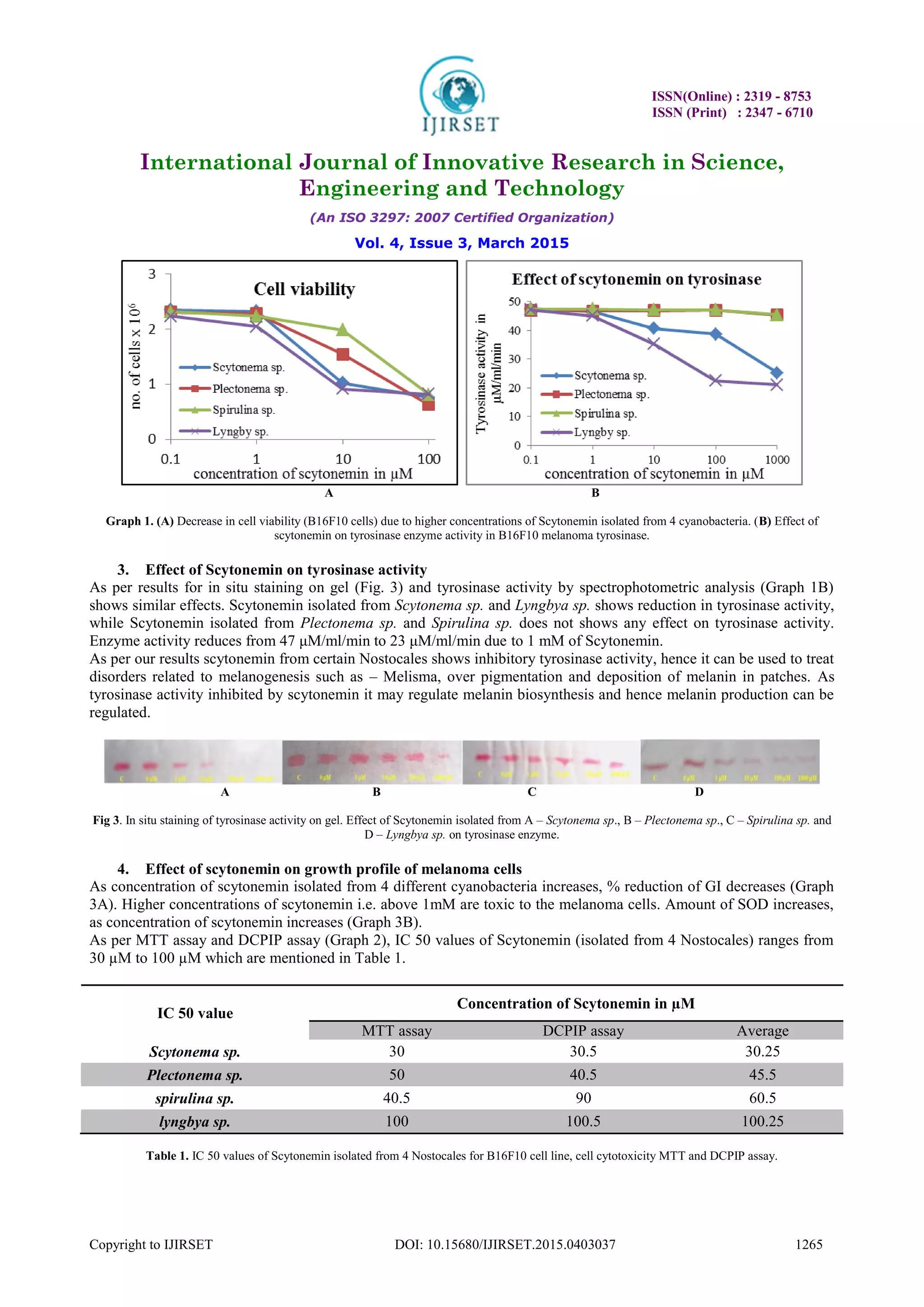
IV. CONCLUSION
Major role of scytonemin in cyanobacteria is to protect cells from UV rays. As it is a natural UV screening compound
having radical scavenging activity, it can be used in cosmetic industries. As per our results scytonemin isolated from
Scytonema sp. and Lyngbya sp. inhibit tyrosinase activity, hence it can be used to treat disorders related to
melanogenesis such as – Melisma, over pigmentation and deposition of melanin in patches. As tyrosinase activity
inhibited by scytonemin it may regulate melanin biosynthesis and hence melanin production can be regulated.
ACKNOWLEDGEMENT
Authors are thankful to Dr. R. G. Pardeshi, Principal Fergusson College, Pune for provision of laboratory and
chemicals.
REFERENCES
[1] A. Slominski, Desmond J. Tobin, S. Shibahara, and J. Wortsman. Melanin Pigmentation in Mammalian Skin and Its Hormonal
Regulation. Physiol Rev. 2004; 84:1155-228.
[2] Adriana M. Carvalho, Ana M. P. Neto, Angela P. Tonon, Ernani Pinto, Karina H. M. Cardozo, Maisa RPL Brigagao, Marcelo P. Barros,
Moacir Aluisio Torres, Paula Magalhães, Sara C. G. Campos, Thais Guaratini, Teresa C. S. Sigaud-Kutner, Vanessa R. Falcao and Pio
Colepicolo. Circadian protection against oxidative stress in marine algae, Hypnos. 2004; 1 (Suppl 1): 142-57.
[3] Algal Culturing Techniques, Edited by Robert A. Andersen, Elsevier Academic Press 2005 - ISBN: 0-12-088426-7.
[4] Bradford M. M., A rapid and sensitive method for the quantitation of microgram quantities of protein utilizing the principle of
protein-dye binding, Anal Biochem. 1976; 72, 248–54.
8. ISSN(Online) : 2319 - 8753
ISSN (Print) : 2347 - 6710
International Journal of Innovative Research in Science,
Engineering and Technology
(An ISO 3297: 2007 Certified Organization)
Vol. 4, Issue 3, March 2015
Copyright to IJIRSET DOI: 10.15680/IJIRSET.2015.0403037 1267
[5] Bultel-Ponce V, Felix-Theodose F, Sarthou C, Ponge J F, Bodo B., New pigments from the terrestrial cyanobacterium Scytonema sp.
collected on the Mitaraka inselberg, French Guyana. J Nat Prod. 2004; 67(4):678-81.
[6] Celia Jimenez-Cervantes, MariaMartinez-Esparza, Cristina Perez, Nicole Daum, Francisco Solano and Jose Carlos Garcia-Borron.
Inhibition of melanogenesis in response to oxidative stress: transient downregulation of melanocyte differentiation markers and
possible involvement of microphthalmia transcription factor. Journal of Cell Science. 2001; 114, 2335-44.
[7] Cullen, J. J., Neale, P. J., Lesser, M. P. Biological weighting function for the inhibition of phytoplankton photosynthesis by ultraviolet
radiation. Science. 1992; 258, 646-50.
[8] Culture of animal cell, a manual of basic techniques. By Ion Freshney.
[9] Cyanophyta by T. V. Desikachary. Published by Indian Council of Agricultural Research, New Delhi (1959).
[10] Del Marmol V, Beermann F. Tyrosinase and related proteins in mammalian pigmentation. FEBS Lett. 1996; 381:165-8.
[11] E. P. Samsel, J. C. Aldrich. Application of Anthrone Test to Determination of Cellulose Derivatives in Nonaqueous Media. Anal. Chem.,
1957; 29 (4), 574-6.
[12] Fleming, E. D., and Castenholz, R. W. Effects of periodic desiccation on the synthesis of the UV-screening compound, scytonemin, in
cyanobacteria. Environmental Microbiology. 2007; 9, 1448-55.
[13] Garciapichel, F., and Castenholz, R. W. Characterization and biological implications of scytonemin, a cyanobacterial sheath pigment.
Journal of Phycology. 1991; 27, 395-409
[14] Hearing V j, Tsukamoto K. Enzymatic control of pigmentation in mammals. FASEB J. 1991; 5:2902-9.
[15] Hearing V. J., Ekel T. M., Montague P. M., Nicholson J. M. Mammalin tyrosinase. Stoichiometry and measurement of reaction
products. Biochim. Biophys. Acta. 1980; 611 (2): 251–68.
[16] J. B. Kerr, C. T. McElroy, Evidence for large upward trends of ultraviolet-B radiation linked to ozone depletion, Science. 1993; 262
(5136), 1032-34.
[17] Jimbow K, Hau C, Gomez PF, Hirosaki K, Shinoda K, Salopek T G, et al. Intracellular vesicular trafficking of tyrosinase grne family
protein in eu and pheomelanosome biogenesis. Pigment Cell Res 2000; 13(Suppl.8), 110-7.
[18] Jimenez-Cervantes C, Valverde P, Garcia-Borron J C, Solano F, Lozano JA. Improved tyrosinase activity stains in polyacrylamide
electrophoresis gels. Pigment cell Res 1993; 6, 394-9.
[19] John D. M., B. A. Whitton and A. J. Brook. (2002). The freshwater algal flora of the British Isle: an identification guide to freshwater
and terrestrial algae. Cambridge University Press. New York.
[20] Karl Buch, Tanja Peters, Thomas Nawroth, Markus Sänger, Heinz Schmidberger, and Peter Langguth. Determination of cell survival after
irradiation via clonogenic assay versus multiple MTT Assay – A comparative study. Radiat Oncol. 2012; 7: 1.
[21] Kuwahara, V. S., Toda, T., Hamasaki, K., Kikuchi, T. and Taguchi, S. Variability in the relative penetration of ultraviolet radiation to
photosynthetically available radiation in temperate coastal waters, Japan. J. Oceanog., 2000; 56, 399-408.
[22] Mang, R., Stege, H., and Krutmann, J. (2006) Mechanisms of phototoxic and photoallergic reactions, In Contact Dermatitis (Frosch, P.
J., Menné, T., and Lepoittevin, J.-P., Eds.) pp 97-104, ISBN: 978-3-662-10304-3 Springer-Verlag, Berlin Heidelberg.
[23] Naoki Wada, Toshio Sakamoto and Seiichi Matsugo. Multiple Roles of Photosynthetic and Sunscreen Pigments in Cyanobacteria
Focusing on the Oxidative Stress. Metabolites. 2013; 3, 463-83.
[24] Proteau, P. J., Gerwick, W. H., Garciapichel, F., and Castenholz, R. The structure of scytonemin, an ultraviolet sunscreen pigment from
the sheaths of cyanobacteria. Experientia. 1993; 49, 825-9.
[25] S. Madronich. Implications of recent total atmospheric ozone measurements for biologically active ultraviolet radiation reaching the
Earth’s surface, Geophys. Res. Lett. 1992; 19, 37–40.
[26] S. Nurdjanah, James Hook, J. Paton and J. Paterson. Galacturonic Acid Content and Degree of Esterification of Pectin from Sweet
Potato Starch Residue Detected using 13C CP/MAS Solid State NMR. European Journal of Food Research & Review, 2013; 3(1), 16-37.
[27] Saman Mushir, Satyanarayan Deep, Tasneem Fatma Screening of cyanobacterial strains for UV screening compound Scytonemin –
Environmental Perspectives International Journal of Innovative Research in Science, Engineering and Technology. 2014; Vol. 3, Issue 2,
ISSN: 2319-8753.
[28] Shailendra P. Singh, Sunita Kumari, Rajesh P. Rastogi, Kanchan L. Singh, Richa and Rajeshwar P. Sinha, Photoprotective and
biotechnological potentials of cyanobacterial sheath pigment, scytonemin. African Journal of Biotechnology. 2010; Vol. 9(5), pp. 580-8.
[29] Shazia Suhail, Deboshree Biswas, Alvina farooqui, J. M. Arif and Mohd. Zeeshan, Antibacterial and free radical scavenging potential of
some cyanobacterial strains and their growth characteristics. J. Chem. Pharm. Res. 2011; 3(2):472-478.
[30] Sinha R. P., Klisch M., Gröniger A., Häder D. P. Responses of aquatic algae and cyanobacteria to solar UV-B. Plant. Ecol. 2001; 154,
219–36.
[31] Sinha R. P., Klisch M., Gröniger A., Häder D. P. Ultraviolet absorbing screening substances in cyanobacteria, phytoplankton and
microalgae. J. Photochem. Photobiol. B: Biol. 1998; 47: 83-94.
[32] Stevenson, C. S., Capper, E. A., Roshak, A. K., Marquez, B., Eichman, C., Jackson, J. R., Mattern, M., Gerwick, W. H., Jacobs, R. S., and
Marshall, L. A. Scytonemin – a marine natural product inhibitor of kinases key in hyperproliferative inflammatory diseases.
Inflammation Research, 51, 112-114. The identification and characterization of the marine natural product scytonemin as a novel
antiproliferative pharmacophore. Journal of Pharmacology and Experimental Therapeutics. 2002; 303, 858-866.
[33] Takamatsu, S., Hodges, T. W., Rajbhandari, I., Gerwick, W. H., Hamann, M. T., and Nagle, D. G. Marine natural products as novel
antioxidant prototypes. Journal of Natural Products. 2003; 66, 605-8.
[34] Vincent, W.F., Downes, M.T., Castenholz, R.W., Howard-Williams C. Community structure and pigment organization of
cyanobacteria-dominated microbial mats in Antarctica. Eur. J. Phycol. 1993; 28, 213-21.