2016_Association for Research in Vision and Ophthalmology_MR
•
1 like•51 views

ARVO Presentation_Project Poster

Recommended
More Related Content
What's hot
What's hot (20)
Viewers also liked
Viewers also liked (16)
Similar to 2016_Association for Research in Vision and Ophthalmology_MR
Similar to 2016_Association for Research in Vision and Ophthalmology_MR (20)
Recently uploaded
Recently uploaded (20)
2016_Association for Research in Vision and Ophthalmology_MR
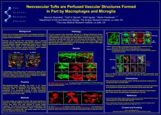
- 1. Neovascular Tufts are Perfused Vascular Structures Formed In Part by Macrophages and Microglia Mauricio Rosenfeld, 1 Faith H. Barnett, 1 Edith Aguilar, 1 Martin Friedlander 1, 2 1 Department of Cell and Molecular Biology, The Scripps Research Institute, La Jolla, CA. 2 The Lowy Medical Research Institute, La Jolla, CA. Contact and Funding Email: mrosen@scripps.edu National Eye Institute to MF (EY11254 ) and the Lowy Medical Research Institute. + + . • Cells expressing the macrophage marker CD11B are structural components of neovascular tufts in the OIR model. • Tufts perfused by systemically injected dye demonstrate a functional connection to the superficial vascular plexus in the neonatal mouse retina after OIR. • Mac/Mg are structurally involved in the formation of perfused, multinucleated tufts in both tumors and pathological angiogenesis in the eye. Conclusions Results Lectin R. Dextran CD11b Hoechst Reference 1. Yoshiaki Kubota, Keiyo Takubo, Takatsune Shimizu, Hiroaki Ohno, Kazuo Kishi, Masabumi Shibuya, Hideyuki Saya, and Toshio Suda. M-CSF inhibition targets pathological angigensis and lymphangiogenesis. J. Exp. Med. 2009; 205(5): 1089-1102. 2. Smith LE, Wesolowski E, McLellan A, Kostyk SK, D'Amato R, Sullivan R, D'Amore PA. Oxygen-induced retinopathy in the mouse. Invest Ophthalmol Vis Sci. 1994; 35(1): 101-11. LSM Image 3D IMARIS After hi O2 induced central vascular obliteration, large and perfused multinucleated retinal neovascular tufts are detectable in areas of the retina where the superficial vascular plexus remains present. This OIR P17 retinal Tuft is a perfused vascular structure. Rhodamine Lectin was administered by intracardiac injection and retinal whole mount was not stained with antibody. The tuft’s body projects towards the vitreous while connected to the superficial vascular plexus by way of a stalk-like structure. Macrophages/Microglia cells (CD11b+ ) are part of the structural composition of a retinal neovascular tuft. These images represent a region of an OIR-P17 retinal superficial vascular plexus (lectin+ ) as it undergoes neovascularization. HistologyBackground During murine postnatal development, macrophages participate in retinal vascular remodeling and pathological neovascularization in oxygen–induced retinopathy 1 . Recently, we have found that macrophages form a perfused vascular mimicry (VM) network under hypoxic conditions in both the subcutaneous matrigel angiogenesis model and in a mouse melanoma tumor model. In the subcutaneous tumor model, macrophages form perfused tuft-like structures (tumor rosettes) within the tumor associated angiogenic vasculature. Methods For these studies we utilized the mouse OIR model described by Smith et al 2 . Briefly, C57BL6/J mice were exposed to 75% O2 from P7-P12, after which mice were returned to normal oxygen conditions. This causes extensive central vaso-obliteration in the retina followed by neovascularization, which is associated with the formation of vascular tufts that extend toward the vitreous from the superficial vascular plexus. P17 mice received a 50ul intracardiac injection of a 50mg/ml Rhodamine Dextran (155KD) solution in sterile saline. Dye was allowed to circulate for 4-5 minutes, mice were euthanized, eyes enucleated and fixed. Retinal whole mounts were either left untouched or stained with Lectin and a rat anti-mouse CD11B antibody. Confocal microscopy was used to visualized neovascular tufts and Z-stack images were gathered using a 63X oil immersion lens. 3D renderings of the original LSM images were prepared using IMARIS software. Hoechst CD11b CD31 Rhodamine Dextran LSM Image A B Lectin CD11b Dextran Merge LSM Image 3D IMARIS Figure A. Confocal image of neovascular tufts associated with the superficial vascular plexus of a lectin stained OIR P17 retina whole mount. Figure B. 3D rendition of a confocal retinal Z-stack depicting a small and perfused multinucleated neovascular tuft in an OIR P17 retina. This particular tuft is positive for both lectin and Cd11b. A subcutaneous tumor bearing mouse was intravenously injected with low molecular weight rhodamine dextran (3KD). Tumor tissue was harvested, fixed, stained and imaged. CD11B+ Tumor associated macrophages form a perfused VM network containing multinucleated neovascular rosettes that are therefore connected to the traditional endothelial vasculature. Purpose Macrophages/microglia (Mac/Mg) are known to be closely associated with retinal neovascular tufts during oxygen induced retinopathy (OIR). To determine if the Mac/Mg were simply juxtaposed to or structurally involved in these neovascular structures, we studied the role of Mac/Mg in retinal neovascular regions in a murine model of OIR. lectin CD11b Merge 3D IMARIS rosettes rosettes * * * * * * * * * * stalk stalk * * ** stalk stalk Primary Vascular Plexus Original LSM Primary Vascular Plexus Primary Vascular Plexus Body of Tuft Stalk Body of Tuft Stalk IMARIS 3D IMARIS 3DVitreous VitreousVitreous lectin CD11b Merge Mergelectin NV Tuft NV Tuft NV Tuft NV Tuft NV Tuft