2016_Association for Research in Vision and Ophthalmology_2
•
0 likes•40 views

ARVO Presentation_Poster
Recommended

Recommended
More Related Content
What's hot
What's hot (19)
Viewers also liked
Viewers also liked (20)
Similar to 2016_Association for Research in Vision and Ophthalmology_2
Similar to 2016_Association for Research in Vision and Ophthalmology_2 (20)
Recently uploaded
Recently uploaded (20)
2016_Association for Research in Vision and Ophthalmology_2
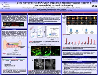
- 1. Bone T H E S C R I P P S R E S E A R C H I N S T I T U T E Bone marrow derived CX3CR1+ progenitors facilitate vascular repair in a murine model of ischemic retinopathy Results Acknowledgements Conclusions This work was supported by grants to M.F. from the National Eye Institute (RO1 EY011254) and the Lowy Medical Research Institute. Results (continued) Edith Aguilar, Susumu Sakimoto, Salomé Murinello, Peter D. Westenskow, Yoshihiko Usui, Felicitas Bucher, Maki Kitano, Daniel Feitelberg, Mauricio Rosenfeld, Martin Friedlander Department of Cell and Molecular Biology, The Scripps Research Institute, La Jolla, CA References 1. Smith LE, et al . Oxygen –induced retinopathy in the mouse. Invest Ophthalmol. Vis. Sci. 1994; 35:101-111. 2. Ritter MR et al. Myeloid progenitors differentiate into microglia and promote vascular repair in a model of Ischemic Retinopathy. J Clin Invest. 2006 116:3266-76. 3. Banin E, et al.T2-TrpRS Inhibits Preretinal Neovascularization and Enhances Physiological Vascular Regrowth in OIR as Assessed by a New Method of Quantification Invest Ophthalmol Vis Sci. 2006 May;47(5):2125-34. 4. Marchetti V,et al. Differential macrophage polarization promotes tissue remodeling and repair in a model of ischemic retinopathy. Sci Rep. 2011;1:76. 5. Ginhoux F, et al. Monocytes and macrophages: developmental pathways and tissue homeostasis. Nat Rev Immunol. 2014;14:392-404. 6. Kumar AH, et al. Bone marrow derived CX3CR1 progenitors contribute to neointimal smooth muscle cells via fractalkine CX3CR1 interaction. FASEB J. 2010; 24: 81-92. BM-derived CX3CR1+ progenitors potentiate vascular repair in an ischemic retinopathy mouse model. Injection of myeloid progenitor cells may provide a potential clinical application for the treatment of retinal vascular disease. Bone marrow (BM) derived cells have been reported to serve as proangiogenic microglia or macrophage in various ischemic conditions. We previously reported a subpopulation of myeloid progenitor cells that differentiate into microglia. These cells promoted vascular repair in oxygen-induced retinopathy (OIR). In this study, we wanted to further characterize the differentiation state of these myeloid progenitor cells. Purpose Figure 4 . Flow Cytometry analysis. 3.1 % of BM cells were CX3CR1+/CD34+ and 2.3 % were CX3CR1+/CD34-. Microglia and/or macrophages play an important role in the formation of the normal superficial and intermediate retinal vascular plexuses by releasing pro-angiogenic factors. The chemokine receptor CX3CR1 is associated with the monocyte/macrophage/DC lineage, while CD34 is associated with their progenitor cells. In this study, we show that intravitreal injections of undifferentiated CX3CR1+/CD34+ BM cells migrate to the retina and facilitate normalization of retinal vasculature in OIR. Methods Figure 3. C57Bl/6J mice were exposed to the oxygen-induced retinopathy model (OIR). Animals were subjected to 75% oxygen from P7 to P12, injected with fluorescently labeled cells at P7, and subsequently analyzed at P17. Intravitreal Injection: Right Eye: CX3CR1+/CD34+ cells Left Eye: CX3CR1+/CD34- cells 1.0x10⁵ cells / eye P7 P12 P17 Hyperoxia (75% O2) NormoxiaNormoxia Introduction Figure 5. Continued HSC GMP MDP Common Monocyte Progenitor Monocyte ↓ (Macrophage, DC) CD34 CX3CR1 HSC: Hematopoietic Stem Cells GMP: Granulocyte-macrophage progenitor MDP: Macrophage and Dendritic Cell (DC) precursor Control Lin- HSC Ritter et al. 2006 CX3CR1+ CD34+ 2.99% CX3CR1+ CD34- 6.23% Bone Marrow isolation CX3CR1 CD34 CX3CR1+ CD34+CX3CR1+ CD34- Isolectin-B4 Bone Marrow (BM) cells from 6 weeks old transgenic CX3CR1GFP/+ mice were isolated by flow cytometry. C57Bl/6J OIR mice were sacrificed at P17, evaluated by whole mount preparation, and stained with Isolectin Griffonia Simplicifolia I-B4 Alexa Fluor 568 (Invitrogen). We quantified the areas of pathological neovascularization and obliteration as previously described (Banin et al 2006). Also we performed RT-PCR with an array of 84 angiogenesis-related genes for CX3CR1+CD34+ cells and CX3CR1+CD34-. Figure 5 . (A) In the OIR model, injected CX3CR1+/CD34+ BM cells homed to the retinal vessels more than CX3CR1+/34-; and (B and C) injection of CX3CR1+/CD34+ BM cells decreased both the area of vascular obliteration and neovascular tufts compared to CX3CR1+/CD34- BM cells injection (*p=0.019 and p=0.018, respectively). Bar 500um. Methods (continued) A B C CX3CR1+ CD34+ CX3CR1+ CD34- Figure 6 . Results of qPCR based array analysis showing angiogenic gene upregulated in CX3CR1+/CD34+ BM cells compared to CX3CR1+/CD34-. CX3CR1Isolectin-B4 CX3CR1+/CD34+ Injected OIR * * 0 2 4 6 8 10 12 ANGPT1 ENG F3 MMP14 PF4 PLG SPHK1 TIE1 TYMP VEGFA CX3CR1+CD34- CX3CR1+CD34+ CX3CR1+ CD34+ CX3CR1+ CD34- CX3CR1+ CD34+ CX3CR1+ CD34- Figure 2 . Figure 1 . pix 3652 B03243