
Recommended
More Related Content
Similar to Anatomy of heart ,it's components and circulation
Similar to Anatomy of heart ,it's components and circulation (20)
Recently uploaded
Recently uploaded (20)
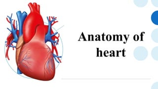
Anatomy of heart ,it's components and circulation
- 2. Introduction The heart is a muscular organ that serves to collect deoxygenated blood from all parts of the body, carries it to the lungs to be oxygenated and release carbon dioxide. Then, it transports the oxygenated blood from the lungs and distributes it to all the body parts.
- 3. The heart pumps around 7,200 litres of blood in a day throughout the body. The heart is situated at the centre of the chest and points slightly towards the left. On average, the heart beats about 100,000 times a day, i.e., around 3 billion beats in a lifetime. An adult heart beats about 60 to 100 per minute. Newborn babies heart beats faster than an adult which is about 70 to 180 beats per minute.
- 4. Anatomy The heart is a conical hollow muscular organ situated in the middle mediastinum and is enclosed within the pericardium The heart measures 12 x 8.5 x 6 cm weighs ~310 g (males) and ~255 g (females) The Greek name for the heart is cardia from which we have the adjective cardia
- 5. Relations Anteriorly: the body of the sternum, and adjoining costal cartilages; left lung, and pleura Posteriorly: esophagus, descending thoracic aorta, azygos, hemiazygos veins, and thoracic duct Superficially : bifurcation of the main pulmonary trunk Inferiorly: diaphragm Laterally: lungs, pleura
- 6. Coverings of the Heart Walls Pericardium: The heart is enclosed in a double-walled sac called the pericardium. It consists of two main parts: The fibrous pericardium - The fibrous pericardium is composed of tough, inelastic, dense irregular connective tissue. o Prevent overstretching of the heart o Protection of heart o Anchors the heart in the mediastinum The serous pericardium - The serous pericardium is a thinner membrane that forms a double layer around the heart. oThe outer parietal layer: it is fused to the fibrous pericardium. oThe inner visceral layer is also called the epicardium helps the layers of the heart wall to adheres tightly to the surface of the heart
- 8. Layers of the Heart • The heart wall consists of three layers enclosed in the pericardium: 1. Epicardium - the outer layer of the wall of the heart and is formed by the visceral layer of the serous pericardium. 2. Myocardium - the muscular middle layer of the wall of the heart and has excitable tissue and the conducting system. 3. Endocardium- A middle concentric layer A subendocardial layer.
- 9. Structure and Function • Right atrium • Left atrium • Right ventricle • Left ventricle
- 12. Blood Supply • Left main coronary artery carries 80% of the flow to the heart muscle. Left anterior descending artery that supplies anterior two-thirds of the inter-ventricular septum and adjoining part of the left ventricular anterior wall Circumflex coronary artery that supplies blood to the lateral and posterior portions of the left ventricle. • Right coronary artery: branches supply the right ventricle, right atrium, and left ventricle's inferior wall.
- 13. Venous drainage and Lymphatics Brachiocephalic nodes, in front of brachiocephalic veins Tracheobronchial nodes, located at the distal end of the trachea Venous drainage is via the variable coronary veins and the coronary sinus. The lymphatic vessels drain mainly into:
- 14. Nerve Supply The main control of the heart resides with the medulla oblongata. There is an area called the cardio acceleratory centre, or pressor centre, in the upper part of the medulla oblongata, and an area called the cardioinhibitory centre, or depressor centre, in the lower part. Together they are called the cardio regulatory centre, since they interact to control heart rate, etc.
- 15. Nerve Supply The sympathetic nervous system acts on the sinoatrial node, speeding up the depolarization rate, and therefore increasing the heart rate The parasympathetic system works in reverse in order to slow the heart rate down The heart itself has a natural pacemaker, the sinoatrial node, which does not need a nervous supply to function The nervous supply to the heart is autonomic, consisting of both sympathetic and parasymp athetic parts. The sympathetic fibres arise from the pressor centre, while the parasympathetic fibres arise in the depressor centre.
- 16. Conduction system of heart Your cardiac conduction system contains specialized cells and nodes that control your heartbeat. These are the: • Sinoatrial node: Your sinoatrial node is sometimes called your heart’s natural pacemaker. It sends the electrical impulses that start the heartbeat. Your autonomic nervous system controls how fast or slowly your SA node sends electrical signals. This part of the nervous system directs hormones that control your heart rate based on what you are doing. For example, your heart rate increases during exercise and slows when you are asleep.
- 18. • Atrioventricular node The atrioventricular node delays the SA node’s electrical signal. It delays the signal by a consistent amount of time (a fraction of a second) each time. The delay ensures that your atria are empty of blood before the contraction stops. The atria are the heart’s upper chambers. They receive blood from your body and empty it into the ventricles. • Bundle of His (atrioventricular bundle) It is a branch of fibers (nerve cells) that extends from your AV node. This fiber bundle receives the electrical signal from the AV node and carries it to the Purkinje fibers. The bundle of His runs down the length of the interventricular septum, the structure that separates your right and left ventricles.
- 19. • Purkinje fibers The Purkinje fibers are branches of specialized nerve cells. They send electrical signals very quickly to your right and left heart ventricles. Your Purkinje fibers are in the subendocardial surface of your ventricle walls. The subendocardial surface is part of the endocardium, the inner layer of tissue that lines your heart’s chambers. When the Purkinje fibers deliver electrical signals to your ventricles, the ventricles contract. As they contract, blood flows from your right ventricle to your pulmonary arteries and from your left ventricle to your aorta. The aorta is the body’s largest artery. It sends blood from your heart to the rest of your body
- 20. Recapitulation • What is the weight of the human heart? • Explain the blood supply of heart. • What are the layers of heart? • What are the components involved in conduction system of heart? • Name the atrioventricular valves.
- 21. Conclusion The heart is an essential, powerful organ that constantly pumps oxygen and nutrients around the body. If a person is born with congenital heart disease, or if damage occurs due to illness or other factors, the heart’s function may diminish, and this can lead to life threatening complications, such as heart failure. If the heart stops, a person cannot survive for long. Staying active and maintaining a healthful diet are two ways to protect the heart
- 22. Bibliography 1. Ross and Wilson. Anatomy and physiology.12th edition. Toronto; Elsevier:2014. p. 87-92 2. https://www.slideshare.net/binuenchappanal/anatomy-of-heart-65381385 3. https://www.physio-pedia.com/Anatomy_of_the_Human_Heart 4. https://my.clevelandclinic.org/health/body/21704-heart 5. https://www.texasheart.org/heart-health/heart-information- center/topics/heart-anatomy/
- 23. Thank you!