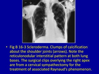
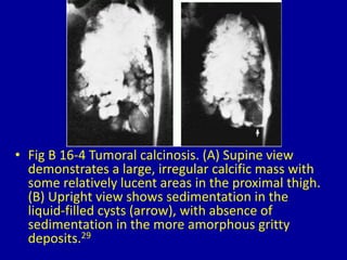
16Periarticular Calcification
CLINICAL IMAGAGING
AN ATLAS OF DIFFERENTIAL DAIGNOSIS
EISENBERG
DR. Muhammad Bin Zulfiqar
PGR-FCPS III SIMS/SHL
• Fig B 16-1 Calcific tendinitis. Frontal view of
the shoulder demonstrates amorphous
calcium deposits (arrows) in the supraspinatus
tendon.
• Fig B 16-2 Hyperparathyroidism. Dense mass of tumoral
calcification in joint capsules and periarticular soft tissues of the
lateral aspect of the foot in a patient with renal osteodystrophy.
• Fig B 16-3 Scleroderma. Clumps of calcification
about the shoulder joints (arrows). Note the
reticulonodular interstitial pattern at both lung
bases. The surgical clips overlying the right apex
are from a cervical sympathectomy for the
treatment of associated Raynaud's phenomenon.
• Fig B 16-4 Tumoral calcinosis. (A) Supine view
demonstrates a large, irregular calcific mass with
some relatively lucent areas in the proximal thigh.
(B) Upright view shows sedimentation in the
liquid-filled cysts (arrow), with absence of
sedimentation in the more amorphous gritty
deposits.29
• Fig B 16-5 Calcinosis universalis. Dense calcific
deposits in the soft tissues on the ulnar aspect
of the thumb.
Fig B 16-6 Gout. (A) Frontal and (B) lateral views demonstrate
massive deposition of calcium in a long-standing tophaceous
lesion about the elbow.
• Fig B 16-7 Myositis ossificans. Marked heterotopic
bone formation about the hip joint in a patient with
paralysis.
Fig B 16-8 Pellegrini-Stieda disease. Post-traumatic ossification
(arrows) along the femoral condyle.
16 periarticular calcification

16 periarticular calcification

  • 1.
  • 2.
    CLINICAL IMAGAGING AN ATLASOF DIFFERENTIAL DAIGNOSIS EISENBERG DR. Muhammad Bin Zulfiqar PGR-FCPS III SIMS/SHL
  • 3.
    • Fig B16-1 Calcific tendinitis. Frontal view of the shoulder demonstrates amorphous calcium deposits (arrows) in the supraspinatus tendon.
  • 4.
    • Fig B16-2 Hyperparathyroidism. Dense mass of tumoral calcification in joint capsules and periarticular soft tissues of the lateral aspect of the foot in a patient with renal osteodystrophy.
  • 5.
    • Fig B16-3 Scleroderma. Clumps of calcification about the shoulder joints (arrows). Note the reticulonodular interstitial pattern at both lung bases. The surgical clips overlying the right apex are from a cervical sympathectomy for the treatment of associated Raynaud's phenomenon.
  • 6.
    • Fig B16-4 Tumoral calcinosis. (A) Supine view demonstrates a large, irregular calcific mass with some relatively lucent areas in the proximal thigh. (B) Upright view shows sedimentation in the liquid-filled cysts (arrow), with absence of sedimentation in the more amorphous gritty deposits.29
  • 7.
    • Fig B16-5 Calcinosis universalis. Dense calcific deposits in the soft tissues on the ulnar aspect of the thumb.
  • 8.
    Fig B 16-6Gout. (A) Frontal and (B) lateral views demonstrate massive deposition of calcium in a long-standing tophaceous lesion about the elbow.
  • 9.
    • Fig B16-7 Myositis ossificans. Marked heterotopic bone formation about the hip joint in a patient with paralysis.
  • 10.
    Fig B 16-8Pellegrini-Stieda disease. Post-traumatic ossification (arrows) along the femoral condyle.