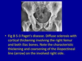
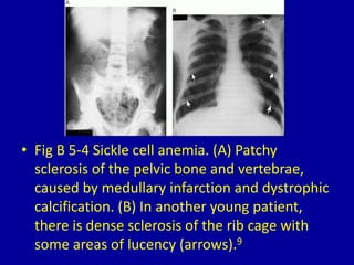
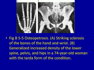
5Generalized Osteosclerosis
CLINICAL IMAGAGING
AN ATLAS OF DIFFERENTIAL DAIGNOSIS
EISENBERG
DR. Muhammad Bin Zulfiqar
PGR-FCPS III SIMS/SHL
• Fig B 5-1 Myelosclerosis. Diffuse uniform
sclerosis of the bones of the thorax produces
an appearance of jail bars.
• Fig B 5-2 Osteoblastic metastases. (A)
Carcinoma of the prostate. (B) Carcinoma of
the breast.
• Fig B 5-3 Paget's disease. Diffuse sclerosis with
cortical thickening involving the right femur
and both iliac bones. Note the characteristic
thickening and coarsening of the iliopectineal
line (arrow) on the involved right side.
• Fig B 5-4 Sickle cell anemia. (A) Patchy
sclerosis of the pelvic bone and vertebrae,
caused by medullary infarction and dystrophic
calcification. (B) In another young patient,
there is dense sclerosis of the rib cage with
some areas of lucency (arrows).9
• Fig B 5-5 Osteopetrosis. (A) Striking sclerosis
of the bones of the hand and wrist. (B)
Generalized increased density of the lower
spine, pelvis, and hips in a 74-year-old woman
with the tarda form of the condition.
• Fig B 5-6 Pyknodysostosis. Generalized
increase in density with cortical thickening of
the bones of the hand. The distal phalanges
are hypoplastic, and the terminal tufts are
absent.
Fig B 5-7 Melorheostosis. Dense cortical sclerosis involves the
proximal femur and the lower portion of the ilium.
• Fig B 5-8 Fluorosis. (A) Dense skeletal sclerosis
with obliteration of individual trabeculae
causes the pelvis and proximal femurs to
appear chalky white. (B) Diffuse vertebral
sclerosis in another patient.10
• Fig B 5-9 Progressive diaphyseal dysplasia. Dense
endosteal and periosteal cortical thickening causes
fusiform enlargement and increased density of the
midshafts of the radius and ulna.
• Fig B 5-10 Polyostotic fibrous dysplasia. The
bones of the feet show a smudgy, ground-glass
appearance of the medullary cavities with failure
of normal modeling.
• Fig B 5-11 Renal osteodystrophy. Sclerosis of the long bones in a boy with chronic
glomerulonephritis, renal rickets, and secondary hyperparathyroidism. In addition to the increased
skeletal density, note the widened zone of provisional calcification at the ankles and the
subperiosteal resorption along the medial margins of the upper tibial shafts (arrow).
Fig B 5-12 Congenital syphilis. Diffuse sclerosis with transverse bands of
lucency (arrows) in the diaphyses of the femurs and tibias.
• Fig B 5-13 Sclerotic myeloma. Views of (A) the leg and
(B) the femur demonstrate diffuse and nodular
sclerosis. Cortical thickening of the tibia encroaches on
the medullary canal. Similar changes were evident in
the pelvis.11
• Fig B 5-14 Hereditary hyperphosphatasia. Areas of
sclerosis about the metacarpals and middle phalanges
associated with thinning of the cortices. The proximal
phalanges show diffuse deossification.
5 generalized osteosclerosis

5 generalized osteosclerosis

  • 1.
  • 2.
    CLINICAL IMAGAGING AN ATLASOF DIFFERENTIAL DAIGNOSIS EISENBERG DR. Muhammad Bin Zulfiqar PGR-FCPS III SIMS/SHL
  • 3.
    • Fig B5-1 Myelosclerosis. Diffuse uniform sclerosis of the bones of the thorax produces an appearance of jail bars.
  • 4.
    • Fig B5-2 Osteoblastic metastases. (A) Carcinoma of the prostate. (B) Carcinoma of the breast.
  • 5.
    • Fig B5-3 Paget's disease. Diffuse sclerosis with cortical thickening involving the right femur and both iliac bones. Note the characteristic thickening and coarsening of the iliopectineal line (arrow) on the involved right side.
  • 6.
    • Fig B5-4 Sickle cell anemia. (A) Patchy sclerosis of the pelvic bone and vertebrae, caused by medullary infarction and dystrophic calcification. (B) In another young patient, there is dense sclerosis of the rib cage with some areas of lucency (arrows).9
  • 7.
    • Fig B5-5 Osteopetrosis. (A) Striking sclerosis of the bones of the hand and wrist. (B) Generalized increased density of the lower spine, pelvis, and hips in a 74-year-old woman with the tarda form of the condition.
  • 8.
    • Fig B5-6 Pyknodysostosis. Generalized increase in density with cortical thickening of the bones of the hand. The distal phalanges are hypoplastic, and the terminal tufts are absent.
  • 9.
    Fig B 5-7Melorheostosis. Dense cortical sclerosis involves the proximal femur and the lower portion of the ilium.
  • 10.
    • Fig B5-8 Fluorosis. (A) Dense skeletal sclerosis with obliteration of individual trabeculae causes the pelvis and proximal femurs to appear chalky white. (B) Diffuse vertebral sclerosis in another patient.10
  • 11.
    • Fig B5-9 Progressive diaphyseal dysplasia. Dense endosteal and periosteal cortical thickening causes fusiform enlargement and increased density of the midshafts of the radius and ulna.
  • 12.
    • Fig B5-10 Polyostotic fibrous dysplasia. The bones of the feet show a smudgy, ground-glass appearance of the medullary cavities with failure of normal modeling.
  • 13.
    • Fig B5-11 Renal osteodystrophy. Sclerosis of the long bones in a boy with chronic glomerulonephritis, renal rickets, and secondary hyperparathyroidism. In addition to the increased skeletal density, note the widened zone of provisional calcification at the ankles and the subperiosteal resorption along the medial margins of the upper tibial shafts (arrow).
  • 14.
    Fig B 5-12Congenital syphilis. Diffuse sclerosis with transverse bands of lucency (arrows) in the diaphyses of the femurs and tibias.
  • 15.
    • Fig B5-13 Sclerotic myeloma. Views of (A) the leg and (B) the femur demonstrate diffuse and nodular sclerosis. Cortical thickening of the tibia encroaches on the medullary canal. Similar changes were evident in the pelvis.11
  • 16.
    • Fig B5-14 Hereditary hyperphosphatasia. Areas of sclerosis about the metacarpals and middle phalanges associated with thinning of the cortices. The proximal phalanges show diffuse deossification.