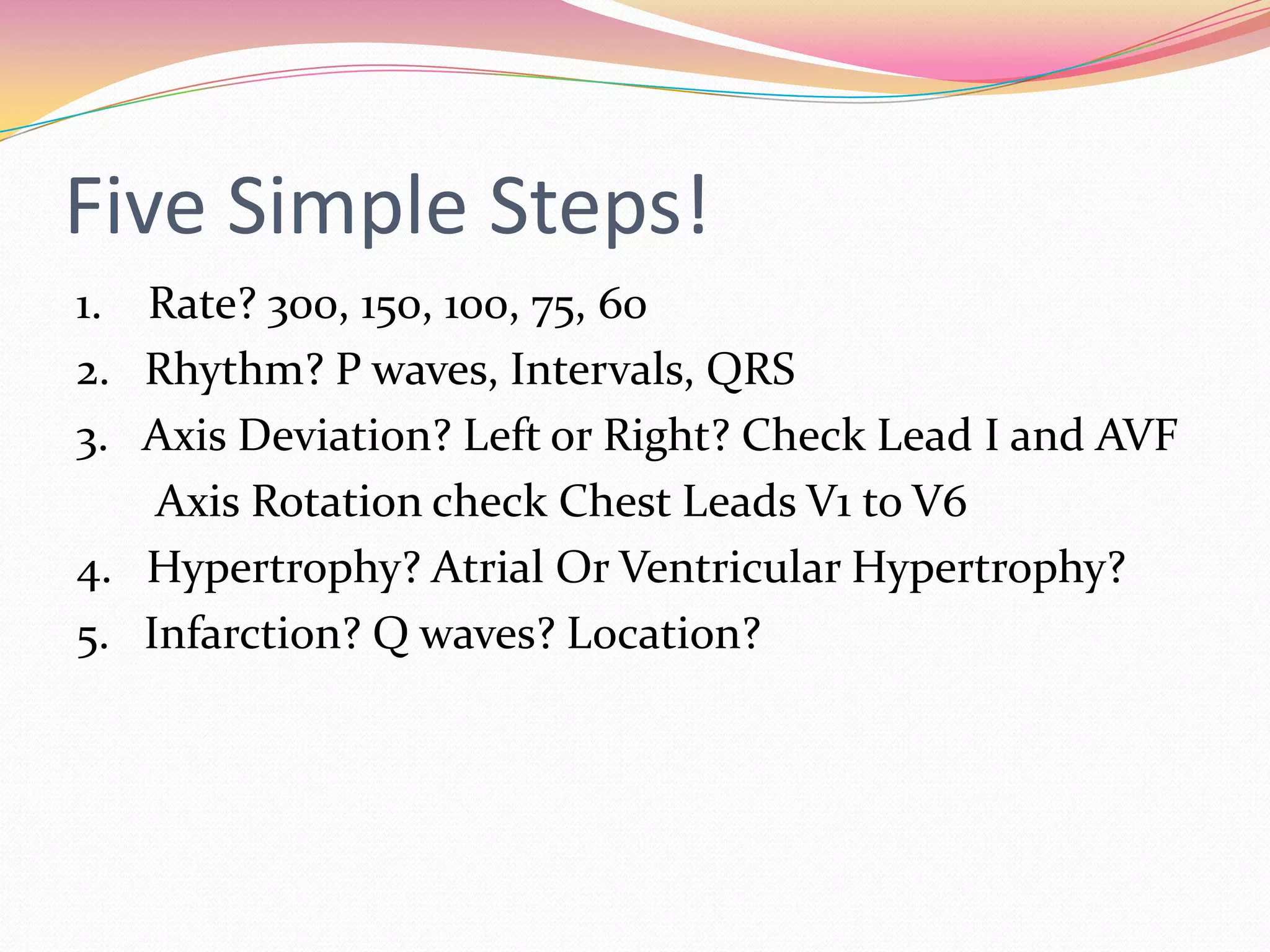
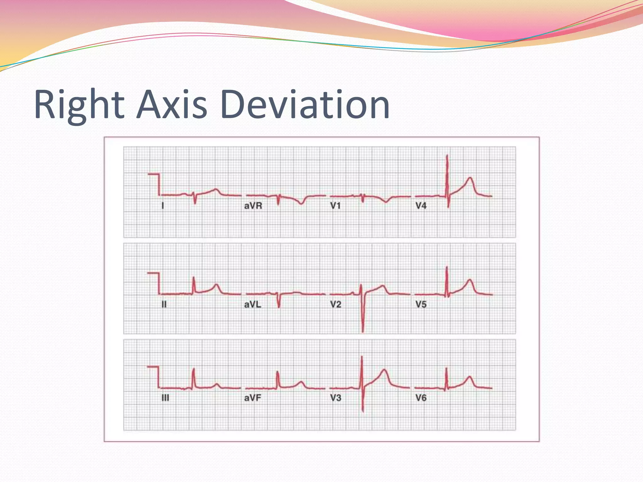
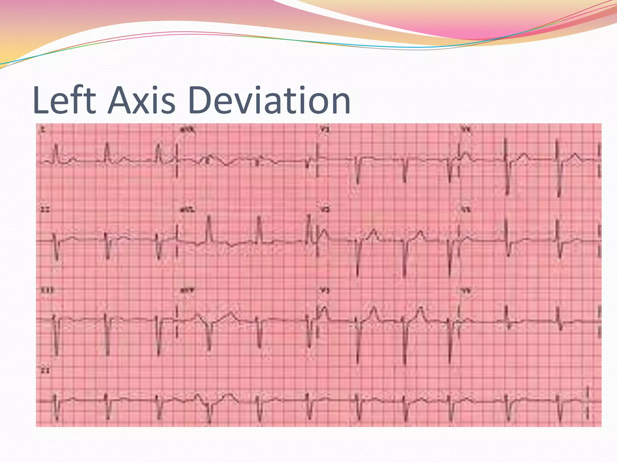
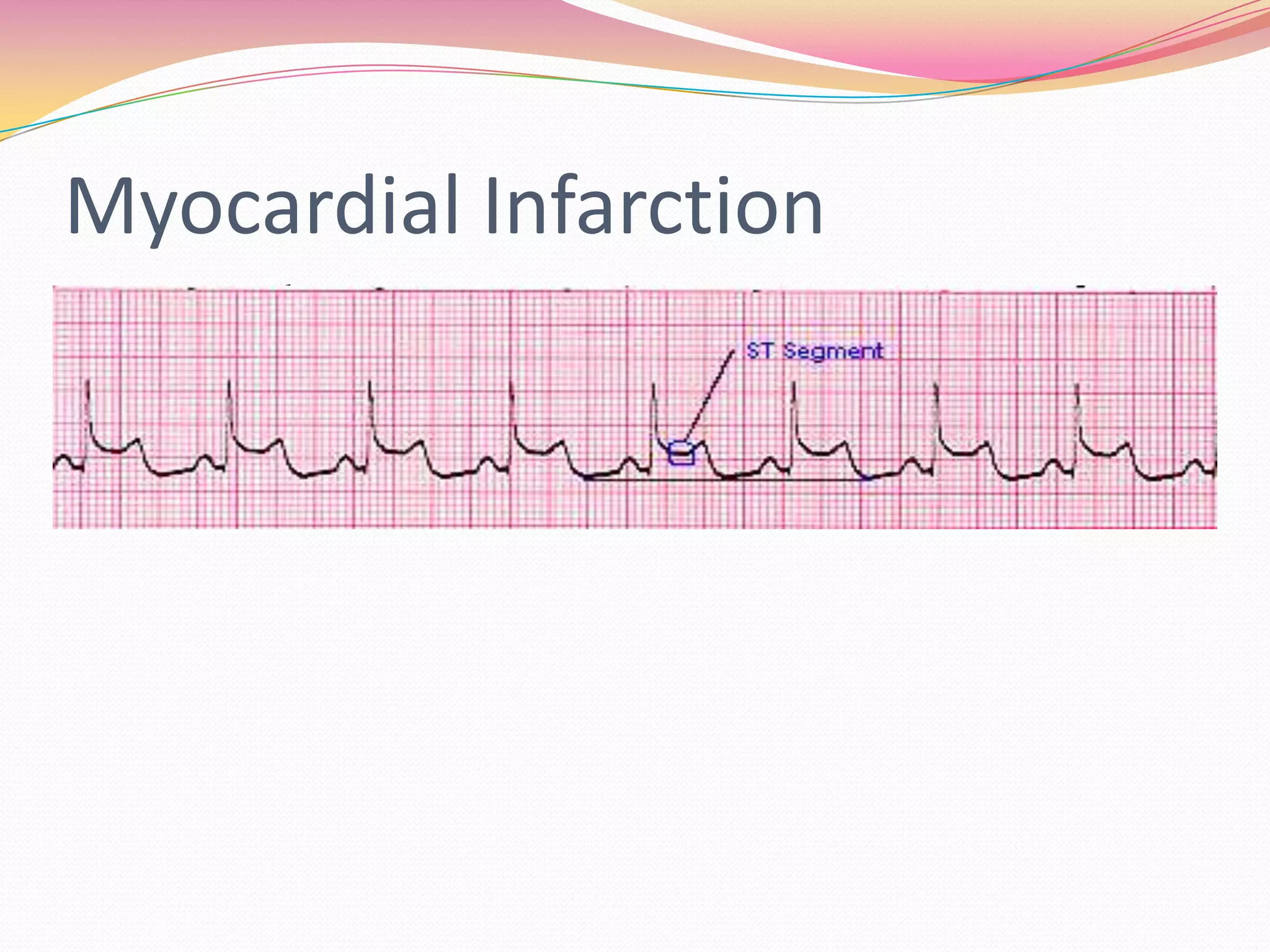
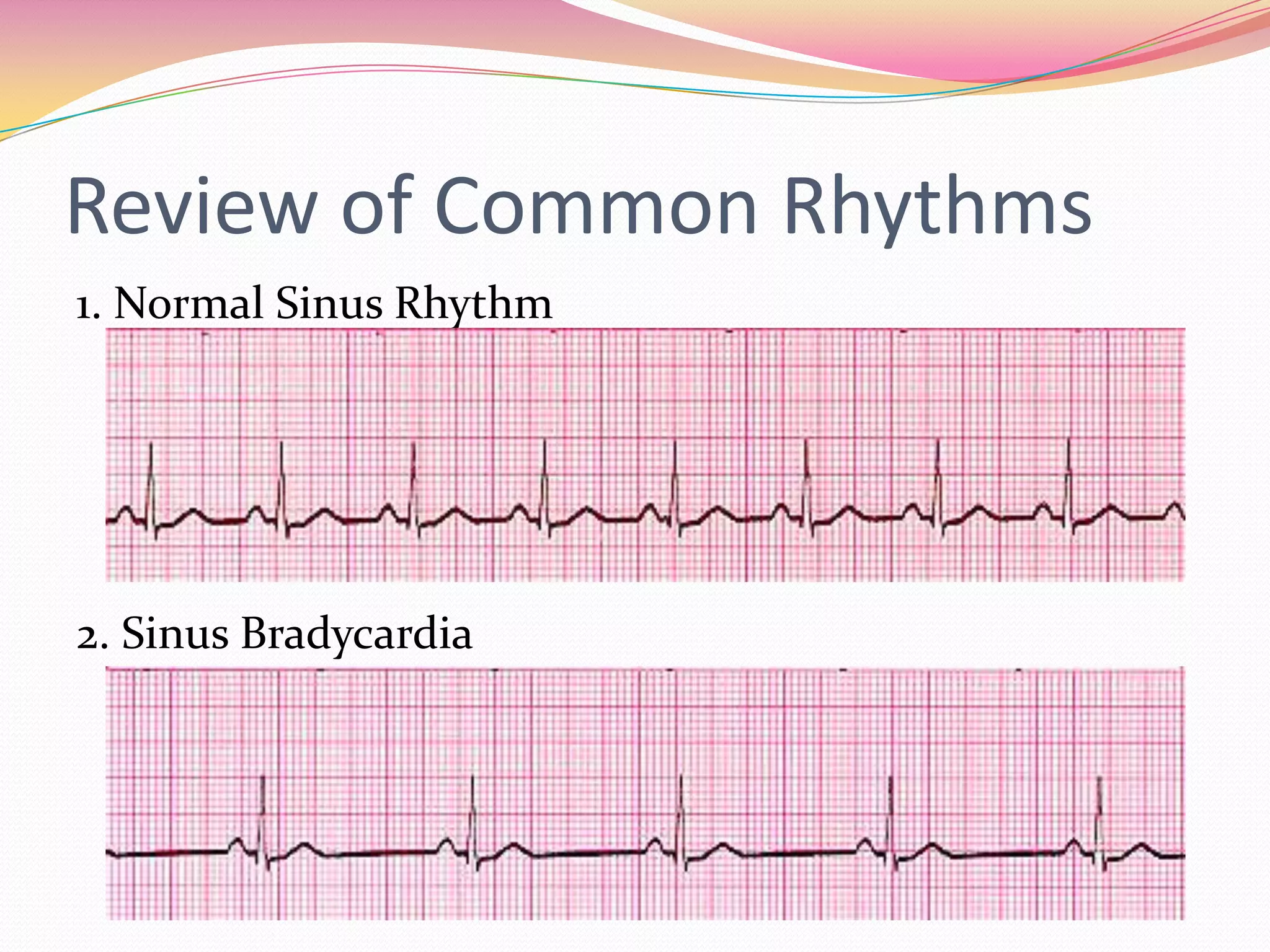
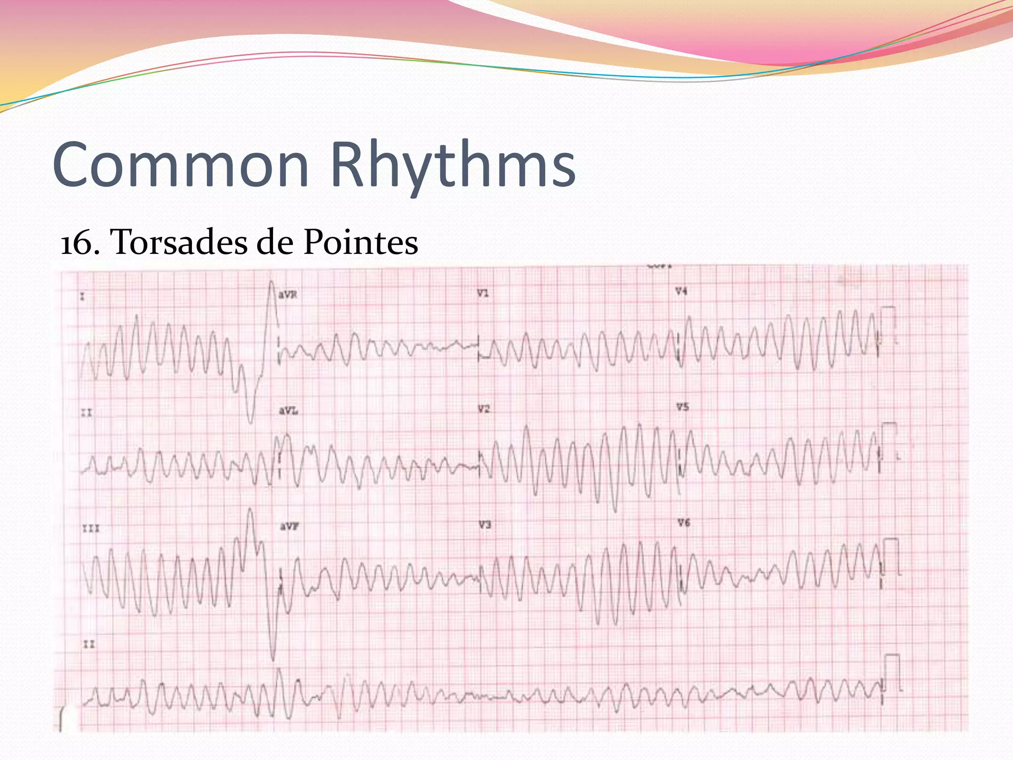
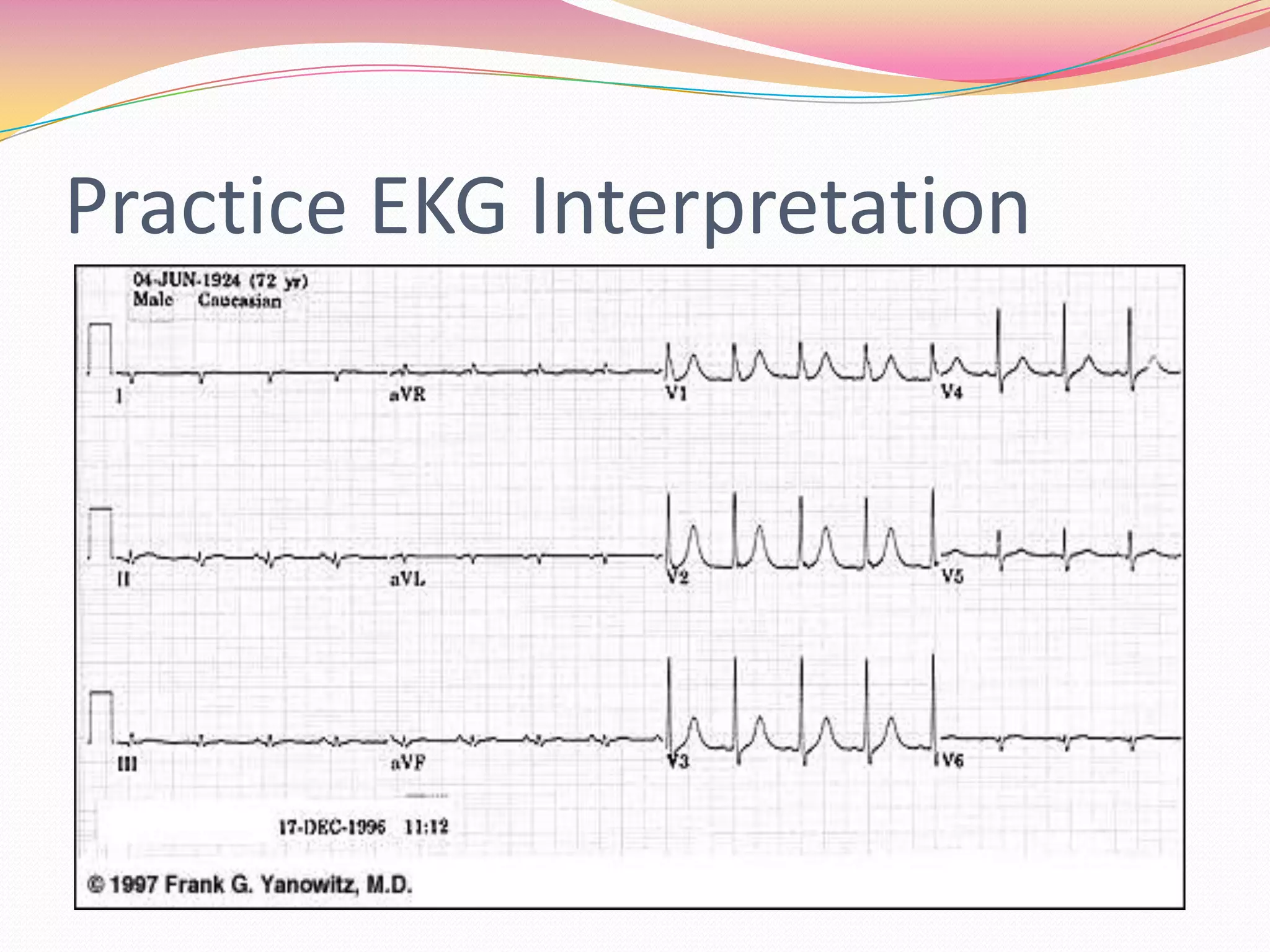
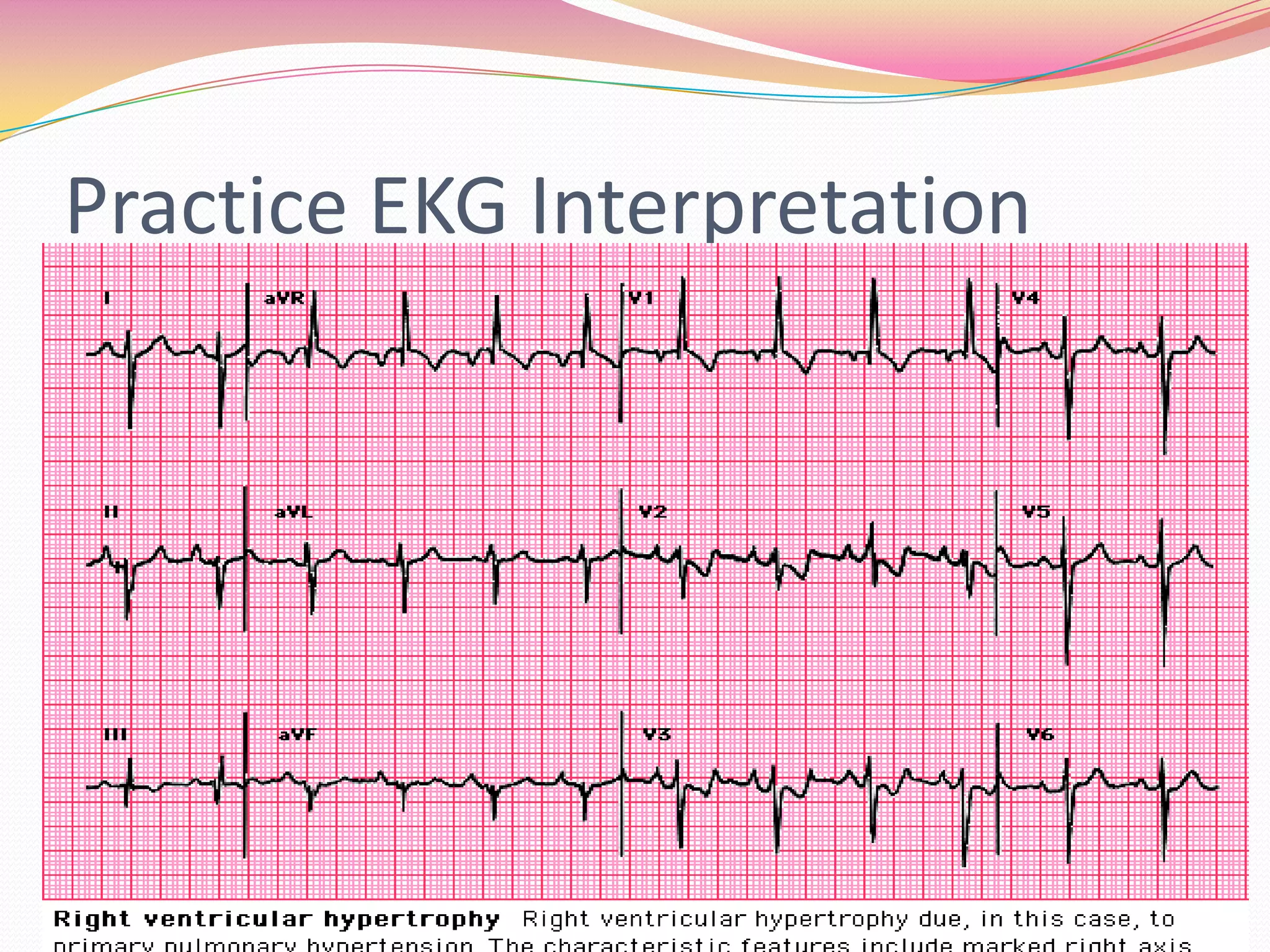
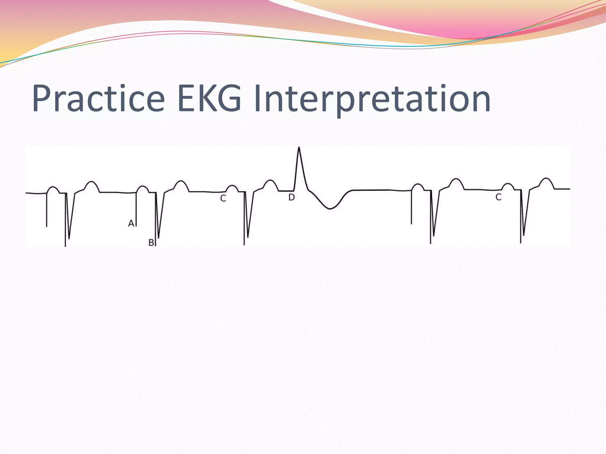
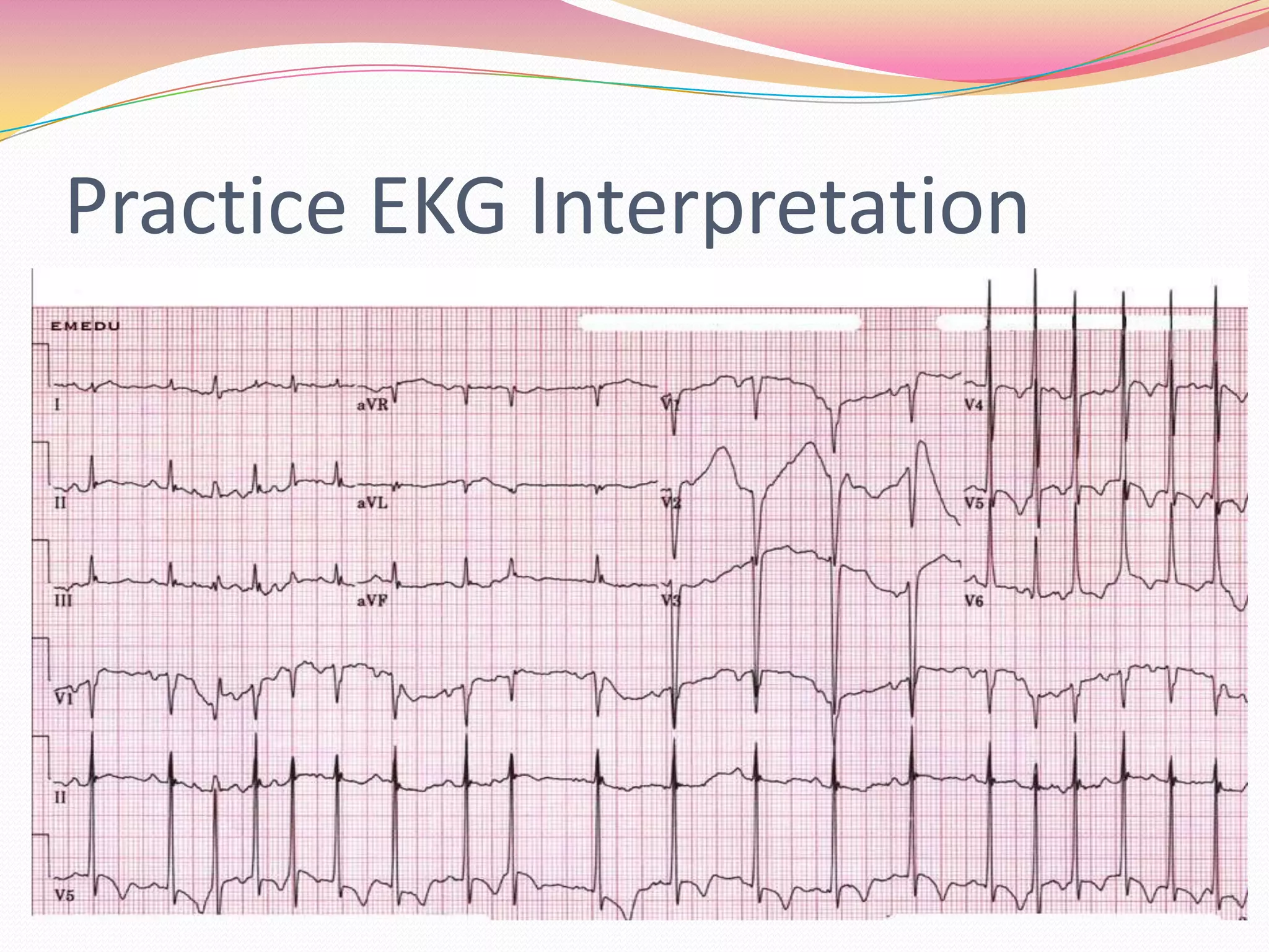
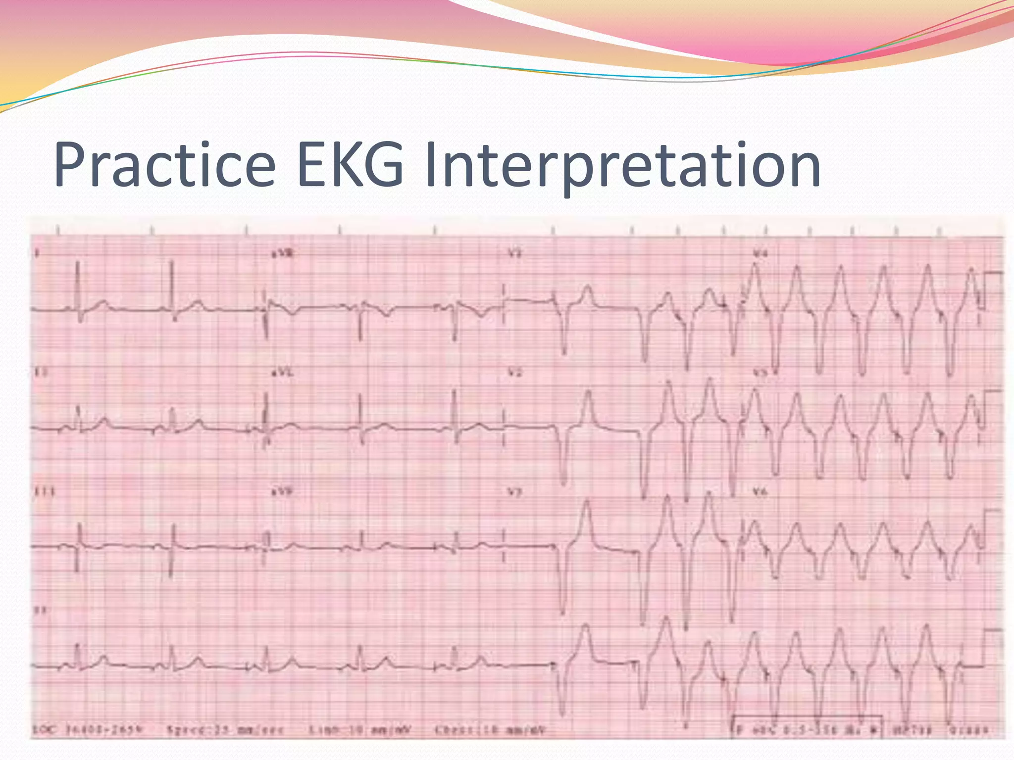
12 Lead EKG InterpretationReading with Confidence!!!!!Presented by: Herbert Hanter
Five Simple Steps!1.    Rate? 300, 150, 100, 75, 602.   Rhythm? P waves, Intervals, QRS3.   Axis Deviation? Left or Right? Check Lead I and AVF       Axis Rotation check Chest Leads V1 to V64.   Hypertrophy? Atrial Or Ventricular Hypertrophy?5.   Infarction? Q waves? Location?
When Interpreting a 12 lead EKGForget about AVR!!!!
Location of Infarction and EnlargementLateral Infarction- Q waves in I and AVL, V5 and V6Inferior Infarction- Q waves in II, III, and AVFAnterior Infarction- Q waves in  V3 V4 plus ST elevationPosterior Infarction- Large R wave In V1 V2 V3 plus ST depression, plus Q wave in V6Septal Infarction- Q waves V1 V2Atrial Enlargement- Check P wave in V1Right Ventricular Enlargement- large R wave and small S wave in V1 then R wave becomes smaller and smaller from V2, V3, and V4
ContinuationLeft Ventricular Hypertrophy- tall R wave in V5 and deep S wave in V1 (add mm V1 and V5..if total 35 mm there is LVH!)
Right Axis Deviation
Left Axis Deviation
Right Atrial Hypertrophy
Left Atrial Hypertrophy
Myocardial InfarctionST Wave Elevation
Review of Common Rhythms1. Normal Sinus Rhythm2. Sinus Bradycardia
Common Rhythms3. Sinus Tachycardia4. Supraventricular Tachycardia
Common Rhythms5. Atrial Fibrillation6. Atrial Flutter
Common Rhythms7. First Degree AV Block8. 2nd Degree AV Block Type 1 (Wenckebach)
Common Rhythms9. 2nd Degree AV Block Type 210. 3rd Degree AV Block
Common Rhythms11. Bundle Branch Block
Common Rhythms12. Premature Ventricular ComplexesSingle Multiple
Common Rhythms13. Junctional RhythmsAccelerated Junctional Rhythm
Common Rhythms14. Ventricular Tachycardia15. Ventricular Fibrillation
Common Rhythms16. Torsades de Pointes
Common Rhythms17. Asystole
Practice EKG Interpretation

12 Lead EKG Interpretation

Editor's Notes

  • #3 Axis refers to the direction of the depolarization, which spreads throughout the heart to stimulate the myocardium to contract. Depolarization means an advancing wave of Na positive ions! Avis deviation is frontal plane, Axis rotation is horizontal plane
  • #9 Bi phasic p wave looks like a letter M
  • #10 Biphasic P wave
  • #11 ST elevation of above 2 mm at least from baseline
  • #23 Inferior Wall MI: check Q waves II, III, AVF…Normal axis deviation (check I and AVF), normal axis rotation (checKIsoelectric QRS V1 to V6)
  • #24 Postero-Lateral Wall MI check Q waves in I, AVL, V5, V6 plus large R wave in V1 V2 V3
  • #25 Postero-Inferior Wall MI with RBBB: Large R wave in V1 V2 V3 V4; Q waves in II, III, AVF; V1 Rabbit ears RBBB
  • #26 Right Ventricular Hypertrophy: Large R wave in V1 then R wave becomes smaller and smaller from V2 V3 V4
  • #27 WPW Rhythm
  • #29 Wandering Atrial Pacemaker
  • #31 Atrial Pacemaker