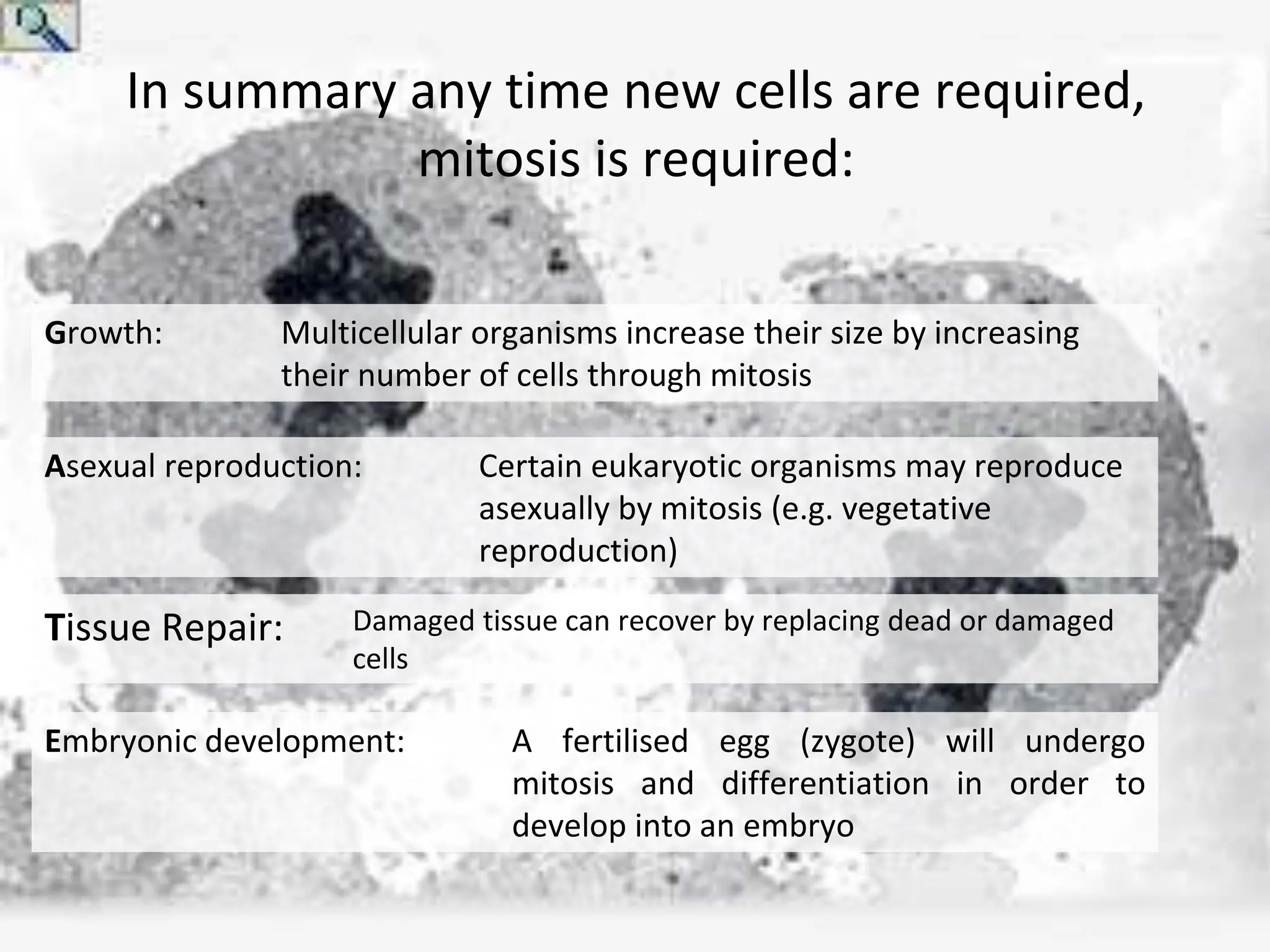
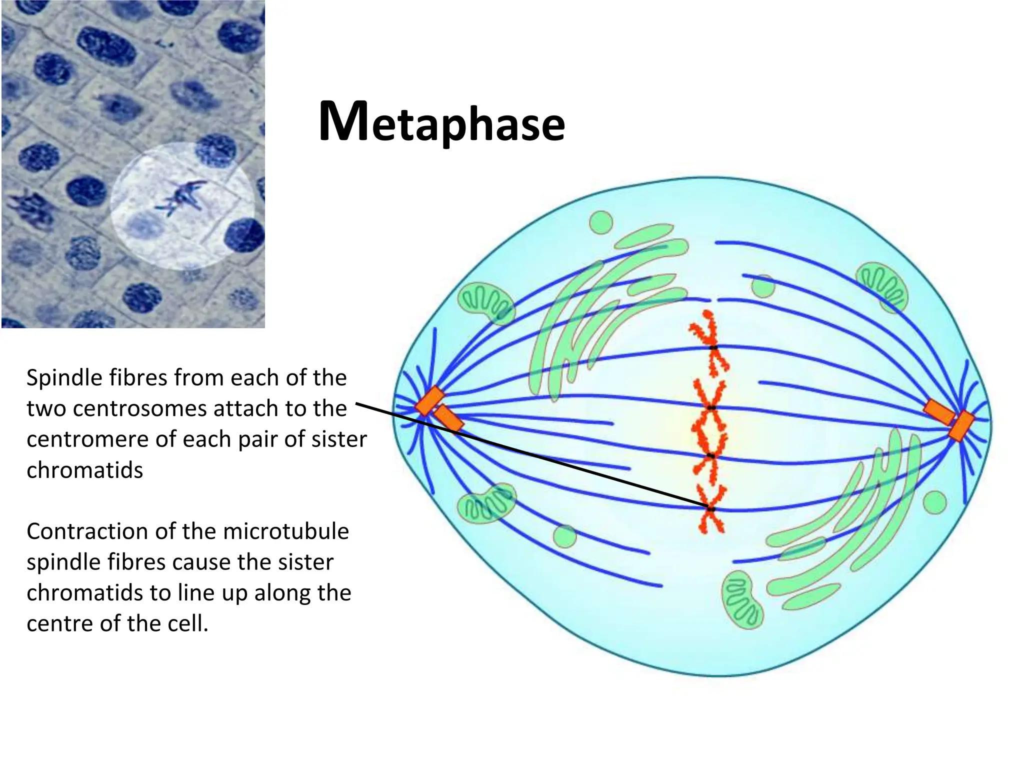
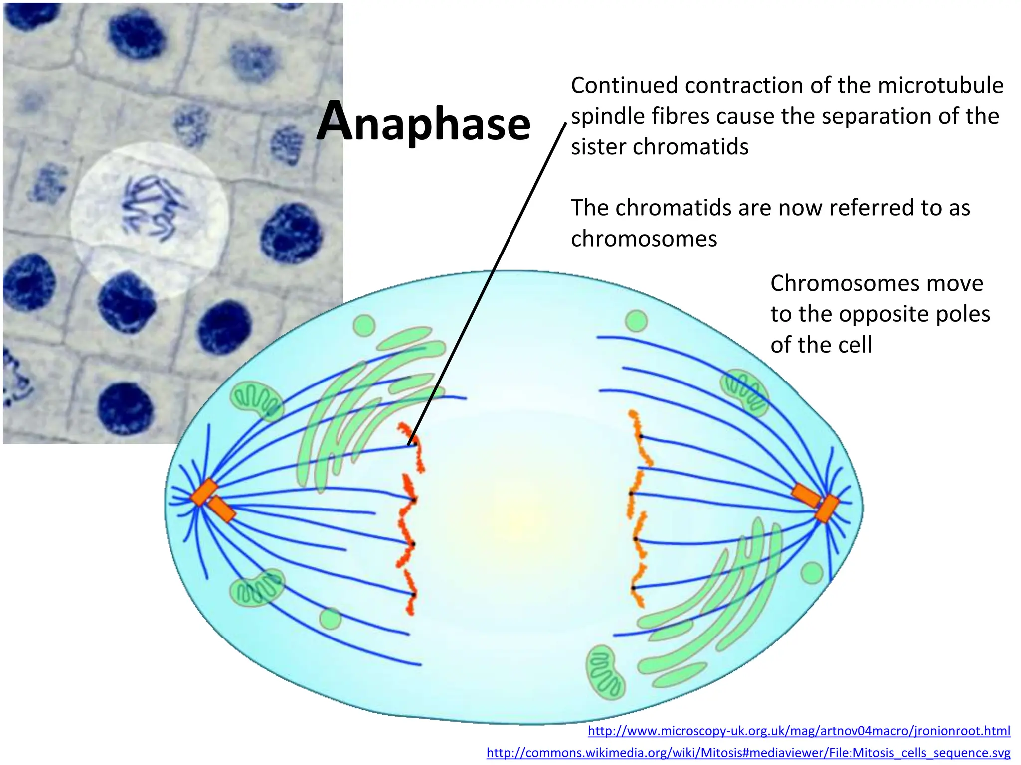
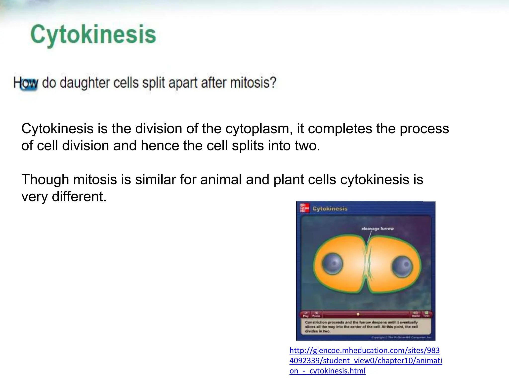
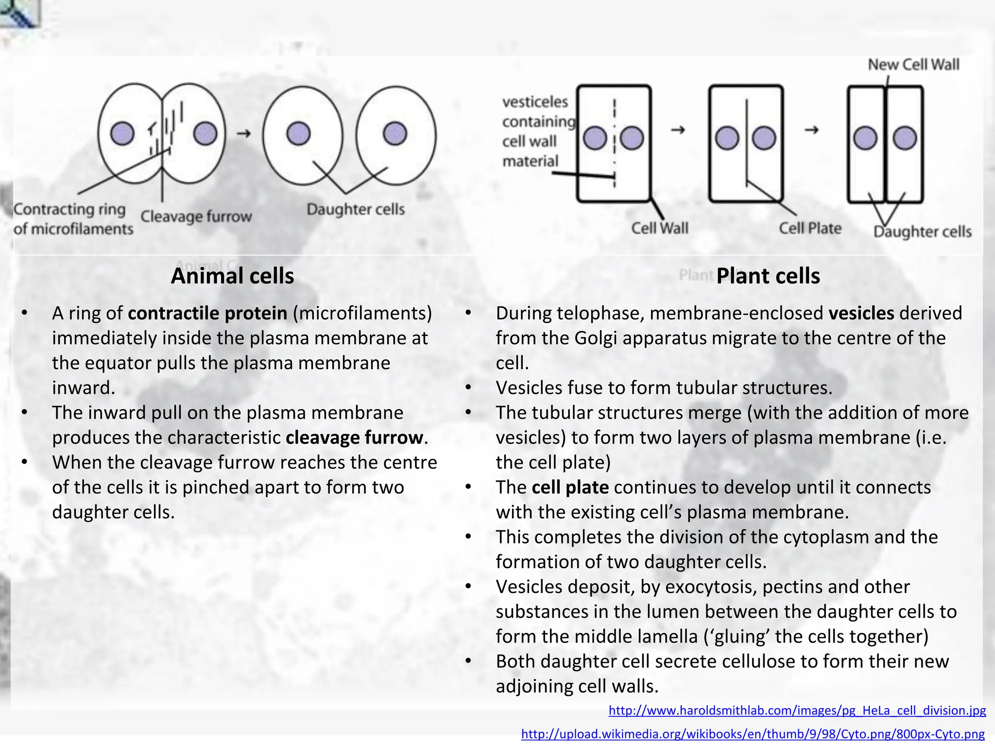
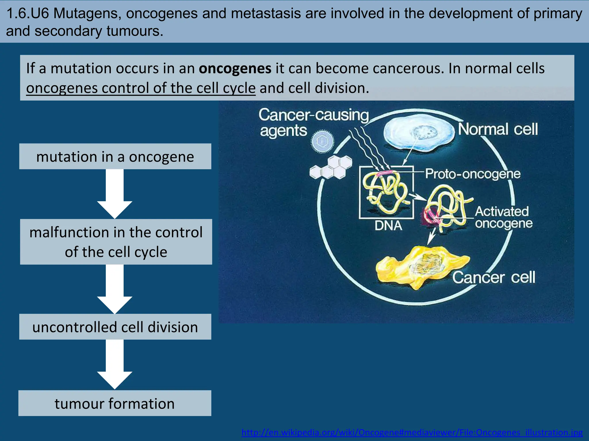
By Chris Paine
https://bioknowledgy.weebly.com/
1.6 Cell division
Essential idea: Cell division is essential but
must be controlled.
The background image shows a cancerous tumor in the lungs. Tumors are caused by
uncontrolled cell division.
By Chris Paine
https://bioknowledgy.weebly.com/
http://www.flickr.com/photos/pulmonary_pathology/4388301142/
http://youtu.be/s1ylUTbXyWU
In summary any time new cells are required,
mitosis is required:
Embryonic development: A fertilised egg (zygote) will undergo
mitosis and differentiation in order to
develop into an embryo
Growth: Multicellular organisms increase their size by increasing
their number of cells through mitosis
Asexual reproduction: Certain eukaryotic organisms may reproduce
asexually by mitosis (e.g. vegetative
reproduction)
Tissue Repair: Damaged tissue can recover by replacing dead or damaged
cells
http://upload.wikimedia.org/wikipedia/commons/thumb/7/71/Diagram_of_mitosis.svg/800px-Diagram_of_mitosis.svg.png
The cell cycle is the series of events
through which cells pass to divide
and create two identical daughter
cells.
Chromosomes make it possible to separate DNA
precisely during cell division
The genetic information that is passed to one generation
to another is carry out by chromosomes
Every cell must copy its genetic information before cell
division begins
Cells of every organism have a specific number od
chromosomes
Chromatin is composed of DNA and histone proteins
1.6.U1 Mitosis is division of the nucleus into two genetically identical daughter nuclei.
http://commons.wikimedia.org/wiki/File:Chromosome.svg
centromere is the part
of a chromosome that
links sister chromatids
Sister chromatids are duplicated
chromosomes attached by a centromere
Get the terminology right centrioles
organise spindle
microtubules
Spindle
microtubules
(also referred to
as spindle
fibres)
In animal cells two centrioles are held by a
protein mass referred to as a centrosome
After anaphase when the sister chromatids
separate they should then be referred to as
chromosomes
It is easy to misuse the terms chromatid and
chromosome. It is even easier to confuse the terms
centromere, centriole and centrosome due to their
similar spelling. Keep the terms clear in your mind
to avoid losing marks.
http://commons.wikimedia.org/wiki/Mitosis#mediaviewer/File:Mitosis_cells_sequence.svg
1.6.U4 Interphase is a very active phase of the cell cycle with many processes occurring
in the nucleus and cytoplasm.
Interphase consists of the parts of the cell cycle that don’t involve
cell division.
G1 (Gap 1)
• Increase the volume of cytoplasm
• Organelles produced
• Proteins synthesized
S (Synthesis)
• DNA replicated
G2 (Gap 2)
• Increase the volume of
cytoplasm
• Organelles produced
• Proteins synthesised
n.b. cells can also be said to be
in G0 (Gap 0). This is a ‘resting’
phase where the cell has left
the cycle and has stopped
dividing. Cells in G0 still carry
out all their normal functions.
Interphase
This when the cell carries out it’s normal functions
Metabolic reactions (e.g. respiration to produce ATP) are
necessary for the life of the cell
Protein synthesis - proteins and enzymes are necessary to allow
cell grow
Organelles numbers are increased to first support the enlarged
cell
DNA is replicated to ensure a second copy is available to enable
mitosis
Cells spend the majority of their time in interphase. It is a
very active phase of the cycle.
Mr
P
O
D
http://botit.botany.wisc.edu/Resources/Botany/Mitosis/Allium/Various%20views/Interphase%20prophase.JPG
1.6.U2 Chromosomes condense by supercoiling during mitosis.
How are chromosomes
supercoiled? Strain is placed on a DNA helix by overwinding or
underwinding of the helix
This causes the DNA molecule to coil back on itself
becoming shorter and wider
n.b. in eukaryotes proteins called histones aid the process
http://www.maths.uq.edu.au/~infinity/Infinity7/images/supercoiling.gif
http://vanat.cvm.umn.edu/mMeiosis/images/chromosome-X.jpg
http://www.microscopy-uk.org.uk/mag/artnov04macro/jronionroot.html
http://commons.wikimedia.org/wiki/Mitosis#mediaviewer/File:Mitosis_cells_sequence.svg
DNA supercoils* chromatin
condenses and becomes
sister chromatids, which are
visible under a light
microscope
The centrosomes
move to opposite
poles of the cell and
spindle fibres begin
to form between
them Nuclear
membrane
breaks down
and the
nucleolus
disappears
Prophase
*supercoling is dealt with in more detail by 1.6.U2
Metaphase
Spindle fibres from each of the
two centrosomes attach to the
centromere of each pair of sister
chromatids
Contraction of the microtubule
spindle fibres cause the sister
chromatids to line up along the
centre of the cell.
Continued contraction of the microtubule
spindle fibres cause the separation of the
sister chromatids
The chromatids are now referred to as
chromosomes
http://www.microscopy-uk.org.uk/mag/artnov04macro/jronionroot.html
http://commons.wikimedia.org/wiki/Mitosis#mediaviewer/File:Mitosis_cells_sequence.svg
Anaphase
Chromosomes move
to the opposite poles
of the cell
Now cytokinesis begins!
http://www.microscopy-uk.org.uk/mag/artnov04macro/jronionroot.html
http://commons.wikimedia.org/wiki/Mitosis#mediaviewer/File:Mitosis_cells_sequence.svg
Telophase
New nuclear membranes reform
around each set of chromosomes
Microtubule
spindle fibers
disappear
The chromosomes uncoil de-
condense to chromatin (and
are no longer visible under a
light microscope).
Chromosomes arrive at
the poles.
Cytokinesis is the division of the cytoplasm, it completes the process
of cell division and hence the cell splits into two.
Though mitosis is similar for animal and plant cells cytokinesis is
very different.
http://glencoe.mheducation.com/sites/983
4092339/student_view0/chapter10/animati
on_-_cytokinesis.html
http://upload.wikimedia.org/wikibooks/en/thumb/9/98/Cyto.png/800px-Cyto.png
Animal cells Plant cells
• A ring of contractile protein (microfilaments)
immediately inside the plasma membrane at
the equator pulls the plasma membrane
inward.
• The inward pull on the plasma membrane
produces the characteristic cleavage furrow.
• When the cleavage furrow reaches the centre
of the cells it is pinched apart to form two
daughter cells.
• During telophase, membrane-enclosed vesicles derived
from the Golgi apparatus migrate to the centre of the
cell.
• Vesicles fuse to form tubular structures.
• The tubular structures merge (with the addition of more
vesicles) to form two layers of plasma membrane (i.e.
the cell plate)
• The cell plate continues to develop until it connects
with the existing cell’s plasma membrane.
• This completes the division of the cytoplasm and the
formation of two daughter cells.
• Vesicles deposit, by exocytosis, pectins and other
substances in the lumen between the daughter cells to
form the middle lamella (‘gluing’ the cells together)
• Both daughter cell secrete cellulose to form their new
adjoining cell walls.
http://www.haroldsmithlab.com/images/pg_HeLa_cell_division.jpg
The mitotic index is a measure of the proliferation status of a cell
population (i.e. the proportion of dividing cells).
The mitotic index may be elevated during processes that promote
division, such as normal growth or cellular repair.
It also functions as an important prognostic tool for predicting the
response of cancer cells to chemotherapy
Mitotic Index
1.6.U5 Cyclins are involved in the control of the cell cycle.
Cyclinsare a family of proteins that control the progression of
cells through the cell cycle
Cells cannot progress to the next
stage of the cell cycle unless the
specific cyclin reaches it threshold.
http://upload.wikimedia.org/wikipedia/commons/thumb/9/99/Protein_CCNE1_PDB_1w98.png/800px-Protein_CCNE1_PDB_1w98.png
Cyclins bind to enzymes called
cyclin-dependent kinases
These kinases then become active and
attach phosphate groups to other
proteins in the cell.
The attachment of phosphate triggers the other proteins to become active
and carry out tasks (specific to one of the phases of the cell cycle).
4
3
2
1
1.6.U5 Cyclins are involved in the control of the cell cycle.
http://upload.wikimedia.org/wikipedia/commons/thumb/9/99/Protein_CCNE1_PDB_1w98.png/800px-Protein_CCNE1_PDB_1w98.png
Triggers cells to
move from G0 to
G1 and from G1
into S phase.
prepares the cell
for DNA
replication in S
phase.
activates DNA
replication inside
the nucleus in S
phase.
promotes the assembly
of the mitotic spindle
and other tasks in the
cytoplasm to prepare
for mitosis.
Progression through parts of the cell cycle are affected in various
ways by specific cyclins
1.6.U6 Mutagens, oncogenes and metastasis are involved in the development of primary
and secondary tumours.
Tumours are abnormal growth of tissue that develop at any stage of life in any part of
the body. A cancer is a malignant tumour and is named after the part of the body
where the cancer (primary tumour) first develops. Use the links to find out:
• most common types of cancer
• what causes cancer and associated risk factors
• how cancer can be treated
http://www.e-learningforkids.org/health/lesson/cancer/
http://www.cancer.gov/cancertopics/types/commoncancers
http://youtu.be/8BJ8_5Gyhg8
http://www.cancerresearchuk.org/cancer-
info/cancerandresearch/all-about-cancer/what-is-cancer/
What causes
cancer?
1.6.U6 Mutagens, oncogenes and metastasis are involved in the development of primary
and secondary tumours.
Mutagens are agents that
cause gene mutations. Not all
mutations result in cancers,
but anything that causes a
mutation has the potential to
cause a cancer.
Mutagens can be:
• chemicals that cause mutations are
referred to as carcinogens
• high energy radiation such as X-rays
• short-wave ultraviolet light
• Some viruses
A mutation is a change in an organisms genetic code. A mutation/change in the
base sequence of a certain genes can result in cancer.
http://en.wikipedia.org/wiki/Oncogene#mediaviewer/File:Oncogenes_illustration.jpg
1.6.U6 Mutagens, oncogenes and metastasis are involved in the development of primary
and secondary tumours.
mutation in a oncogene
If a mutation occurs in an oncogenes it can become cancerous. In normal cells
oncogenes control of the cell cycle and cell division.
http://en.wikipedia.org/wiki/Oncogene#mediaviewer/File:Oncogenes_illustration.jpg
uncontrolled cell division
tumour formation
malfunction in the control
of the cell cycle
1.6.U6 Mutagens, oncogenes and metastasis are involved in the development of primary
and secondary tumours.
Factors (other than exposure to
mutagens) that increase the probability
of tumour development include:
• The vast number of cells in a human
body – the greater the number of cells
the greater the chance of a mutation.
• The longer a life span the greater the
chance of a mutation.
Several mutations must occur in the same cell for it to become a tumour
causing cell. The probability of this happening in a single cell is extremely small.
http://en.wikipedia.org/wiki/Oncogene#mediaviewer/File:Oncogenes_illustration.jpg
1.6.U6 Mutagens, oncogenes and metastasis are involved in the development of primary
and secondary tumours.
The development of a primary tumours (cancers) have been outlined. Below is
how a primary tumor can become a secondary tumour.
A primary tumor is a malignant tumor
growing at the site where the
abnormal growth first occurred.
http://en.wikipedia.org/wiki/Neoplasm#mediaviewer/File:Colon_cancer_2.jpg
Cancerous cells can detach from the
primary tumour.
The circulating cancerous cells invade
tissues at a different locations and
develop, by uncontrolled cell division,
into a secondary tumours.
Metastasis is the movement of cells
from a primary tumour to set up
secondary tumours in other parts of
the body.
Some cancerous cells gain the ability
to penetrate the walls of lymph or
blood vessels and hence circulate
around the body
1.6.A1 The correlation between smoking and incidence of cancers.
A significant body of scientific literature exists which provides a
strong link between smoking and the incidence of cancers.
Cigarette smoke contains over 4,000 chemical compounds, over 60
of which are known to be carcinogenic
There appears to be a strong positive
correlation between the frequency of
smoking and the development of cancer
The risk of lung cancer is strongly
correlated with smoking, with ~90% of lung
cancers attributable to tobacco use
Smoking also increases the risk of over a
dozen other cancers, including mouth,
stomach, liver, pancreas and bowel.
1.6.A1 The correlation between smoking and incidence of cancers.

1.6 Cell division (CANCER,MITOSIS, DIVISION)

  • 1.
    By Chris Paine https://bioknowledgy.weebly.com/ 1.6Cell division Essential idea: Cell division is essential but must be controlled. The background image shows a cancerous tumor in the lungs. Tumors are caused by uncontrolled cell division. By Chris Paine https://bioknowledgy.weebly.com/ http://www.flickr.com/photos/pulmonary_pathology/4388301142/
  • 2.
  • 3.
    In summary anytime new cells are required, mitosis is required: Embryonic development: A fertilised egg (zygote) will undergo mitosis and differentiation in order to develop into an embryo Growth: Multicellular organisms increase their size by increasing their number of cells through mitosis Asexual reproduction: Certain eukaryotic organisms may reproduce asexually by mitosis (e.g. vegetative reproduction) Tissue Repair: Damaged tissue can recover by replacing dead or damaged cells
  • 4.
    http://upload.wikimedia.org/wikipedia/commons/thumb/7/71/Diagram_of_mitosis.svg/800px-Diagram_of_mitosis.svg.png The cell cycleis the series of events through which cells pass to divide and create two identical daughter cells.
  • 5.
    Chromosomes make itpossible to separate DNA precisely during cell division The genetic information that is passed to one generation to another is carry out by chromosomes Every cell must copy its genetic information before cell division begins Cells of every organism have a specific number od chromosomes
  • 6.
    Chromatin is composedof DNA and histone proteins
  • 10.
    1.6.U1 Mitosis isdivision of the nucleus into two genetically identical daughter nuclei. http://commons.wikimedia.org/wiki/File:Chromosome.svg centromere is the part of a chromosome that links sister chromatids Sister chromatids are duplicated chromosomes attached by a centromere Get the terminology right centrioles organise spindle microtubules Spindle microtubules (also referred to as spindle fibres) In animal cells two centrioles are held by a protein mass referred to as a centrosome After anaphase when the sister chromatids separate they should then be referred to as chromosomes It is easy to misuse the terms chromatid and chromosome. It is even easier to confuse the terms centromere, centriole and centrosome due to their similar spelling. Keep the terms clear in your mind to avoid losing marks. http://commons.wikimedia.org/wiki/Mitosis#mediaviewer/File:Mitosis_cells_sequence.svg
  • 12.
    1.6.U4 Interphase isa very active phase of the cell cycle with many processes occurring in the nucleus and cytoplasm. Interphase consists of the parts of the cell cycle that don’t involve cell division. G1 (Gap 1) • Increase the volume of cytoplasm • Organelles produced • Proteins synthesized S (Synthesis) • DNA replicated G2 (Gap 2) • Increase the volume of cytoplasm • Organelles produced • Proteins synthesised n.b. cells can also be said to be in G0 (Gap 0). This is a ‘resting’ phase where the cell has left the cycle and has stopped dividing. Cells in G0 still carry out all their normal functions.
  • 13.
    Interphase This when thecell carries out it’s normal functions Metabolic reactions (e.g. respiration to produce ATP) are necessary for the life of the cell Protein synthesis - proteins and enzymes are necessary to allow cell grow Organelles numbers are increased to first support the enlarged cell DNA is replicated to ensure a second copy is available to enable mitosis Cells spend the majority of their time in interphase. It is a very active phase of the cycle. Mr P O D http://botit.botany.wisc.edu/Resources/Botany/Mitosis/Allium/Various%20views/Interphase%20prophase.JPG
  • 14.
    1.6.U2 Chromosomes condenseby supercoiling during mitosis. How are chromosomes supercoiled? Strain is placed on a DNA helix by overwinding or underwinding of the helix This causes the DNA molecule to coil back on itself becoming shorter and wider n.b. in eukaryotes proteins called histones aid the process http://www.maths.uq.edu.au/~infinity/Infinity7/images/supercoiling.gif http://vanat.cvm.umn.edu/mMeiosis/images/chromosome-X.jpg
  • 15.
    http://www.microscopy-uk.org.uk/mag/artnov04macro/jronionroot.html http://commons.wikimedia.org/wiki/Mitosis#mediaviewer/File:Mitosis_cells_sequence.svg DNA supercoils* chromatin condensesand becomes sister chromatids, which are visible under a light microscope The centrosomes move to opposite poles of the cell and spindle fibres begin to form between them Nuclear membrane breaks down and the nucleolus disappears Prophase *supercoling is dealt with in more detail by 1.6.U2
  • 16.
    Metaphase Spindle fibres fromeach of the two centrosomes attach to the centromere of each pair of sister chromatids Contraction of the microtubule spindle fibres cause the sister chromatids to line up along the centre of the cell.
  • 17.
    Continued contraction ofthe microtubule spindle fibres cause the separation of the sister chromatids The chromatids are now referred to as chromosomes http://www.microscopy-uk.org.uk/mag/artnov04macro/jronionroot.html http://commons.wikimedia.org/wiki/Mitosis#mediaviewer/File:Mitosis_cells_sequence.svg Anaphase Chromosomes move to the opposite poles of the cell
  • 18.
    Now cytokinesis begins! http://www.microscopy-uk.org.uk/mag/artnov04macro/jronionroot.html http://commons.wikimedia.org/wiki/Mitosis#mediaviewer/File:Mitosis_cells_sequence.svg Telophase Newnuclear membranes reform around each set of chromosomes Microtubule spindle fibers disappear The chromosomes uncoil de- condense to chromatin (and are no longer visible under a light microscope). Chromosomes arrive at the poles.
  • 19.
    Cytokinesis is thedivision of the cytoplasm, it completes the process of cell division and hence the cell splits into two. Though mitosis is similar for animal and plant cells cytokinesis is very different. http://glencoe.mheducation.com/sites/983 4092339/student_view0/chapter10/animati on_-_cytokinesis.html
  • 20.
    http://upload.wikimedia.org/wikibooks/en/thumb/9/98/Cyto.png/800px-Cyto.png Animal cells Plantcells • A ring of contractile protein (microfilaments) immediately inside the plasma membrane at the equator pulls the plasma membrane inward. • The inward pull on the plasma membrane produces the characteristic cleavage furrow. • When the cleavage furrow reaches the centre of the cells it is pinched apart to form two daughter cells. • During telophase, membrane-enclosed vesicles derived from the Golgi apparatus migrate to the centre of the cell. • Vesicles fuse to form tubular structures. • The tubular structures merge (with the addition of more vesicles) to form two layers of plasma membrane (i.e. the cell plate) • The cell plate continues to develop until it connects with the existing cell’s plasma membrane. • This completes the division of the cytoplasm and the formation of two daughter cells. • Vesicles deposit, by exocytosis, pectins and other substances in the lumen between the daughter cells to form the middle lamella (‘gluing’ the cells together) • Both daughter cell secrete cellulose to form their new adjoining cell walls. http://www.haroldsmithlab.com/images/pg_HeLa_cell_division.jpg
  • 21.
    The mitotic indexis a measure of the proliferation status of a cell population (i.e. the proportion of dividing cells). The mitotic index may be elevated during processes that promote division, such as normal growth or cellular repair. It also functions as an important prognostic tool for predicting the response of cancer cells to chemotherapy Mitotic Index
  • 22.
    1.6.U5 Cyclins areinvolved in the control of the cell cycle. Cyclinsare a family of proteins that control the progression of cells through the cell cycle Cells cannot progress to the next stage of the cell cycle unless the specific cyclin reaches it threshold. http://upload.wikimedia.org/wikipedia/commons/thumb/9/99/Protein_CCNE1_PDB_1w98.png/800px-Protein_CCNE1_PDB_1w98.png Cyclins bind to enzymes called cyclin-dependent kinases These kinases then become active and attach phosphate groups to other proteins in the cell. The attachment of phosphate triggers the other proteins to become active and carry out tasks (specific to one of the phases of the cell cycle). 4 3 2 1
  • 23.
    1.6.U5 Cyclins areinvolved in the control of the cell cycle. http://upload.wikimedia.org/wikipedia/commons/thumb/9/99/Protein_CCNE1_PDB_1w98.png/800px-Protein_CCNE1_PDB_1w98.png Triggers cells to move from G0 to G1 and from G1 into S phase. prepares the cell for DNA replication in S phase. activates DNA replication inside the nucleus in S phase. promotes the assembly of the mitotic spindle and other tasks in the cytoplasm to prepare for mitosis. Progression through parts of the cell cycle are affected in various ways by specific cyclins
  • 24.
    1.6.U6 Mutagens, oncogenesand metastasis are involved in the development of primary and secondary tumours. Tumours are abnormal growth of tissue that develop at any stage of life in any part of the body. A cancer is a malignant tumour and is named after the part of the body where the cancer (primary tumour) first develops. Use the links to find out: • most common types of cancer • what causes cancer and associated risk factors • how cancer can be treated http://www.e-learningforkids.org/health/lesson/cancer/ http://www.cancer.gov/cancertopics/types/commoncancers http://youtu.be/8BJ8_5Gyhg8 http://www.cancerresearchuk.org/cancer- info/cancerandresearch/all-about-cancer/what-is-cancer/ What causes cancer?
  • 25.
    1.6.U6 Mutagens, oncogenesand metastasis are involved in the development of primary and secondary tumours. Mutagens are agents that cause gene mutations. Not all mutations result in cancers, but anything that causes a mutation has the potential to cause a cancer. Mutagens can be: • chemicals that cause mutations are referred to as carcinogens • high energy radiation such as X-rays • short-wave ultraviolet light • Some viruses A mutation is a change in an organisms genetic code. A mutation/change in the base sequence of a certain genes can result in cancer. http://en.wikipedia.org/wiki/Oncogene#mediaviewer/File:Oncogenes_illustration.jpg
  • 26.
    1.6.U6 Mutagens, oncogenesand metastasis are involved in the development of primary and secondary tumours. mutation in a oncogene If a mutation occurs in an oncogenes it can become cancerous. In normal cells oncogenes control of the cell cycle and cell division. http://en.wikipedia.org/wiki/Oncogene#mediaviewer/File:Oncogenes_illustration.jpg uncontrolled cell division tumour formation malfunction in the control of the cell cycle
  • 27.
    1.6.U6 Mutagens, oncogenesand metastasis are involved in the development of primary and secondary tumours. Factors (other than exposure to mutagens) that increase the probability of tumour development include: • The vast number of cells in a human body – the greater the number of cells the greater the chance of a mutation. • The longer a life span the greater the chance of a mutation. Several mutations must occur in the same cell for it to become a tumour causing cell. The probability of this happening in a single cell is extremely small. http://en.wikipedia.org/wiki/Oncogene#mediaviewer/File:Oncogenes_illustration.jpg
  • 28.
    1.6.U6 Mutagens, oncogenesand metastasis are involved in the development of primary and secondary tumours. The development of a primary tumours (cancers) have been outlined. Below is how a primary tumor can become a secondary tumour. A primary tumor is a malignant tumor growing at the site where the abnormal growth first occurred. http://en.wikipedia.org/wiki/Neoplasm#mediaviewer/File:Colon_cancer_2.jpg Cancerous cells can detach from the primary tumour. The circulating cancerous cells invade tissues at a different locations and develop, by uncontrolled cell division, into a secondary tumours. Metastasis is the movement of cells from a primary tumour to set up secondary tumours in other parts of the body. Some cancerous cells gain the ability to penetrate the walls of lymph or blood vessels and hence circulate around the body
  • 29.
    1.6.A1 The correlationbetween smoking and incidence of cancers. A significant body of scientific literature exists which provides a strong link between smoking and the incidence of cancers. Cigarette smoke contains over 4,000 chemical compounds, over 60 of which are known to be carcinogenic
  • 30.
    There appears tobe a strong positive correlation between the frequency of smoking and the development of cancer The risk of lung cancer is strongly correlated with smoking, with ~90% of lung cancers attributable to tobacco use Smoking also increases the risk of over a dozen other cancers, including mouth, stomach, liver, pancreas and bowel. 1.6.A1 The correlation between smoking and incidence of cancers.