
Recommended
More Related Content
What's hot
What's hot (20)
Similar to Cns i brain tumours
Similar to Cns i brain tumours (20)
More from M Ridhwan Abd Razak
More from M Ridhwan Abd Razak (20)
Recently uploaded
Recently uploaded (20)
Cns i brain tumours
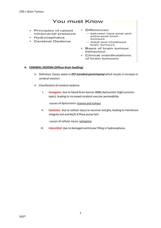
- 1. CNS I: Brain Tumour 1 SG5® A. CEREBRAL OEDEMA (Diffuse Brain Swelling) Definition: Excess water in ECF (cerebral parenchyma) which results in increase in cerebral volume! Classification of cerebral oedema: I. Vasogenic: due to blood-brain barrier (BBB) dysfunction (tight junction open), leading to increased cerebral vascular permeability -causes of dysfunction: trauma and tumour II. Cytotoxic: due to cellular injury to neurone and glia, leading to membrane integrity lost and Na/K ATPase pump fails -causes of cellular injury: ischaemia III. Interstitial: due to damaged ventricular filling in hydrocephalus
- 2. CNS I: Brain Tumour 2 SG5® B. HYDROCEPHALUS Definition: Excess water in ventricular system Classification of hydrocephalus: I. Primary hydrocephalus - Due to: i. CSF overproduction – due to choroid plexus papilloma ii. CSF circulation blockage – at foramen of Monro, aqueduct of Sylvius and foramen of Luschka iii. CSF reabsorption failure - due to arachnoid granulations II. Secondary hydrocephalus - Due to: i. Loss of brain tissue (hydrocephalus ex vacuo) –due to stroke or injury which results in shrinkage of brain III. Congenital hydrocephalus -Arnold-Chiari malformation: Tonsilar herniation Elongated brainstem Small 4th ventricle Tectal beaking (fusion of midbrain) Hydrocephalus
- 3. CNS I: Brain Tumour 3 SG5® C. BRAIN TUMOUR (all brain tumours are lethal!) Never metastasis Always recur locally! Small volume of tumour is lethal Normal brain function until tumour reaches large size! All brain tumours potentially lethal! Irrespective of the grade Causes: (causes mostly unknown!) I. Genetic: Tuberous sclerosis Neurofibromatosis Von Hippel-Lindau syndrome II. Radiation III. Familial -relatives IV. Viral V. Chemical VI. Immunosuppresion Clinical presentations: I. Cerebral oedema II. Neurological deficit- progressive and focal III. Seizure IV. Behavioural change V. RIP (Raised intracranial pressure!)
- 4. CNS I: Brain Tumour 4 SG5® Classification of CNS tumour: Cells of origin CNS tumour Glial cells -Diffusely infiltrating -Blend with surrounding brain -Grow to large size before symptoms appear -Prognosis: i. Histological grade ii. Level of patient disability iii. Adequacy of resection astrocytoma, oligodendroglioma (1p19q deletion), ependymoma, glioblastoma Arachnoidal cells Meningioma Nerve sheath cells Schwannoma (unilateral nerve deafness), neurofibroma -if bilateral deafness, think of neurofibromatosis Lymphoreticular cells Lymphoma Primitive neuroectodermal cells (PNET) Medulloblastoma, neuroblastoma Pinealoma and pituitary adenoma! Development of normal brain tissues and neoplasm of brain tissue
- 5. CNS I: Brain Tumour 5 SG5® a) GLIOMA 1) Astrocytoma Classifications: Non-infiltrating -Juvenile pilocytic -Subependymal giant cell astrocytoma -Subependymoma Infiltrating -Astrocytoma (low grade) (No mitoses or necrosis) -Anaplastic astrocytoma (Mitoses) -Glioblastoma multiforme (Mitoses and necrosis) Tx: Surgery (resection) -Craniotomy + maximum safe debulking -Stereotactic volumetric resection Radiotherapy -Conventional external beam -Stereotactic radiation -Proton beam Chemotherapy -Systemic -Direct (into the tumour):- Gliadel Therapeutic resistances: Radical resection is difficult Radiotherapy constraints Tumour heterogeneity Blood tumour barrier
- 6. CNS I: Brain Tumour 6 SG5® b) TUMOUR OF ARACHNOIDAL CELLS 1) Meningioma Do not invade the brain Relatively easy to resect Lethal if not treated due to RIP!!! c) PITUITARY ADENOMA Classifications: Macroadenoma -Compress optic nerve bilateral hemianopia Microadenoma -Hypo and hyper secretions d) CHILDHOOD BRAIN TUMOURS more devastating than adult 70% below tentorium Treatments have negative impacts on developing nervous system Endocrine effects Psychological effects Behavioural effects Development effects Classifications: i. Cerebellar astrocytoma (morning headache and vomiting) Cystic Easily resected! No need additional therapy Excellent long term survival ii. Brain stem astrocytoma Impossible to resect! Craniopharyngioma/Rathke’s pouch tumour – tumour of pituitary gland of embryonic tissue 4C’s Childhood -growth retardation and blindness Cystic Calcified Cholesterol in cyst
- 7. CNS I: Brain Tumour 7 SG5® Consequences: -Major neurological defects -Obstructive hydrocephalus Low grade Palliative care indicated iii. Medulloblastoma (PNET) Pluripotent stem cell Divergent differentiation