11 respiratory failure
•Download as PPT, PDF•
7 likes•1,241 views

patholophysiology of RespiratoryFailure Pathophysiology(病理生理学) Pathology uploaded by prabesh 杰诗

Recommended
More Related Content
What's hot
What's hot (20)
Viewers also liked
Similar to 11 respiratory failure
Similar to 11 respiratory failure (20)
More from Prabesh Raj Jamkatel
More from Prabesh Raj Jamkatel (20)
Recently uploaded
Recently uploaded (20)
11 respiratory failure
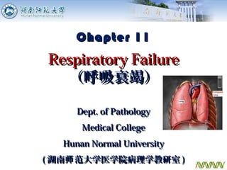
- 1. Dept. of PathologyDept. of Pathology Medical CollegeMedical College Hunan Normal UniversityHunan Normal University (( 湖南 范大学医学院病理学教研室师湖南 范大学医学院病理学教研室师 )) 1 Chapter 11Chapter 11 Respiratory FailureRespiratory Failure (呼吸衰竭)(呼吸衰竭)
- 2. 22 Respiratory FailureRespiratory Failure a.a. IntroductionIntroduction b.b. Etiology and ClassificationEtiology and Classification c.c. PathogenesisPathogenesis d.d. Alterations of Metabolism andAlterations of Metabolism and FunctionFunction e.e. Pathophysiological Basis ofPathophysiological Basis of Prevention and TreatmentPrevention and Treatment
- 3. Normal Process of Respiration Air Lungs Blood Tissue External respiration Internal respiration Transportation Ventilation Diffusion Perfusion 3
- 4. Respiratory Failure: Definition Respiratory failure (RF) is a syndrome in which the respiratory system fails to adequately oxygenate the venous blood w/ or w/o retention of carbon dioxide. PaO2: ≤ 60 mmHg (when breathing room air) PaCO2: Normal (type I) or ≥ 50 mmHg (type II) 7
- 5. Running a race at 12,000 feet Is This Respiratory Failure? 8
- 6. 99 Respiratory FailureRespiratory Failure a.a. IntroductionIntroduction b.b. Etiology and ClassificationEtiology and Classification c.c. PathogenesisPathogenesis d.d. Alterations of Metabolism andAlterations of Metabolism and FunctionFunction e.e. Pathophysiological Basis ofPathophysiological Basis of Prevention and TreatmentPrevention and Treatment
- 7. Common Causes of Respiratory Failure 10
- 8. Etiology Respiratory pump damage Brain disease (trauma or tumor) Cephalitis Pleural Effusion Pneumothorax ( 气胸 ) Lung solid lesion Pneumonia Emphysema ( 肺气肿 ) Atelectasis ( 肺不张 ) Airway obstruction Laryngeal edema Chronic bronchitis Foreign body (or tumor) Gas-exchanging problem Pulmonary edema 11
- 9. ( 气胸 )( 胸腔积液 ) Pleural Effusion Pneumothorax Emphysema ( 肺气肿 ) 12
- 10. Classification According to blood gas changes Type I: PaO2 ≤ 60 mmHg Type II: PaO2 ≤ 60 mmHg + PaCO2 ≥ 50 mmHg According to pathogenesis Ventilation Gas-exchanging According to duration Acute Chronic According to primary site Central Peripheral 13
- 11. 1414 Respiratory FailureRespiratory Failure a.a. IntroductionIntroduction b.b. Etiology and ClassificationEtiology and Classification c.c. PathogenesisPathogenesis d.d. Alterations of Metabolism andAlterations of Metabolism and FunctionFunction e.e. Pathophysiological Basis ofPathophysiological Basis of Prevention and TreatmentPrevention and Treatment
- 12. Pathogenesis of Respiratory Failure a.a. Dysfunction in ventilationDysfunction in ventilation Restrictive hypoventilationRestrictive hypoventilation Obstructive hypoventilationObstructive hypoventilation a.a. Gas-exchange dysfunctionGas-exchange dysfunction Diffusion impairmentDiffusion impairment Ventilation/perfusion imbalanceVentilation/perfusion imbalance Increase of anatomic shuntIncrease of anatomic shunt
- 13. a.a. Dysfunction in ventilationDysfunction in ventilation Restrictive hypoventilationRestrictive hypoventilation a)a) Dysfunction of respiratory pump activityDysfunction of respiratory pump activity b)b) Decrease of lung compliance (solid lesions)Decrease of lung compliance (solid lesions) Pathogenesis
- 14. Pathogenesis of Respiratory Failure a.a. Dysfunction in ventilationDysfunction in ventilation Restrictive hypoventilationRestrictive hypoventilation Obstructive hypoventilationObstructive hypoventilation a.a. Gas-exchange dysfunctionGas-exchange dysfunction Diffusion impairmentDiffusion impairment Ventilation/perfusion imbalanceVentilation/perfusion imbalance Increase of anatomic shuntIncrease of anatomic shunt
- 15. Causes: Asthma Chronic Obstructive Pulmonary Disease (COPD) (Chronic bronchitis) Types: Central airway obstruction Peripheral airway obstruction Obstructive Hypoventilation 22
- 16. Outside of chest Inside of chest Central Airway Obstruction Trachea crotch 23
- 17. Inspiration Obstruction Located Outside the Chest Inspiratory dyspnea Expiration 24
- 18. ExpirationInspiration Expiratory dyspnea Obstruction Located Inside the Chest 25
- 19. Alveolus Inspiration Bronchiole Elastic tissue Expiration Expiratory dyspnea Alveolus Bronchiole Elastic tissue Peripheral airway obstruction 27
- 20. Pathogenesis of Respiratory Failure a.a. Dysfunction in ventilationDysfunction in ventilation Restrictive hypoventilationRestrictive hypoventilation Obstructive hypoventilationObstructive hypoventilation a.a. Gas-exchange dysfunctionGas-exchange dysfunction Diffusion impairmentDiffusion impairment Ventilation/perfusion imbalanceVentilation/perfusion imbalance Increase of anatomic shuntIncrease of anatomic shunt
- 21. Pathogenesis Diffusion ImpairmentDiffusion Impairment a)a) Increase of thicknessIncrease of thickness b)b) Decrease of gas-exchange areaDecrease of gas-exchange area c)c) Shortening of diffusion timeShortening of diffusion time
- 22. Structure of Alveolar-Capillary Membrane (Diffusion Membrane) Diffusion Speed∝ Surface Area Thickness Alveolus R B C Capillary Normal: Thickness : ~1µm Surface area: 80 m2 O2 CO2 Epithelium Surfactant Endothelium
- 23. Normal Gas Exchange Blood Total blood flow time from the arteriole end to the venule end: 0.75 s 32
- 24. Prolonged Time for Gas Exchange PO2 100 80 60 40 20 0s 0.25s 0.50s 0.75 s PCO2 46 PaO2 PaCO2 40 Time of Blood Flow Through Capillary Dotted lines showing thickened diffusion membrane. C O 2 Epithelium Surfactant 33
- 25. Increase of Thickness of Diffusion Membrane Normal Edema C O 2 34
- 26. Decrease of Gas Exchange Area Atelectasis ( 肺不张 ) of the whole left lung caused by cancer 35
- 27. Pathogenesis of Respiratory Failure a.a. Dysfunction in ventilationDysfunction in ventilation Restrictive hypoventilationRestrictive hypoventilation Obstructive hypoventilationObstructive hypoventilation a.a. Gas-exchange dysfunctionGas-exchange dysfunction Diffusion impairmentDiffusion impairment Ventilation/perfusion imbalanceVentilation/perfusion imbalance Increase of anatomic shuntIncrease of anatomic shunt
- 28. Normal Ventilation/Blood Flow Balance V: Alveolar ventilation (N: 4 L/min) Q: Pulmonary blood flow (N: 5 L/min) Normal V/Q : 0.8 (bottom) V/Q ratio = Ventilation (V) Blood flow (Q)
- 29. Pathological V/Q Imbalance Hypoventilation (↓V) - V /Q? (< 0.8) - Also called “Functional shunt” or “Venous admixture” Hypoperfution (↓Q) - V /Q ? (> 0.8) - Also called “Dead space-like ventilation” 40
- 30. Normal Functional Shunt Hypoventilation (functional shunt) : V/Q ↓ Seen in asthma, COPD, edema, fibrosis
- 31. Pathological V/Q Imbalance Hypoventilation (↓V) - V /Q? (< 0.8) - Also called “Functional shunt” or “Venous admixture” Hypoperfution (↓Q) - V /Q ? (> 0.8) - Also called “Dead space-like ventilation” 42
- 32. Normal Dead Space-like Hypoperfusion (dead space-like ventilation) : V/Q ↑ Seen in pulmonary artery embolism, pulmonary vasoconstriction, pulmonary DIC
- 33. Pathogenesis of Respiratory Failure
- 34. Anatomic Shunt (True Shunt) Part of venous blood directly flows into the pulmonary vein through the bronchial vein or arterio-venous fistula. Airway Capillary Alveolus vein Artery Arterio-venous fistulas
- 35. Functional vs. Anatomical Shunt? Distinquish : Inspire Pure O2 PaOPaO22 ↑ ↑ ↑↑ ↑ ↑ PaOPaO22 ↑↑ Anatomical Functional
- 36. 47
- 37. Summary of Pathogenesis of RF Caused by dysfunction of external respiration. Dysfunction inDysfunction in ventilationventilation Restrictive Obstructive Type Ⅱ RF Dysfunction in gas-exchange Diffusion V/Q Ratio Anatomic shunt TypeⅠRF
- 38. 4949 Respiratory FailureRespiratory Failure a.a. IntroductionIntroduction b.b. Etiology and ClassificationEtiology and Classification c.c. PathogenesisPathogenesis d.d. Alterations of Metabolism andAlterations of Metabolism and FunctionFunction e.e. Pathophysiological Basis ofPathophysiological Basis of Prevention and TreatmentPrevention and Treatment
- 39. Alterations of Metabolism and Function a.a. Acid-base imbalanceAcid-base imbalance b.b. Electrolyte disturbanceElectrolyte disturbance c.c. Organ system dysfunctionOrgan system dysfunction Pulmonary systemPulmonary system Circulatory systemCirculatory system Central nervous systemCentral nervous system Urinary and digestive systemUrinary and digestive system
- 40. Acid-Base Imbalance Metabolic acidosis (seen in both types) Respiratory acidosis Respiratory alkalosis Metabolic alkalosis (Iatrogenic) Mixed acid-base disturbances Type Ⅰ RF accompanied with hyperventilation: Metabolic acidosis + Respiratory alkalosis Type Ⅱ RF: Metabolic acidosis + Respiratory acidosis Simple acid-base disturbances
- 41. Hyperkalemia (↑ K+ ) Acidosis Increased tissue catabolism Hypochloremia (↓ Cl- ) or Hyperchloremia (↑ Cl- ) Depending on types of acid-base disturbance RAc: Hypochloremia RAl: Hyperchloremia Electrolyte Disturbance 52
- 42. Cl- -HCO3 - Exchange + H +Hb(O2)-HHb(O2) RBC HCO3 - CO2 H2CO3+ H2O Cl- Cl- HCO3 - CO2 H2CO3 + H2O Hypochloremia Occurring in Type II RF 53
- 43. Alterations of Respiratory System PaO2 <60 mmHg: ↑ respiratory movement <30 mmHg:↓respiratory center PaCO2 >50 mmHg: ↑ respiratory movement >80 mmHg:↓respiratory center 54
- 44. Alterations of Circulatory System Compensatory responses Hypoxia and hypercapnia → ↑ vasomotor center Increase HR, CO, myocardial contraction, BP; blood redistribution Injurious responses Hypoxia and hypercapnia → ↓vasomotor center decrease HR, CO, myocardial contraction, BP; cor pulmonale
- 45. Pulmonary Heart Disease (cor pulmonale) 58
- 46. Alterations of central nervous system CNS is the most sensitive organ to hypoxia. PO2<60 mmHg: gentle impairment of intelligence and vision PaCO2>80 mmHg: CO2 narcosis PO2<50 mmHg: appearance of nervous and psychiatric symptoms 61
- 47. Alterations of urinary and digestive system Functional acute renal insufficiency: Excitement of sympathetic nerve leads to renal vessel constriction and RBF and GFR reduction. Gastro-intestinal insufficiency: Excitement of sympathetic nerve leads to GI organ vessel constriction → erosion, necrosis, hemorrhage, ulcer. 62
- 48. 6363 Respiratory FailureRespiratory Failure a.a. IntroductionIntroduction b.b. Etiology and ClassificationEtiology and Classification c.c. PathogenesisPathogenesis d.d. Alterations of Metabolism andAlterations of Metabolism and FunctionFunction e.e. Pathophysiological Basis ofPathophysiological Basis of Prevention and TreatmentPrevention and Treatment
- 49. Prevention and Treatment 1. Remove the factors that cause RF 2. Raise PaO2 via oxygen therapy 3. Reduce PaCO2 through improving ventilation 4. Others: Correct acid-base imbalance Correct electrolyte disturbance Protect against heart and brain failure 64
- 50. Type II RF: Low concentration (30% O2) Low flow (1 - 2 L/min) - Avoid too rapid correction of hypoxia Oxygen Therapy Type I RF: High concentration (40% O2) 65
- 51. Introduction Etiology and Pathogenesis ARDS Alterations of Metabolism and Function Principle for Treatment Contents 66
- 52. Acute Respiratory Distress Syndrome Definition Clinical concept defined it as a spectrum of ALI - Acute onset - bilateral infiltrates on CXR (“White lung”) - PCWP =< 18 mmHg - Hypoxia and PaO2/FiO2 =< 200 ( ALI if P/F ratio =< 300 ) -No cardiovascular lesion 67
- 53. 返 68
- 54. 69
- 55. ARDS is a severe lung syndrome (not a disease) caused by a variety of direct and indirect issues. It is characterized by inflammation of the lung parenchyma leading to impaired gas exchange Pathophysiological concept Alveolar-capillary membrane injury 70
- 57. ARDS Acute lung injury Diffuse alveolar damage (DAD)/Acute respiratory distress syndrome 72
- 58. ARDS 73
- 59. White lung 75
- 60. Causes Sepsis and Shock Severe multiple trauma Aspiration of gastric contents Inhalation of toxic gases and fumes etc. Insults involved in alveolar capillary membrane injury 76
- 61. Pathogenesis A-Cm injury Pathogenic factors PMN activation Protease release ROS generation Inflammatory mediators Plt activation aggregation Thrombosis Pulmonary edema Bleeding hyaline membrane Atelectasis Bronchial spasm Vaso- constriction Diffusion dysfunction Shunt Dead space Hypoxemia 77
Editor's Notes
- Respiratory failure relates to external respiration.
- Acute respiratory failure can defined as a state in which the pulmonary system is no longer able to meet the metabolic demands of the body. It can be divided into hypoxaemic respiratory failure and hypercapnic respiratory failure. Hypoxaemic respiratory failure is defined as an arterial partial pressure of oxygen of less than or equal to 6.7 kPa when breathing room air and hypercapnic respiratory failure is defined as an arterial partial pressure of carbon dioxide of more than or equal to 6.7 kPa
- 12,000 feet = 3,780 m. No, because the external respiratory system functions normally.
- Cephalitis: 脑炎
- pleural Effusion 胸腔积液; 胸膜渗出(液) On the left is a diagram of the lungs and airways with an inset showing a detailed cross-section of normal bronchioles and alveoli. On the right is lungs damaged by COPD with an inset showing a cross-section of damaged bronchioles and alveoli
- (2) A measure of the distensibility of lung using the following formula: C = ΔV / ΔP, where ΔV is the change in volume and ΔP is the change in pressure, typically expressed in L/cm HOH. Lung compliance can be influenced by disease states. For instance, fibrosis in lungs makes the lungs stiffer, thereby, decreasing lung compliance. In Emphysema, where many alveolar walls are lost resulting in the lungs becoming loose and floppy that only a small pressure difference is necessary to maintain a large volume., there will be an increase in lung compliance.
- The greater surface tension, the greater 弹性回缩力, the lower the lung compliance。
- dipalmitoyl lecithin: 二棕榈酰卵磷脂(磷脂酰胆碱)
- Pulmonary surfactant decreases surface tension. The greater surface tension, the greater 弹性回缩力, the lower the lung compliance.
- Obstruction mainly comes from big airways (not small airways).
- Diffusion impairment = Impairment of diffusion membrane
- Normal surface area of respiratory membrane is about 70-80 m^2. Normally, only 50% (40 m^2) respiratory membrane is used under resting conditions. Respiratory membrane surface area decrease by half, respiratory failure may occur.
- &lt;0.25 s is required for O2 and CO2 diffusion.
- In case of shortening of diffusion time (as short as 0.25 s), only PaO2 (not PaCO2) will be affected. CO2 diffuses much faster than O2. Normal case, PaCO2 changes from venous to arterial blood at 0.13 s.
- At rest, only about half of alveolus is involved in the respiratory process, while in motion the number will increase.
- V/Q at top can be as high as 3.0.
- Normal: 3% COPD: 30-50%
- Normal:30% Lung vessel disease:60%~70%
- Normally, anatomic shunt accounts for 2-3% of total cardiac output. Distinguishment of true or functional shunt by breathing 100% oxygen
- Inspire pure O2 for 15-30 min to distinguish. Patients with anatomical shunt can’t be treated with pure O2, but with mechanical ventilation.
- 肺泡通气与血流比例失调模式图
- Why in gas-exchange dysfunction, PCO2 can be normal or down, just because 1) overventilation stimulated by low O2 level; 2) CO2 diffuses faster than O2.
- Why acidosis leads to hyperkalemia? Across cells: H+-K+ interchange; Kidney: H+-Na+ exchange increase, K+-Na+ exchange decrease. In acidosis, 1) HCO3- goes out from the cell, and Cl- goes in; 2) NH4Cl secretion is increased (therefore lower Cl).
- Decrease of Cl- in type II respiratory failure because of respiratory acidosis.
- Hypoxia inhibits the central respiratory center and excites peripheral respiratory center. &lt;60 mmHg: Respiration faster and deeper.
- This was talked about in Hypoxia.
- Cor pulmonale is a condition in which the right ventricle of the heart enlarges (with or without right-sided heart failure) as a result of diseases that affect the structure or function of the lung or its vasculature. Any disease affecting the lungs and accompanied by hypoxemia may result in cor pulmonale.
- CNS is activated to control our body, so, CNS need large energy to maintain its activation status.
- Too rapid correction of hypoxia will repress respiration (not good for expiration of CO2).
- ARDS was defined as the ratio of arterial partial oxygen tension as fraction of inspired oxygen below 200 mmHg in the presence of bilateral infiltrates on the chest x-ray, and PAWP is less than 18 mmHg .
- A-Cm injury: Alveolar-capillary membrane.