

Recommended
More Related Content
What's hot
What's hot (20)
Similar to Heart
Similar to Heart (20)
Recently uploaded
Recently uploaded (20)
Heart
- 1. ANATOMY OF THE HEART 1
- 2. 2
- 3. INTRODUCTION The Heart is the pressure regenerator for the circulation of blood. In fact, each day, the average heart beats 100,000times, pumping about 7,571 liters of blood. The Heart and all Blood vessels make up the Cardiovascular System. 3
- 5. HEART • A hollow muscular organ. • Locate in the thorax between 2 lungs. • 4 chamber. • 4 valves. • 2 atriums & 2 ventricles. • 2 separate pump(R&L sides). • Right side receives blood from the body and sends it to the lungs(pulmonary). • Left side receives blood from lungs and sends it to body(systemic). 5
- 6. BASIC ANATOMY OF THE HEART There are three kinds of blood vessels are: Artery, Vein and Capillary. • Arteries carry rich oxygen in blood back away from your heart. • Veins carry poor oxygen in blood back to your heart. • Capillaries are kinds of blood vessel which gas and nutrition take place. The process of flow we call Circulation. Artery Arteriole Capillary Venule Vein 6
- 7. LOCATION • The heart is located in the thoracic cavity between the lungs. This area is called the Mediastinum. • the heart size varies with body size. • The heart weights between 200 to 425 gram(female average 225g and average 310g) and is a little larger than the size of the fist. • The average size of the heart: - 14 cm long - 9 cm wide 7
- 8. LOCATION The heart is : • Posterior to the sternum • Medial to the lungs • Anterior to the vertebral column • The base lies beneath the 2nd rib • The apex at the 5th inter costal space • It lay just above the diaphragm 8
- 9. PERICARDIUM • The heart is surrounded by membrane called Pericardium. • Its function is to restrict excessive movements of the heart as a whole and to serve as a lubricates container in which the different parts of the heart can contact. • Pericardium divided into two: 1. parietal pericardium 2. visceral pericardium= epicardium 9
- 10. HEART WALL 10
- 11. THE HEART CHAMBERS • Four chambers: 1.Two atrium are separated by interatrial septum. 2.Two ventricles are separated by interventricular septum. 11
- 12. THE HEART CHAMBER • Atrium: Receiving Chamber and Ventricle: Pumping Chamber • There are four valves of the heart: 1. Tricuspid Valve 2. Pulmonary Valve 3. Mitral Valve 4. Aortic Valve ( All Physician Taste Milk) 12
- 13. VAlVES OF THE HEART 13
- 14. VALVES OF THE HEART 14
- 15. RIGHT ATRIUM & LEFT ATRIUM • Right atrium received blood from Superior Vena Cava and Inferior Vena The tricuspid ( Right AV Valve) Valve Prevents backflow of blood from the right ventricle to the right ventricle contraction. • The left atrium receives blood from the lungs, by the way of four Pulmonary veins. Then the blood flow from left atrium to through left atrioventricular valve, also called the Mitral valve or bicuspid(two flaps) Valves. The mitral valve prevents backflow of blood from the left ventricle to the left ventricle contracts. 15
- 16. RIGHT VENTRICLE & LEFT VENTRICLE • The right ventricle contracts the tricuspid valve close and the blood is pumped through Pulmonary Semilunar Valve to the pulmonary artery (or trunk) then to lungs. • The walls of the left ventricle are thicker than those of the right ventricle. At the junction of the aorta and the left ventricle is the aortic semilunar valve. This valve is opened by the force of contraction of the left ventricle, which also closes the mitral valve. • The aortic valve closes when the left ventricle relaxes, to prevent backflow of blood from the aorta to the left ventricle. When the mitral(left AV valve) Valve closes, It prevents backflow of blood to the left atrium. 16
- 17. បែេះដូងបែងបែកជា០៤ថតគឺថតប ើស្ត ាំនិងថត ប ើបវេងថតបរោមស្ត ាំនិងថតបរោមបវេង។ (Right and Left Atrium, Right and Left Ventricle). 1)Right Atrium: Receives blood from -Inferior Vena Cava. -Superior Vena Cava. 2)Left Atrium: Receives blood from -Pulmonary Vein. 3)Right Ventricle: Receives blood from -Right Atrium 4)Left Ventricle : Receives blood from -Left Atrium
- 19. 19
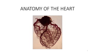
- 21. ARTERIAL SUPPLY OF THE HEART • The arterial supply of the heart is provided by the right and left coronary arteries, which arise from the ascending aorta immediately above the aortic valve. 21
- 22. RIGHT CORONARY ARTERY Branches Right marginal arteries. (acute marginal artery) Posterior interventricular artery. Sinoatrial nodal artery. Atrioventricular nodal artery. 22
- 23. LEFT CORONARY ARTERY Branches Left anterior descending(LAD) or anterior interventricular artery.(lies in anterior IV sulcus) - Septal branches. - Diagonal branches. Left marginal artery. (Obtuse marginal artery) Left circumflex artery. 23
- 24. 24
- 25. 25
- 26. 26
- 28. VENOUS DRAINAGE OF THE HEART • Most blood from the heart wall drains into the right atrium through the coronary sinus ,which lies in the posterior part of the arteriorventricular groove. • It is a continuation of the great cardiac vein. • It opens into the right atrium to the left of the inferior vena cava 28
- 29. VENOUS DRAINAGE OF THE HEART 29
- 30. NERVE SUPPLY OF THE HEART • The heart is innervated by sympathetic and parasympathetic fibers of the autonomic nervous system via the cardiac plexuses situated below the arch of the aorta. 30