Deep Vein thrombosis.pptx
•
1 like•709 views

This presentation is about Deep vein Thrombosis which includes in-depth information including anatomy and physiology, introduction, causes, pathophysiology, s/s/,management (medical and nursing) which is very important for PG and UG nurses.

Recommended
More Related Content
What's hot
What's hot (20)
Similar to Deep Vein thrombosis.pptx
Similar to Deep Vein thrombosis.pptx (20)
Recently uploaded
Recently uploaded (20)
Deep Vein thrombosis.pptx
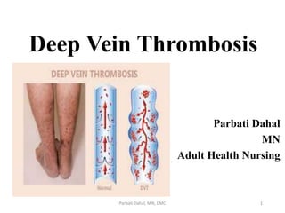
- 1. Deep Vein Thrombosis Parbati Dahal MN Adult Health Nursing 1 Parbati Dahal, MN, CMC
- 2. Anatomy of Venous system • Arteries are the blood vessels that carry oxygen-rich blood from the heart to all other parts of the body. Veins return the oxygen-depleted blood back to the heart. There are two types of veins in the body: • Superficial veins lie just below the skin's surface • Deep veins are located deep within the muscles Parbati Dahal, MN, CMC 2
- 3. 3 Parbati Dahal, MN, CMC
- 4. 4 Parbati Dahal, MN, CMC
- 5. 5 Parbati Dahal, MN, CMC
- 6. Introduction Related Terminology: • Venous thrombosis: involves the formation of a thrombus in association with inflammation of the vein. It is most common disorder of vein and is classified either superficial vein thrombosis or deep vein thrombosis. • Superficial vein thrombosis (SVT): is the formation of a thrombus in a superficial vein usually the greater or lesser saphenous vein. 6 Parbati Dahal, MN, CMC
- 7. Deep vein thrombosis (DVT) It is a disorder involving a thrombus in a deep vein, most commonly the iliac and femoral vein. Venous thromboembolism (VTE): It is the preferred terminology and represents the spectrum of pathology from DVT to pulmonary embolism(PE). 7 Parbati Dahal, MN, CMC
- 8. Introduction of DVT • Deep vein thrombosis (DVT) is a blood clot that forms in a vein deep in the body. • Blood clots occur when blood thickens and clumps together. • Most deep vein blood clots occur in the lower leg or thigh. They also can occur in other parts of the body. 8 Parbati Dahal, MN, CMC
- 9. Introduction Cont’d… • It is also called as phlebothrombosis. • A blood clot in a deep vein can break off and travel through the bloodstream. • The loose clot is called an embolus. • It can travel to an artery in the lungs and block blood flow. This condition is called pulmonary embolism. 9 Parbati Dahal, MN, CMC
- 10. Epidemiology In United States • Deep venous thrombosis (DVT) and thromboembolism remain a common cause of morbidity and mortality in bedridden or hospitalized patients, as well as generally healthy individuals. • Existing data that probably underestimate the true incidence of DVT suggest that about 80 cases per 100,000 population occur annually. 10 Parbati Dahal, MN, CMC
- 11. Epidemiology Cont’d… • Approximately 1 person in 20 develops a DVT in the course of his or her lifetime. • About 600,000 hospitalizations per year occur for DVT in the United States. 11 Parbati Dahal, MN, CMC
- 12. Epidemiology Cont’d… Age distribution • In elderly persons the incidence is increased four-fold. • Deep venous thrombosis usually affects individuals older than 40 years. Sex: • The male-to-female ratio is 1.2:1, indicating that males have a higher risk of DVT than females (Patel, 2019). 12 Parbati Dahal, MN, CMC
- 13. Etiology / Risk factors • Three main factors contribute to the development of DVT: • These represent the Virchow triad 13 Parbati Dahal, MN, CMC
- 14. Risk factor Venous stasis • Advanced age • Immobilization longer than 3 days • Previous DVT • Chronic heart failure • Stroke • Acute myocardial infarction (AMI) • Congestive heart failure (CHF) • Sepsis 14 Parbati Dahal, MN, CMC
- 15. Risk factor Cont’d… Hypercoagulability of blood • Polycythemia rubra vera • Thrombocytosis • Inherited disorders of coagulation/fibrinolysis • Protein C deficiency • Protein S deficiency • Antithrombin III deficiency • Heparin-induced thrombocytopenia (HIT) 15 Parbati Dahal, MN, CMC
- 16. Risk factor Cont’d… Endothelial Damage • Major surgery in previous 4 weeks • CNS/spinal cord injury • Burns • Lower extremity fractures • History of previous venous thromboembolism • IV drug abuse • Trauma 16 Parbati Dahal, MN, CMC
- 17. 17 Parbati Dahal, MN, CMC
- 18. Pathophysiology Virchow's triad: A major theory delineating the pathogenesis of venous thromboembolism (VTE), often called Virchow's triad, proposes that VTE occurs as a result of: • Alterations in blood flow (i.e., stasis) • Vascular endothelial injury • Alterations in the constituents of the blood (i.e., inherited or acquired hypercoagulable state) 18 Parbati Dahal, MN, CMC
- 19. Pathophysiology Virchow's triadTriad • Platelets aggretes (especially at vein valve cups) • Clotting factors stimulated to produce fibrin • Fibrin entraps RBC, WBC and platelets and begins to adhere to vein wall Endothelial damage • Release of clotting factors • Activation of platelets Blood hypercoagulability • Imbalance in clotting mechanism • Increase in fibrin production Venous Stasis • Dysfunctional vein valves • Inactive extremity muscle • Change in unidirectional blood flow Thrombus formation Clinical manifestation • Unilateral leg edema, pain and erythema • Chronic venous insufficiency • Embolism of thrombotic fragments 19 Parbati Dahal, MN, CMC
- 20. Sign and Symptoms • Asymptomatic • Fever –earliest symptoms • Swelling of leg or long vein in the leg (70% of patient) • Leg pain occurs in 50% of patients, but this is entirely nonspecific. Pain can occur on dorsiflexion of the foot (positive Homan’s sign). 20 Parbati Dahal, MN, CMC
- 21. Sign and Symptoms Cont’d… • Mose’s sign: Gentle squeezing of lower part of the calf muscle from side to side is painful. Gentleness is important otherwise it may dislodge a thrombus to form an embolus. 21 Parbati Dahal, MN, CMC
- 22. Sign and Symptoms Cont’d… • Neuhof’s sign: Thickening and deep tenderness elicited while palpating deep calf muscles. • Linton’s sign: After applying tourniquet at saphenofemoral junction, patient is made to walk and without removing the tourniquet, limb is elevated which shows persisting prominent superficial veins will be observed in DVT. 22 Parbati Dahal, MN, CMC
- 23. 23 Parbati Dahal, MN, CMC
- 24. 24 Parbati Dahal, MN, CMC
- 25. Sign and Symptoms Cont’d… • Tenderness occurs in 75% of patients but is also found in 50% of patients without objectively confirmed DVT. • Red discolored skin on the leg • Leg cramps (especially at night and/or in the calf) 25 Parbati Dahal, MN, CMC
- 26. Warning sign of DVT 1. Swelling 2. Gradual onset of pain 3. Redness 4. Warmth to the touch 5. Worsening leg pain when bending the foot 6. Leg cramps, especially at night, and often starting in the calf 7. Bluish or whitish discoloration of skin 26 Parbati Dahal, MN, CMC
- 27. Diagnosis History taking • Pain (50% of patients) • Redness • Swelling (70% of patients) Physical Examination • Limb edema may be unilateral or bilateral if the thrombus is extending to pelvic veins • Red and hot skin, with dilated veins • Tenderness • Pain on dorsiflexion of the foot (the Homans sign) 27 Parbati Dahal, MN, CMC
- 28. Diagnosis cont’d… • Duplex ultrasonography: is an imaging test that uses sound waves to look at the flow of blood in the veins. It can detect blockages or blood clots in the deep veins. It is the standard imaging test to diagnose DVT. • D-dimer blood test: Fragment of fibrin formed as result of fibrin degradation and clot lysis. Elevated results suggest VTE. • Normal Level- <250ng/ml 28 Parbati Dahal, MN, CMC
- 29. Assess clinical risk Measure D – dimmer levels D- dimer –ve Risk low D-dimer +ve D-dimer –ve Risk high Not DVT/PE Risk low Treat • USG leg veins • CT pulmonary angiography Confirm diagnosis Risk high Parbati Dahal, MN, CMC 29
- 30. Diagnosis cont’d… • Contrast venography: is a special type of X-ray where contrast material (dye) is injected into a large vein so that can visualize the deep veins in the leg and hip. • It is the most accurate test for diagnosing blood clots but it is an invasive procedure, Therefore this test has been largely replaced by duplex ultrasonography, and it is used only in certain patients. 30 Parbati Dahal, MN, CMC
- 31. Diagnosis cont’d… • Magnetic resonance imaging (MRI) and CT scan • Chest X-ray: Pulmonary edema • Blood test: CBC,PT, INR, BT, CT 31 Parbati Dahal, MN, CMC
- 32. 32 Parbati Dahal, MN, CMC
- 33. Management The main goal of treating DVT are to: • Stop the blood clot getting bigger • Prevent the blood clot from breaking off and moving to lungs (risking pulmonary embolism). • Reduce the reoccurrence 33 Parbati Dahal, MN, CMC
- 34. Medical Management Anticoagulation Therapy: • Measures for preventing or reducing blood clotting within the vascular system are indicated in patients with thrombophlebitis, recurrent embolus formation, and persistent leg edema from heart failure. • They are also indicated in elderly patients with a hip fracture that may result in lengthy immobilization. 34 Parbati Dahal, MN, CMC
- 35. Unfractionated Heparin: • Unfractionated heparin (heparin) is administered subcutaneously to prevent development of deep vein thrombosis, or by intermittent intravenous infusion or continuous infusion for 5 to 7 days to prevent the extension of a thrombus and the development of new thrombi. • Oral anticoagulants, such as warfarin (Coumadin), are administered with heparin therapy. • Medication dosage is regulated by monitoring the partial thromboplastin time, the international normalized ratio (INR), and the platelet count. 35 Parbati Dahal, MN, CMC
- 36. Low-Molecular-Weight Heparin • Subcutaneous low-molecular weight heparin (LMWH) is an effective treatment for some cases of deep vein thrombosis. • It has a longer half-life than unfractionated heparin, so doses can be given in one or two subcutaneous injections each day. • Doses are adjusted according to weight. LMWH prevents the extension of a thrombus and development of new thrombi and is associated with fewer bleeding complications than unfractionated heparin. 36 Parbati Dahal, MN, CMC
- 37. Medical Management Cont’d… Thrombolytic Therapy: • Unlike the heparins, thrombolytic (fibrinolytic) therapy causes the thrombus to lyse and dissolve in 50% of patients. • Thrombolytic therapy (eg, tissue plasminogen activator [t-PA, alteplase, Activase], reteplase [r-PA, Retavase], tenecteplase [TNKase], staphylokinase, urokinase, streptokinase) is given within the first 3 days after acute thrombosis. 37 Parbati Dahal, MN, CMC
- 38. Management Cont’d… Applying Elastic compression stockings • Elastic compression stockings usually are prescribed for patients with venous insufficiency. • These stockings exert a sustained, evenly distributed pressure over the entire surface of the calves, reducing the caliber of the superficial veins in the legs and resulting in increased flow in the deeper veins. 38 Parbati Dahal, MN, CMC
- 39. 39 Parbati Dahal, MN, CMC
- 40. Cont’d… Exercise: • walking and calf exercise reduce venous stasis because leg muscle contraction compress the veins and pump blood up towards the heart. 40 Parbati Dahal, MN, CMC
- 41. Surgical Management • Surgery is necessary for deep vein thrombosis when anticoagulant or thrombolytic therapy is contraindicated the danger of pulmonary embolism is extreme, or the venous drainage is so severely compromised that permanent damage to the extremity will probably result. 41 Parbati Dahal, MN, CMC
- 42. • Vena cava Filter: The filter is inserted inside the venacava. The filter catches blood clots before they travel to the lungs, which prevents pulmonary embolism. However, the filter doesn’t stop new blood clots from forming. 42 Parbati Dahal, MN, CMC
- 43. Thrombectomy/ Embolectomy • In rare cases, a surgical procedure to remove the clot may be necessary. • Thrombectomy involves removal of the clot in a patient with DVT. • Embolectomy involves removal of the blockage in the lungs caused by the clot in a patient with PE. 43 Parbati Dahal, MN, CMC
- 44. Assessment History taking Physical examination Nursing Management 44 Parbati Dahal, MN, CMC
- 45. • Acute pain related to venous congestion, impaired venous return and inflammation • Ineffective health maintenance related to lack of knowledge about disorder and its treatment • Risk for impaired skin integrity related to altered peripheral tissue perfusion • Potential complication: bleeding related to anticoagulation therapy • Potential complication: PE related to embolization of thrombus, dehydration and immobility Nursing Diagnosis 45 Parbati Dahal, MN, CMC
- 46. Pain management • Assess the patient’s pain, including location, quality and intensity, respiratory rate, and level of conscious. • Provide psychological support to the patient. • Provide comfortable position. • Administration analgesics as prescribed. • Diversional Therapy and relaxation technique • Reassess the pain to evaluate the effectiveness of interventions Cont’d… 46 Parbati Dahal, MN, CMC
- 47. Emotional and psychological care • Provide a safe environment for the expression of the ful range of feeling. • Encourage to identify and learn individual coping strengths. • Provide accurate and complete information about disease process. • Acknowledge patient that this could be a fearful situation to any one and others have expressed similar fears Cont’d… 47 Parbati Dahal, MN, CMC
- 48. • Monitor the partial thromboplastin time, prothrombin time, hemoglobin and hematocrit values, platelet count, and fibrinogen level. • Close observation is also required to detect bleeding; if bleeding occurs, it must be reported immediately and anticoagulant therapy discontinued. Minimize Risk of Bleeding 48 Parbati Dahal, MN, CMC
- 49. Cont’d… • To prevent inadvertent infusion of large volumes of heparin, which could cause hemorrhage, continuous intravenous infusion by electronic infusion device is the preferred method of administering unfractionated heparin. • Dosage calculations are based on the patient’s weight, and any possible bleeding tendencies are detected by a pretreatment clotting profile 49 Parbati Dahal, MN, CMC
- 50. • If renal insufficiency exists, lower doses of heparin are required. • Periodic coagulation tests and hematocrit levels are obtained. • Heparin is in the effective, or therapeutic, range when the partial thromboplastin time is 1.5 times the control. Cont’d… 50 Parbati Dahal, MN, CMC
- 51. • Oral anticoagulants, such as warfarin, are monitored by the prothrombin time or INR. Because their effect is delayed for 3 to 5 days, they are usually administered with heparin until desired anticoagulation has been achieved (ie, when the prothrombin time is 1.5 to 2 times normal or the INR is 2.0 to 3.0). Contd… 51 Parbati Dahal, MN, CMC
- 52. Monitoring and managing potential complications Bleeding • The principal complication of anticoagulant therapy is spontaneous bleeding anywhere in the body. • Bleeding from the kidneys is detected by microscopic examination of the urine and is often the first sign of anticoagulant toxicity from excessive dosage. 52 Parbati Dahal, MN, CMC
- 53. • Bruises, nosebleeds, and bleeding gums are also early signs. • To reverse the effects of heparin promptly, intravenous injections of protamine sulfate may be administered. • Reversing the effects of warfarin, a coumarin derivative, is more difficult, but effective measures that may be prescribed include vitamin K and possibly transfusion of fresh frozen plasma. Cont’d… 53 Parbati Dahal, MN, CMC
- 54. Thrombocytopenia • which may develop in patients who receive heparin for more than 5 days or on re-administration after a brief interruption of heparin therapy. • Beginning warfarin concurrently with heparin can provide a stable INR or prothrombin time by day 5 of heparin treatment. Cont’d… 54 Parbati Dahal, MN, CMC
- 55. • The use of LMWH is less frequently associated with heparin induced thrombocytopenia. • The thrombocytopenia is thought to result from an immunologic mechanism that causes aggregation of platelets. Cont’d… 55 Parbati Dahal, MN, CMC
- 56. • This serious complication results in thromboembolic manifestations, and the prognosis is extremely guarded. • Prevention of thrombocytopenia depends on regular monitoring of platelet counts. Cont’d… 56 Parbati Dahal, MN, CMC
- 57. • Early signs of thrombocytopenia are a falling platelet count to less than 100,000/mL, a decrease. • if thrombocytopenia does occur, platelet aggregation studies are conducted, the heparin is discontinued, and protamine sulfate is administered to reverse heparin’s effects. Cont’d… 57 Parbati Dahal, MN, CMC
- 58. Providing Comfort • Bed rest, elevation of the affected extremity, elastic compression stockings, and analgesics for pain relief are adjuncts to therapy. • They help to improve circulation and increase comfort. • Depending on the extent and location of a venous thrombosis, bed rest may be required for 5 to 7 days after diagnosis. 58 Parbati Dahal, MN, CMC
- 59. • This is approximately the time necessary for the thrombus to adhere to the vein wall, preventing embolization. • Warm, moist packs applied to the affected extremity reduce the discomfort associated with deep vein thrombosis, as do mild analgesics prescribed for pain control. Cont’d… 59 Parbati Dahal, MN, CMC
- 60. • When the patient begins to ambulate, elastic compression stockings are used. • Walking is better than standing or sitting for long periods. • Bed exercises, such as dorsiflexion of the foot, are also recommended. Cont’d… 60 Parbati Dahal, MN, CMC
- 61. Applying elastic compression stockings • Elastic compression stockings usually are prescribed for patients with venous insufficiency. • These stockings exert a sustained, evenly distributed pressure over the entire surface of the calves, reducing the caliber of the superficial veins in the legs and resulting in increased flow in the deeper veins. • The stockings may be knee-high, thigh-high, or panty hose. 61 Parbati Dahal, MN, CMC
- 62. • Thigh-high stockings are difficult for the patient to wear, because they have a tendency to roll down. • The roll of the stocking further restricts blood flow rather than the stocking providing evenly distributed pressure over the thigh. Cont’d… 62 Parbati Dahal, MN, CMC
- 63. • When the stockings are off, the skin is inspected for signs of irritation, and the calves are examined for possible tenderness. • Any skin changes or signs of tenderness are reported. Stockings are contraindicated in patients with severe pitting edema because they can produce severe pitting at the knee. Cont’d… 63 Parbati Dahal, MN, CMC
- 64. Complication • Chronic venous occlusion • Pulmonary emboli from dislodged thrombi • Valvular destruction oChronic venous insufficiency oIncreased venous pressure o Varicosities o Venous ulcers Parbati Dahal, MN, CMC
- 65. Complication Cont’d… •Venous obstruction oIncreased distal pressure oFluid stasis o Edema oVenous gangrene 65 Parbati Dahal, MN, CMC
- 66. Prevention • Lose weight if overweight or obese • Avoid periods of prolonged immobility. • Keep the legs elevated while sitting down or in bed. • After surgery, get out of bed several times a day during the recovery period, use compression devices on the legs or elastic compression socks/stockings. • Take regular dose of heparin or warfarin if prescribed to prevent clot formation. 66 Parbati Dahal, MN, CMC
- 67. Prognosis • Most cases of deep venous thrombosis (DVT) is occult and usually resolves spontaneously without complication. • The principal long-term morbidity from DVT is post thrombotic syndrome (PTS), which complicates about a quarter of cases of symptomatic proximal DVT; most cases develop within 2 years afterward. • Death from DVT is attributed to massive pulmonary embolism (PE), which causes as many as 300,000 deaths annually in the United States. 67 Parbati Dahal, MN, CMC
- 68. • Black, J. M. & Hawks, J. N. (2009). Medical- surgical nursing (8th ed.). New Delhi: Elsevier India Pvt. Ltd. • Lewis, S.L., Dirksen, S.R., Heitkemper, M.M., & Bucher, L. (2015). Medical surgical nursing: assessment and management of clinical problems (2nd ed.). South Asia Edition, Vol 2. New Delhi: Reed Elsevier India Pvt. Ltd • Bhat, S. (2013). SRB’s Manual of surgery (4th ed.). Jaypee brother medical publisher. References 68 Parbati Dahal, MN, CMC
- 69. • Patel, K. (2019). Deep venous thrombosis (DVT). Medescape. Retrieved from https://emedicine.medscape.com/article/1911303- overview#a6 • Smeltzer, S. C., Bare, B. G., Hinkle, J. L. & Cheever, K. H. (2008). Brunner & suddarth’s textbook of medical-surgical nursing (11th ed.). New Delhi: Lippincott Williams & Wilkins , a Wolter Kluwer business. • Walker, B. R., Colledge, N. R., Ralston, S. H., & Penman, I. D. (2014). Davidson's Principles & Practice of Medicine (22nd ed.). China: Churchill Livingstone Elsevier. References 69 Parbati Dahal, MN, CMC
- 70. 70 Parbati Dahal, MN, CMC