
Recommended
More Related Content
Similar to AW Tugas PPT DVT, diagnosis dan tatalaksananya.pptx
Similar to AW Tugas PPT DVT, diagnosis dan tatalaksananya.pptx (20)
Recently uploaded
Recently uploaded (20)
AW Tugas PPT DVT, diagnosis dan tatalaksananya.pptx
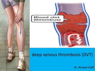
- 1. deep venous thrombosis (DVT) dr. Amwal Halil
- 2. INTRODUCTION • Venous thromboembolism (VTE) encompasses deep venous thrombosis (DVT) and pulmonary embolism (PE) and causes cardiovascular death and disability. • In the United States, the Surgeon General estimates there are 100,000 to 180,000 deaths annually from PE and has declared that PE is the most common preventable cause of death among hospitalized patients.
- 4. • Superficial thrombosis is known as thrombophlebitis or phlebitis a clot that develop on surface of skin. • Deep vein thrombosis is the formation of a blood clot in one of the deep vein of the body usually leg.
- 5. • Deep Venous Thrombosis Lower extremity DVT usually begins in the calf and propagates proximally to the popliteal vein, femoral vein, and iliac veins. • Leg DVT is about 10 times more common than upper extremity DVT, which is often precipitated by placement of pacemakers, internal cardiac defibrillators, or indwelling central venous catheters. • Superficial venous thrombosis usually presents with erythema, tenderness, and a “palpable cord.” Patients are at risk for extension of the thrombosis to the deep venous system.
- 6. PATHOPHYSIOLOGY Virchow’s triad Endothelial injury Altered blood flow Hypercoagulability of blood.
- 8. INFLAMMATION AND PLATELET ACTIVATION • These microparticles contain proinflammatory mediators that bind neutrophils, stimulating them to release their nuclear material and form web-like extracellular networks called neutrophil extracellular traps. • These prothrombotic networks contain histones that stimulate platelet aggregation and promote platelet-dependent thrombin generation. Harsh mohan, text book of pathology 6th edition, pg. 117
- 9. ALTERATION OF BLOOD FLOW Turbulence means unequal flow while stasis means slowing. i) Normally, there is axial flow of blood in which the most rapidly-moving central stream consists of leucocytes and red cells. The platelets are present in the slow-moving laminar stream adjacent to the central stream while the peripheral stream consists of most slow-moving cell-free plasma close to endothelial layer. ii) Turbulence and stasis occur in thrombosis in which the normal axial flow of blood is disturbed. When blood slows thrombi is facilitated by turbulence in the blood flow, while stasis initiates the venous thrombi even without evidence of endothelial injury. Harsh mohan, text book of pathology 6th edition, pg. 117
- 10. Stasis • Surgery, trauma, immobility, paresis • Increasing age • Pregnancy and postpartum • Heart or respiratory failure • Obesity Geerts WH, Pineo GF, Heit JA, Bergqvist D, Lassen MR, Colwell CW, et al. Prevention of venous thromboembolism: the seventh ACCP conference on antithrombotic and thrombolytic therapy. Chest2004
- 12. HYPERCOAGULABILITY OF BLOOD • The effect of hypercoagulability on thrombosis is favoured by advancing age, smoking, use of oral contraceptives and obesity. Hypercoagulability may occur by the following changes in the composition of blood: i) Increase in coagulation factors e.g. fibrinogen, prothrombin, factor VIIa, VIIIa and Xa. ii) Increase in platelet count and their adhesiveness. iii) Decreased levels of coagulation inhibitors e.g. antithrombin III Harsh Mohan, text book of pathology 6th edition, pg. 117
- 13. • Increasing age • Malignancy or cancer therapy • Oestrogen therapy (contraception or hormone replacement) • Acute medical illness • Inflammatory bowel disease • Nephrotic syndrome • Myeloproliferative disorders • Inherited or acquired thrombophilia Geerts WH, Pineo GF, Heit JA, Bergqvist D, Lassen MR, Colwell CW, et al. Prevention of venous thromboembolism: the seventh ACCP conference on antithrombotic and thrombolytic therapy. Chest2004
- 14. ENDOTHELIAL INJURY • Trauma • Surgery • Invasive procedure may distrupt venous integrity • Iatrogenic cause of venous thrombosis like CVC cause upper limb DVT.
- 15. PRESENTATION AND CLINICAL EXAMINATION • PAIN AND TENDERNESS • SWELLING (USUALLY IN ONE LIMB) • REDNESS • WARMTH • EDEMA • CYANOSIS • HOMANS SIGN ( dorsiflexion of foot while knee is extended)
- 19. DIAGNOSIS • PE is known as “the Great Masquerader.” Diagnosis is difficult because symptoms and signs are nonspecific. • With DVT, the most common symptom is a cramp or “charley horse” in the lower calf that persists and intensifies over several days.
- 20. • Sudden, severe calf discomfort suggests a ruptured Baker’s cyst. Fever and chills usually cellulitis rather than DVT. • However, massive DVT often presents with marked thigh swelling, tenderness, and erythema. If the leg is diffusely edematous, DVT is unlikely. More probable is an acute exacerbation of venous insufficiency due to postthrombotic syndrome
- 21. Differential Diagnosis • DVT • Ruptured Baker’s cyst • Cellulitis • Postphlebitic syndrome/venous insufficiency Harrison principle of internal medicine 19th edition, pg. 1367
- 22. D-DIMER ASSAY • The quantitative plasma d-dimer enzyme- linked immunosorbent assay (ELISA) rises in the presence of DVT or PE because of the breakdown of fibrin by plasmin. Elevation of D-dimer indicates endogenous although often clinically ineffective thrombolysis. • The sensitivity of the D-dimer is >80% or DVT (including isolated calf DVT) and >95% for PE.
- 23. • However, the D-dimer assay is not specific. Levels increase in patients with myocardial infarction, pneumonia, sepsis, cancer, and the postoperative state and those in the second or third trimester of pregnancy. • Therefore, D-dimer rarely has a useful role among hospitalized patients, because levels are frequently elevated due to systemic illness.
- 24. Clinical Variable DVT Score Active cancer 1 Paralysis, paresis, or recent cast 1 Bedridden for >3 days; major surgery <12 weeks 1 Tenderness along distribution of deep veins 1 Entire leg swelling 1 Unilateral calf swelling >3 cm 1 Pitting edema 1 Collateral superficial nonvaricose veins 1 Alternative diagnosis at least as likely as DVT -2
- 25. Low Clinical Likelihood of DVT if Point Score Is Zero or Less; Moderate Likelihood if Score Is 1 to 2; High Likelihood if Score Is 3 or Greater Harrison principle of internal medicine 19th edition, pg. 1366 Wells' Criteria for DVT
- 26. Noninvasive Imaging Modalities • Venous ultrasonography Ultrasonography of the deep venous system relies on loss of vein compressibility as the primary criterion for DVT. When a normal vein is imaged in cross-section, it readily collapses with gentle manual pressure from the ultrasound transducer. This creates the illusion of a “wink.” With acute DVT, the vein loses its compressibility because of passive distention by acute thrombus. • The diagnosis of acute DVT is even more secure when thrombus is directly visualized. It appears homogeneous and has low echogenicity.
- 27. • The vein itself often appears mildly dilated, and collateral channels may be absent. Venous flow dynamics can be examined with Doppler imaging. Normally, manual calf compression causes augmentation of the Doppler flow pattern. Loss of normal respiratory variation is caused by an Obstructing DVT or by any obstructive process within the pelvis.
- 29. • color-flow Duplex scanning is the imaging test of choice for patients with suspected DVT • inexpensive, • noninvasive, • widely available • Ultrasound can also distinguish other causes of leg swelling, such as tumor, popliteal cyst, abscess, aneurysm, or hematoma.
- 30. LIMITATION • Expensive • reader dependent • Duplex scans are less likely to detect nonoccluding thrombi. • During the second half of pregnancy, ultrasound becomes less specific, because the gravid uterus compresses the inferior vena cava, thereby changing Doppler flow in the lower extremities
- 31. Magnetic resonance (MR) (contrast- enhanced) imaging When ultrasound • MR is equivocal, MR venography with gadolinium contrast is an excellent imaging modality to diagnose DVT • It detects leg, pelvis, and pulmonary thrombi and is 97% sensitive and 95% specific for DVT. • It distinguishes a mature from an immature clot. • MRI is safe in all stages of pregnancy.
- 32. Contrast phlebography • has virtually replaced contrast phlebography as the diagnostic test for suspected DVT.
- 33. TREATMENT Ambulation early and often (simplest and most cost effective means to reduce risk of DVT) Intermittent External Compression Devices: Also called sequential compression devices or SCDs They increase rate/velocity of venous blood and reduce pooling in the peripheral veins Compression should begin pre-operatively and be continued until the patient is fully ambulatory Anticoagulant Medication Therapy
- 36. Compressive stocking pressure • Mild- 8-15 mm/Hg • Moderate 15-20 mm/Hg • Firm 15-30 mm/Hg • Extra firm 30-40 mm/Hg
- 37. Intermittent External Compression Devices: Also called sequential compression devices or SCDs They increase rate/velocity of venous blood and reduce pooling in the peripheral veins
- 38. In nine studies comparing graduated compression stockings with no prophylaxis, rates of DVT were reduced from 27% to 13%, and in seven studies the addition of the stockings to background prophylaxis further reduced DVT rates from 15% to 2% Review Elastic compression stockings for prevention of deep vein thrombosis.Amaragiri SV, Lees TA Cochrane Database Syst Rev. 2000
- 39. Intermittent External Compression Devices Every cycle of compressive dvt pump contain 12 sec inflate 48 sec deflate Total pressure 40 mm/hg
- 41. • ACCP guideline recommendations for early mobilization are also based on small numbers of studies and therefore subject to bias. • Mobilization may be beneficial in reducing pain and edema from DVTs, but larger scale studies or patient numbers are required to validate these outcomes. Kearon C, Akl EA, Comerota AJ, Prandoni P, Bounameaux H, Goldhaber SZ, et al Antithrombotic therapy for VTE disease: antithrombotic therapy and prevention of thrombosis, 9th ed: American College of Chest Physicians evidence-based clinical practice guidelines. Chest. 2012 Feb;141
- 42. Low-Molecular-Weight Heparins • Low-Molecular-Weight Heparins These fragments of UFH exhibit less binding to plasma proteins and endothelial cells and consequently have greater bioavailability, a more predictable dose response, and a longer half-life than does UFH. No monitoring or dose adjustment is needed unless the patient is markedly obese or has chronic kidney disease.
- 43. • Fondaparinux Fondaparinux, an anti-Xa pentasaccharide, is administered as a weight-based once-daily subcutaneous injection in a prefilled syringe. • No laboratory monitoring is required. Fondaparinux is synthesized in a laboratory and, unlike LMWH or UFH, is not derived from animal products. • It does not cause heparin-induced thrombocytopenia. • The dose must be adjusted downward for patients with renal dysfunction.
- 44. Unfractionated Heparin • UFH anticoagulates by binding to and accelerating the activity of antithrombin, thus preventing additional thrombus formation. • UFH is dosed to achieve a target activated partial thromboplastin time (aPTT) of 60–80 s. The most popular nomogram uses an initial bolus of 80 U/kg, followed by an initial infusion rate of 18 U/kg per h. • The major advantage of UFH is its short half-life, which is especially useful in patients in whom hour-to-hour control of the intensity of anticoagulation is desired.
- 45. Warfarin • This vitamin K antagonist prevents carboxylation activation of coagulation factors II, VII, IX, and X. • The full effect of warfarin requires at least 5 days, even if the prothrombin time, used for monitoring, becomes elevated more rapidly. If warfarin is initiated as monotherapy during an acute thrombotic illness, a paradoxical exacerbation of hypercoagulability increases the likelihood of thrombosis.
- 46. Overlapping UFH, LMWH, fondaparinux, or parenteral direct thrombin inhibitors with warfarin for at least 5 days will nullify the early procoagulant effect of warfarin. Warfarin Dosing In an average-size adult, warfarin is often initiated in a dose of 5 mg. The prothrombin time is standardized by calculating the international normalized ratio (INR), which assesses the anticoagulant effect of warfarin. The target INR is usually • 2.5, with a range of 2.0–3.0. The warfarin dose is usually titrated empirically to achieve the target INR.
- 47. Prevention of Venous Thromboembolism Among Hospitalized Patients High-risk non orthopedic surgery • Unfractionated heparin 5000 units SC bid or tid • Enoxaparin 40 mg daily • Dalteparin 2500 or 5000 units daily Harrison principle of internal medicine 19th edition, pg. 1367
- 48. Major orthopedic surgery • Warfarin (target INR 2.0–3.0) • Enoxaparin 40 mg daily • Enoxaparin 30 mg bid • Dalteparin 2500 or 5000 units daily • Fondaparinux 2.5 mg daily • Intermittent pneumatic compression (with or without pharmacologic prophylaxis) Harrison principle of internal medicine 19th edition, pg. 1367
- 49. • Medically ill patients, especially if immobilized, with a history of prior VTE, with an indwelling central venous catheter, or with cancer (but without active gastroduodenal ulcer, major bleeding within 3 months, or platelet count <50,000) • Unfractionated heparin 5000 units bid or tid • Enoxaparin 40 mg daily • Dalteparin 2500 or 5000 units daily • Fondaparinux 2.5 mg daily Harrison principle of internal medicine 19th edition, pg. 1367
- 50. Harrison principle of internal medicine 19th edition, pg. 1367 • Anticoagulation contraindicated Intermittent pneumatic compression devices (but whether graduated compression stockings are effective in medical patients is controversial)
- 51. Summary of Evidence-Based Recommendations for VTE Prevention Strong evidence exists for the following: • For highest risk patients combine pharmacologic and mechanical prevention methods: • All patients admitted to the critical care unit must be assessed for risk of VTE • Most critically ill patients will require thromboprophylaxis • Aspirin alone should not be used for VTE prophylaxis for any patient group • Pharmacologic thromboprophylaxis with low dose UFH or LMEH (SQ), fondaparinus or rivaroxaban
- 52. • Oral vitamin K agonists (warfarin) used to achieve a target INR of 2.5 (INR range, 2 to 3) • Mechanical prophylaxis: graduated-compression stockings or intermittent pneumatic compression devices • Some of the patients at highest risk for VTE include those undergoing open urologic surgery, gynecologic surgery, or total hip or knee procedures; all trauma patients with at least one risk factor; and medical patients with acute heart failure or acute respiratory failure; others included in this group are immobile patients confined to bed who have at least one risk factor Kearon C, et al. Antithrombotic therapy for VTE disease: Antithrombotic Therapy and Prevention of Thrombosis, 9th ed: American College of Chest Physicians Evidence-Based Clinical Practice Guidelines. Chest.2012;141(2 suppl):e419s.