Deep Vein Thrombosis
•Download as PPTX, PDF•
1 like•434 views

DVT, Prophylaxis, Treatment

Recommended
More Related Content
What's hot
What's hot (20)
Similar to Deep Vein Thrombosis
Similar to Deep Vein Thrombosis (20)
Recently uploaded
Recently uploaded (20)
Deep Vein Thrombosis
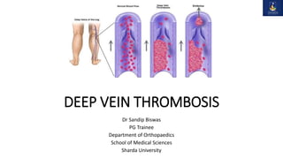
- 1. DEEP VEIN THROMBOSIS Dr Sandip Biswas PG Trainee Department of Orthopaedics School of Medical Sciences Sharda University
- 2. Introduction • Venous thromboembolism (VTE) - one of the commonest complications of lower limb surgery. • includes 2 different clinical entities: deep vein thrombosis (DVT) and pulmonary embolism (PE) • Orthopaedic patients - high risk due to immobility and direct manipulation of veins in long procedures
- 3. Definition • DVT - presence of coagulated blood or thrombus (obstructing or non- obstructing) in one of the deep venous conduits that return blood to the heart. • occurs most commonly in lower limb veins (distal) or pelvic veins (proximal VTE) • In pulmonary embolism, some or all of the thrombus becomes detached and lodge in one or more pulmonary arteries.
- 4. Anatomy • Peripheral Venous System is divided into three primary groups - superficial venous system and deep venous system and perforators • Superficial venous system veins are the primary collecting veins of the lower extremity. (The Greater Saphenous, Lesser Saphenous, Posterior Arch Vein) • Deep venous system veins receive inflow from the superficial system • In calf, there are three groups of deep veins – • The anterior tibial veins, which drain the dorsal aspect of the foot • The posterior tibial veins, which drain the sole of the foot • The peroneal veins, which drain the lateral aspect of the foot
- 6. Together, the calf’s muscles and deep vein system are also known as the “peripheral heart”, that push blood upward from the feet against gravity • With contraction of the calf, proximal migration of the blood occurs due to closure of the feeding perforator vein valves and opening of the outflow valves. • With relaxation, superficial venous system refills the veins and sinusoids via perforating veins, and there is closure of outflow valves, resulting in prevention of retrograde flow. • One contraction of calf drives 40–60% of calf’s venous volume proximally.
- 7. Pathogenesis
- 8. • According to Virchow, 3 critically important factors are required “The Virchow’s triad 1. Venous Blood Stasis- • Most important component as it initiates venous thrombi even without evidence of endothelial injury • In normal blood flow central stream moves fastest and consist of leukocytes and red cells • Platelets are present in slow moving laminar stream adjacent to central stream
- 9. • In stasis, the platelets fall out of the intermediate column and come in contact with the endothelium called pavementation and margination • Due to reduced movement, Adenosine Diphosphate (ADP) gets released further platelet aggregation • formation of temporary plug and then the whole clotting system gets activated propagating the thrombus. • Stasis occurs in- Blood Dyscrasias , Carcinoma, low cardiac output, obesity, advanced age, prolonged bed rest , post partum period, dehydration, polycythaemia, long surgical procedures, esp. under General Anaesthesia, fractures and injury of the lower limbs, left and right ventricular failure, oestrogen use
- 10. 2. Abnormalities of Vessel Wall : • Endothelial injury exposes subendothelial connective tissues (collagen, elastin, fibronectin, laminin, glycosaminoglycans) which are thrombogenic • Play an important role in initiating thrombosis by extrinsic pathway. 3. Abnormal activation of the coagulation pathway or a hypercoagulable state- • Nephritic syndrome, advanced cancers, extensive trauma, burns, during puerperium • Increase in coagulation factors, increase in platelet count and their adhesiveness, and decreased levels of coagulation inhibitors
- 12. ETIOLOGY • Acquired: Previous DVT: The single most powerful risk marker(25% patients) Age (the annual incidence of VTE rises with each decade over the age of forty) Obesity Immobilization longer than 3 days Major surgery (especially orthopaedic) lasting more than 2 hours in previous 4 weeks Long plane or car trips (> 4 hours) in previous 4 weeks
- 13. Mass effect on iliac veins: Cancer Pregnancy Congenital anomaly Stroke Postpartum period Acute myocardial infarction (AMI) Congestive heart failure (CHF) Sepsis Nephrotic syndrome Ulcerative colitis Multiple trauma Burns
- 14. Trauma: Endothelial injury can convert the normally antithrombogenic endothelium to become prothrombotic: • Mechanical injury to veins: Lower extremity fractures—multiple long bone fractures • Severe pelvic fracture (posterior elements) + long bone fracture • Hip and knee arthroplasty (manipulation of nearby major veins) Oral contraceptives Heparin-induced thrombocytopenia (HIT) Intravenous (IV) drug abuse
- 15. • Congenital Systemic lupus erythematosus (SLE) and the lupus anticoagulant Behçet syndrome Homocystinuria Polycythaemia rubra vera Genetic mutations in blood coagulation cascade: Inherited disorders of coagulation/fibrinolysis— altered factor VIII, factor IX, factor XI, and prothrombin, antiphospholipid syndrome Antithrombin III deficiency Protein C deficiency Protein S deficiency Factor V Leiden Dysfibrinogenemia and disorders of plasminogen activation
- 16. Anatomic variants causing venous stasis: Compression of left common iliac vein at the anatomic crossing of the right common iliac artery (May-Thurner syndrome or Cockett syndrome) Anomalous development of inferior vena cava, like duplicated IVC, absence of IVC or azygous vein.
- 17. CLINICAL FEATURES Oedema—most specific sign. Leg and calf pain—in 50% of patients but nonspecific. Calf pain may be reproduced on dorsiflexion of the foot (Homan’s sign). Reddish purple hue of lower limb from venous engorgement and obstruction: • Phlegmasia alba dolens (painful white inflammation), characterized by pale affected extremity, often having poor or even absent distal pulses
- 18. • Phlegmasia cerulea dolens (painful blue inflammation)—the leg is usually markedly oedematous, painful and cyanotic. Calf tenderness—Occurs in 75% of patients, it is usually confined to the calf muscles. Increased temperature or erythema in the area of DVT. Clinical symptoms of pulmonary embolism occur in 10% of patients with confirmed DVT. Superficial thrombophlebitis—Palpable, indurated, cordlike, tender subcutaneous venous segment.
- 19. Diagnosis American College of Physicians (9th edition) provides 4 recommendations for the workup of patients with probable DVT: 1. Well’s Clinical Score to estimate the pretest probability of VTE. 2. D-dimer is obtained in patients with low pretest probability of DVT first and compression ultrasound (PUSG) of proximal veins later. A negative result indicates a low likelihood of VTE and no further testing is necessary. 3. In patients with intermediate to high pretest probability proximal vein ultrasonography (PUSG) is recommended. If positive then treatment for DVT should be initiated rather than confirmation with venography If negative there is no need for further testing or repeat sonography.
- 20. 4. Diagnostic imaging studies are required in patients with intermediate or high pretest probability of pulmonary embolism. E.g.- pulmonary angiography, ventilation-perfusion (V/Q) scan and multidetector helical computed axial tomography (CT).
- 22. Diagnostic Methods Clinical Findings D-Dimer Blood Tests Conventional X-Ray Contrast Venogram Duplex Doppler Ultrasound Magnetic Resonance Imaging Radionuclide Methods
- 23. D-Dimer Testing: when cross-linked fibrin is degraded by plasmin, D-dimer generates High sensitivity (97%) but low specificity (35%) Increase in Myocardial Infarction, pneumonia, sepsis, cancer, post-op state third trimester of pregnancy, trauma With low-to-moderate risk (Wells score< 2), a negative result rules out DVT Further diagnostic study (e.g. duplex ultrasonography is required in all patients with a positive D-dimer and moderate-to-high risk of DVT (Wells score > 2).
- 24. USG of the Deep Leg Veins with Colour Doppler Imaging of the blood flow– ease of use, absence of irradiation or contrast material current first-line imaging examination for DVT High sensitivity and specificity (95%) Lack of vein compressibility is the primary diagnostic criteria Echogenic Thrombus within lumen Abnormal Doppler flow
- 25. MRI (Contrast Enhanced)- Used when usg is equivocal. Utilizes gadolinium contrast agent which unlike iodinated contrast agents used in venography or CT angiography is not nephrotoxic Safe in Renal Insufficiency or contrast dye allergic patients Detects large proximal pulmonary embolism, inferior vena caval or iliac vein thrombois but is not reliable for smaller segmental PE
- 26. Contrast Venography – Gold Standard for imaging DVT when technically adequate Can image entire lower extremities Limitations- Painful Invasive Rarely used now due to accuracy of non-invasive tests
- 27. CT venography— uses venous phase contrast to visualize the IVC, pelvic veins and extremity veins. particularly useful for diagnosis of proximal DVT which is missed on duplex ultrasonography often Impedance plethysmography (IPG) sensitive and specific for proximal vein thrombosis. But may not detect the calf vein thrombosis and non-occluding proximal vein thrombus (limitations of IPG).
- 28. DIFFERENTIAL DIAGNOSIS • DVT is a confusing disease both clinically and even with investigations. • Conditions that should be always looked for while making a diagnosis of DVT include: Ruptured Baker’s cyst Budd-Chiari syndrome Cellulitis Congestive cardiac failure Pulmonary embolism Septic thrombophlebitis Superficial thrombophlebitis.
- 29. TREATMENT Anticoagulation is the mainstay of medical therapy done usually on out-patient basis contraindications for outpatient management Anticoagulation contraindicated Pregnancy Acute of chronic renal failure (S. creatinine level > 2mg/dL) Suspected or proven concomitant pulmonary embolism Iliofemoral DVT Morbid obesity (> 150 kg)
- 30. Familial or inherited disorder of coagulation : antithrombin III (ATIII) deficiency, protein C or protein S deficiency Familial bleeding disorder Close follow-up not possible limitation - inhibits propagation only and does not remove the thrombus that is deemed to resolve by natural physiological process over time. Significant bleeding.
- 31. Contraindications: Ongoing intracranial haemorrhage Malignant hypertension Severe active haemorrhage from wound or in vascular tissues Pregnancy Recent history of brain, eye, or spinal cord surgery
- 32. In general, leg is kept elevated to 15 degrees initiated on a brief course (i.e. less than a week) of heparin(LMWH) treatment And they are continued on 3-6 months of warfarin (direct anti-coagulation can also be begun) • Patient with proximal DVT – anticoagulated for at least 3 months. Also same for patients with isolated distal DVT at high risk of recurrence. • With isolated distal DVT at low risk of recurrence – shorter treatment of 4-6 weeks with lower doses • LMWH is recommended for initial and long term treatment in cancer patient
- 33. For Acute DVT- initial anti-coagulation should be one of the following regimens:- Apixaban 10 mg BD 7 days then 5 mg BD Dabigatran 150 mg bd after a 5 to 10 days lead in course of LMWH Edoxaban 60 mg daily Rivoraxaban 15 mg bd for 21 days then 20 mg daily
- 34. Adjuvant catheter directed thrombolysis – • considered at specialized centres in selected patients with iliac vessel or femoral DVT • life expectancy > 1 year Vena Cava Filters if anticoagulation is contraindicated Compression therapy with early mobilisation and walking exercise – to relieve acute venous symptoms.
- 35. In pregnancy- • USG is 1st line DVT imaging test. • LMWH – for initial and long term treatment. • Continued for at least 6 weeks after delivery. Secondary prophylaxis for recurrent DVT is given with low dose aspirin, apixaban 2.5 mg bd or rivaroxaban 10 mg daily. In general anticoagulation is preferred over aspirin therapy
- 36. PROPHYLAXIS - Mechanical Methods 1. Graduated compression (elastic) knee or thigh- high stockings (GCS) – passive devices and least effective 2. Active [intermittent pneumatic compression (IPC)] devices— good efficacy, serve as external pumps substituting the function of calf pumps 3. Venous foot pumps (VFP) – Foot impulse technology (FIT) and foot impulse devices (FID)
- 38. Pharmacologic Methods • Antithrombotic: Include anticoagulants and antiplatelet Oral vitamin K antagonist, warfarin Oral aspirin (antiplatelet drug) Subcutaneous Pentasaccharide- fondaparinux (selective inhibitor of Xa) Subcutaneous unfractionated heparin (UFH) or LMWH Oral direct factor Xa inhibitors Oral direct thrombin inhibitor—dabigatran etexilate, ximelagatran • Thrombolytic drugs (primary thrombolytic drugs are not used for prophylaxis however there are a few drugs that in addition to anticoagulation effect also have antithrombotic action
- 39. Vitamin K Antagonists (Warfarin) MOA- inhibit carboxylation of coagulation factors II, VII, IX and X which is dependent on vitamin K Optimal INR to be maintained is between 2 and 3, with a target of 2.5 for DVT prophylaxis. Pros- oral drug, known dosage that can be titrated with effect in individual patients Cons- • Long onset of action and also long residual activity after discontinuation • The necessity to monitor INR values frequently • Long half-life that may require vitamin K reversal in incidents of haemorrhage • Narrow therapeutic window • Prominent drug and dietary interactions • Haemorrhagic complications are reported in up to 3–5%
- 40. Antiplatelet Drugs Aspirin (Acetylsalicylic acid) is an effective platelet inhibitor at very low doses (50–100 mg/ day) Pros: • Simple administration • Tested drug • No need for monitoring INR • Low side effects. Cons: • Low efficacy • Dosage still unclear • Need to be combined with other modalities.
- 41. Heparins or Unfractionated Heparin (UFH, 14-16 kDa) acts in conjunction with antithrombin III to catalyse the inactivation of factors IIa, Xa, IXa, and XIIa acting as anticoagulant. While by inactivating thrombin, also inhibits thrombin-induced activation of factor V and factor VIII acting as antithrombotic. Advantage- prompt reversal of the action of heparin with protamine sulphate Disadvantages Variable pharmacokinetics, short half-life and low bioavailability making the consistent dosing difficult. Requirement for aPTT monitoring for adjusted-dose regimens Susceptibility to the development of heparin-induced thrombocytopenia (HIT).
- 42. Low Molecular Weight Heparins (4–8 kDa) MOA: anti-IIa and anti-Xa activity. Pros: • do not require monitoring of either aPTT or INR • half-life is longer (compared to UFH) • effect is more consistent so that lesser dose adjustments are needed • Haemorrhagic complications are less. Cons: • Injection form may not be preferred by patients • Haemorrhage • Haematuria • Hypersensitivity reactions
- 43. • Urticaria • Rash • Hematoma • Anaphylaxis • Prolonged clotting time • Thrombocytopenia • Irritation • Ulceration • Hyperkalaemia • Necrosis • Pain • Osteoporosis • Reversible alopecia.
- 44. Contraindications for the use of LMWH: • Hypersensitivity to drug • Haemophilia • Severely compromised renal function • Severely compromised liver function • Bacterial endocarditis • Gastric ulcer (active disease) • Uncontrolled high blood pressure.
- 45. Examples- • Enoxaparin (Clexane/Lovenox®)—given 20 mg SC every 12 hours, starting 12–24 hours postoperatively for low to moderate risk patients while 40 mg SC BD for high risk patients. • Dalteparin (Fragmin®)—given 5,000 IU SC daily (BDQID), starting 12–24 hours postoperatively. • Reviparin (Cliverin®)—1750 anti-Xa IU, given 2 hours prior to surgery then continued once daily for moderate risk patients. For high risk patients, 4200 anti-Xa IU, given 12 hours before surgery and continued once daily. • Fondaparinux sodium (Arixtra®), a synthetic Pentasaccharide, more effective than enoxaparin in preventing DVT after TKR, but episodes of major bleeding are more frequent
- 46. Factor Xa Direct Inhibitors MOA- direct, selective, reversible factor Xa inhibitor Advantages : • Rapid onset of action, so no need for bridging therapy • Short half-life, so easy control of anticoagulant effect • Little or no food interaction, so no dietary restrictions like warfarin • Predictability of anticoagulation effect without a need for routine coagulation monitoring.
- 47. Disadvantages: • Prohibitively high cost resulting in poor compliance (Apixaban is exception as it is relatively much cheaper) • No monitoring is possible if needed • No specific antidote • Serious bleeding in renal impaired patients and elderly more than 80 years. Examples • Rivaroxaban is administered once daily (10/20 mg) • Apixaban needs a twice daily dosing (2.5 mg).
- 48. Direct Thrombin Inhibitor and Dabigatran Etexilate (Available as Pradaxa® in India) Reversibly binds to the active site on the thrombin molecule, prevent thrombin-mediated activation of coagulation factors. Can inactivate fibrin-bound thrombin and reduce thrombin-mediated inhibition of fibrinolysis and, therefore enhance fibrinolysis Reserve drug to treat DVT or pulmonary embolism where heparin is contraindicated. Contraindications and precautions: • Mechanical prosthetic valves • Pathological bleeding
- 49. • Known hypersensitivity reaction • Caution in elderly patients over 75 years and in patients with active peptic ulcer, bleeding and renal diseases due to risk of bleeding. • Drug interactions may occur with warfarin, clopidogrel, ticlopidine, streptokinase, urokinase, quinidine, rifampicin, antiarrhythmic drugs, calcium-channel blockers, NSAIDs and antifungal drugs. In patients undergoing elective THR or TKR, Dabigatran etexilate is given once daily (150 mg), and bid for secondary prophylaxis for the treatment of VTE
- 51. Choice of Anticoagulant An ideal anticoagulant should be: • Easy to administer (preferably oral) • Effect should be promptly reversible • Effective and safe with a minimum complications or adverse effects • Have a rapid onset of action with therapeutic half-life • Inexpensive • Predictable dose-dependent action with minimal or no drug or dietary interactions • Require no monitoring. Ximelagatran is one such promising drug for future
- 53. DVT Prophylaxis based on Risk Stratification Levels for Orthopaedic Patients In general, the recommendations for cumulative risk assessment : Low-risk (1 or less ) patients require early and aggressive mobilization. No specific prophylaxis is required for these patients. Moderate-risk (2 or less) - LMWH (low-dose), GCS or IPC, antiplatelet drugs (alone) or oral agents (direct Xa inhibitors). High-risk (3 or 4) - LMWH (higher doses), GCS or IPC with antiplatelet drugs, oral agents (GCS or IPC or antiplatelet drugs). Highest-risk (5 or greater) – LMWH (higher doses), fondaparinux, and coumarins (INR 2.5– 3) combined with GCS or IPC and LMWHs
- 54. Guidelines for specific orthopaedic procedures as to the initiation of prophylaxis and type of agent that can be used (modified from ACCP 9th edition) Elective Total Hip Replacement Use prophylaxis for all patients. Administer LMWH 12–24 hours after surgery or 4–6 hours after surgery at one half the dose initially, followed by a full dose on the next day. Continue for 35 days. Alternatives are: • Fondaparinux (2.5 mg) started 6–8 hours postoperatively • Dabigatran etexilate • GCS/IPC ± Aspirin (low-dose) (AAOS recommendation). Unfractionated heparin and warfarin are not recommended unless the latter is used as a therapeutic option already.
- 55. Hip Fracture Surgery Use prophylaxis for all patients. LMWH or fondaparinux immediately administered postoperatively. If pharmacological intervention is contraindicated then IPC or GCS or foot pumps are used LMWH are initiated at the time of admission if the surgery is delayed. They should be discontinued before surgery. Due to their immediate action, reversibility, and short half-life, they are ideal agents for interim prophylaxis during the period between admission and surgery.
- 56. Total Knee Replacement Prophylaxis recommended for all patients. Administer LWMH 12–24 hours postoperatively, and continue for 14 days. Alternative medications are: • Fondaparinux • Dabigatran • GCS/IPC/foot pumps + Aspirin (AAOS) • Foot pumps or GCS or IPC alone can be used for contraindications to pharmacotherapy. Aspirin alone is not recommended as is warfarin unless it has been given otherwise for therapeutic measure (as in previous cardiac surgery).
- 57. Knee Arthroscopy Unless the patient has pre-existing risk factors for VTE or prolonged tourniquet time is anticipated, thromboprophylaxis is not recommended for these patients. LMWH should be given to the patients with the risk factors. Lower Leg Injuries Prophylaxis in the form of LMWH is administered to all patients admitted to hospital with a lower leg fracture or injury with immobilization in a brace or a plaster cast. Continue till mobilization.
- 58. Elective Spine Surgery Same as for knee arthroscopy. It is recommended to use GCS/IPC/foot pumps instead of pharmacotherapy due to high risk area and neural compression in case of bleeding.
- 59. Anaesthesia Regional anaesthesia is associated with lower risk of DVT. Anticoagulant prophylaxis should be used with caution in patients receiving central neural blockade (spinal or indwelling catheter epidural anaesthesia). The risk of spinal hematoma is very small (0.0025% with spinal anaesthesia and 0.03% with epidural anaesthesia) but care should be taken to initiate thromboprophylaxis for at least 2 hours (generally 8 hours) after catheter removal.
- 60. The factors associated with hematoma formation include: • Concomitant use of other drugs like NSAIDs, platelet inhibitors, other anticoagulants • Use of indwelling catheters • History of traumatic or repeated spinal punctures Patients with known bleeding disorders should not receive preoperative prophylaxis if they are to receive spinal anaesthesia.
- 61. Potential complications of DVT- Pulmonary embolism Local complications—phlegmasia cerulea dolens or phlegmasia alba dolens (blanching milk leg) with loss of arterial flow and venous gangrene. Recurrent DVT Post-thrombotic syndrome (PTS)— • chronic complication • manifests months to many years after the initial event. • venous valves are destroyed due to clot formation and organization. • After the clot dissolves the valvular competence is completely lost. • Mild erythema to massive extremity swelling and ulceration • Management- application of compression hose, limb elevation
- 62. CONCLUSION Venous thrombosis is a common complication in hospitalized patients but occurrence of embolization of the thrombus is quite less. Formation of thrombus involves activation of coagulation cascade at site of venous stasis that may involve damage to the venous intima. Evaluation of patients that are bed-ridden should involve regular examination for development of deep vein thrombosis. High-risk patients should receive prophylactic therapy. The true incidence of deep venous thrombosis is not documented in Indian patient population due to lack of unified registry but is possibly much less than reported values for western population. The values for embolism are probably even lower.
- 63. LMW heparins are commonly prescribed for patients that are being replaced by costly oral formulations. But mechanical methods combined with pharmaco-prophylaxis with high dose aspirin would suffice in Indian patients considering low incidence of the disease. Adequate knowledge of the drugs commonly used for DVT prophylaxis is essential before using them anyhow or prescribing for various patient populations.
- 64. References • Apley & Solomon’s system of orthopaedics and trauma, 10th Ed • Bailey & Love's Short Practice of Surgery , 27th Ed • Flevas DA, Megaloikonomos PD, Dimopoulos L, Mitsiokapa E, Koulouvaris P, Mavrogenis AF. Thromboembolism prophylaxis in orthopaedics: an update. EFORT Open Rev. 2018 Apr 27;3(4):136-148. • CAMPBELL'S Operative Orthopaedics - 13th ed