SlideShare a Scribd company logo
1 of 8
Download to read offline
What is Ligament ?
Ans :
Ligament nj GKwU soft tissue hv GKwU joint Gi `yyB ev Z‡ZvwaK bones ‡K a‡i iv‡L Ges H joint Gi
stability maintain K‡i |

Ligament of knee joint :

                 i.          Lateral co-lateral ligament. ( Patella Ges lateral epicondyle Gi gvSvgvwS†Z G ligament
                             Aew¯’Z | )
               ii.           Medial co-lateral ligament. ( Patella Ges medial epicondyle Gi gvSvgvwS†Z G ligament
                             Aew¯’Z | )
               iii.          Anterior cruciate ligament. ( tibial condyle Ges femoral condyle Gi gv‡S AvovAvwofv‡e &Aew¯’Z
                             e‡j G‡K cruciate ligament ejv nq|
               iv.           Posterior cruciate ligament. ( tibial condyle Ges femoral condyle Gi gv‡S AvovAvwofv‡e
                             Aew¯’Z|)
                v.           Medial coronary ligament. ( meniscus ও tibial condyle Gi gv‡S Aew¯’ Z|)
               vi.           Lateral coronary ligament. ( meniscus ও tibial     condyle Gi gv‡S Aew¯’Z|)


What is meniscus ?
Ans :

Meniscus :
                  i.         Medial meniscus.
                 ii.         Lateral meniscus.


Muscles of knee joint :

               i.        Hamstring muscle.
              ii.        Quadriceps muscle.
Bursa :
                        i.      Pre patellar bursa.
                       ii.      Supra patellar bursa.
                      iii.      Infra patellar bursa.
                      iv.       Para patellar bursa.
Muscle injury :
                  Muscle injury n‡j H muscle Gi resisted test positive or painful Ges
opposite passive movement positive n‡e|


                                    Ligament test :

Lateral co-lateral ligament test : ( Varus test )
Knee flex n‡e 5- 10º .

Medial co-lateral ligament test : ( Valgus test )
Knee flex n‡e 5- 10º.

Anterior Cruciate Ligament : ( Anterior Dawer Test ).

Posterior Cruciate Ligament: (Posterior Dawer Test ).

Miniscus Test :
Medial & Lateral Miniscus : ( Mckmarry Test ).
Medial Gi ‡ÿ‡Î tibiaÕi lateral rotation Ges Lateral Gi ‡ÿ‡Î medial rotation কের “D”
Shape Ki‡Z n‡e.


           Sign & Symptoms of Injury ( Knee Joint )

Miniscus injury : Sudden traumatic cause.
Sign :
           Immediate loss of function.
           Swelling.
Symptoms :
           Twings – Sharp pain during knee flexion & extension.
           Locking –flexion I extension G AvuU‡K hvIqv .
           Giving way – tibia mvg‡bi w`‡K P‡j hvIqv.

Ligament Injury :

Main injury nq MCL, M.miniscus & M.coronary ligament G.
  Pain – started after 1-2 hrs.


Healing stage of Soft tissue :
0-5 days – Inflammatory stage.

( Leukocyte,Lymphocyte & macrophage– blood
cell for healing soft tissue.)

5-21 days – Repair stage.
3 complication are founds in this stage –
                      Cross linking.
                      Excessive scar.
                      Adhesion.
21 day -6 month+ – Remodeling stage.

Treatment for reduce this complication
Inflammatory stage :
PRICE – 0-5 days.
P = Protection – by Creps bandage, Plaster of
                   parries, Back slab.
R = Rest.
I = Ice.
C = Compression.
E = Elevation.

Repair stage : 05-21 days.
PRICE- continue.
  GTFM – Gentle Transverse Friction Massage.
  Pain free mobilization. ( Pain limitation )
  Electrical modalities – UST/Wax, & LASER.
  Tapping.
  Gentle stretching.

Remodeling stage :
  DTFM – Deep Transverse Friction Massage.
  Strong stretching.
  ROM mobilization.
  Manipulation.
  UST.
  Balance, co-ordination & proprioception ex’s.
Proprioception : Joint position sense.

Stage   of Injury :
1.      Acute – 2 wk.
2.      Sub-acute – 2-5 wk.
3.      Chronic – 6+ wk.


           Treatment of Knee Joint
     Friction.
     Mulligan.− Caudal (non weight) & lateral
               glide-1st wk.
               Caudal (MWM) after 1st wk.
     Mobilization.
     Manipulation.

  Manipulation of Knee Joint :
        Miniscus manipulation.
        Loose body flexion manipulation.
        Loose body flexion rotation manipulation.
        Loose body extension varus manipulation.
        MCL manipulation towards flexion.
        MCL manipulation towards medial rotation.
        MCL manipulation towards lateal rotation.


 Miniscus Injury :
   Loss of function. ( Immediately )
                 Swelling.
                 Haematoma.

Adhesion – MCL (test) :
  Passive extension + lateral rotation.(Slight pain)
  Valgus strain – Markedly painful.
  Flexion – ROM (5˚-10˚) limited.
  Pain is felt medially.

Examination of patient (
Shoulder Joint


Shoulder Capsulitis :
3 question – symptoms :
     Can you lie on that side at night.
     Is there any pain at rest.
     Has the pain spread below elbow.
Sign :
LAM
L = Lateral rotation.
A = Abduction.
M = Medial rotation.
Causes of Frozen shoulder :
        Capsulitis.
        O.A .
        Traumatic arthritis.
        # of humerus.
        Immobilize – prolong time by POP.


Stage :
3   unfavorable ans – stage ІІ
3   favorable ans – stage І
2   favorable+1 unfavorable ans – stage ІІ-
2   unfavorable+1 favorable ans – stage ІІ+

Stage-I – 15 days below ─ no physiotherapy.
Only pain killer, heat therapy, SWD, UST/IFT.

Management – after 15 days-
   o Mulligan’s mobilization. (continue)
   o Home stretching – after 70%-80%


Functional Examination :
Active abduction/elevation :
Diagnosis :
         Capsular pattern.
         Shoulder fold.
         ROM.
         Tendonitis / Muscle injury.
         Hard end feel. ( Osteo Arthrosis ).


Passive elevation :
Diagnosis :
         Capsular pattern.
         Shoulder fold.
         ROM.
         Tendonitis / Muscle injury.
         Hard end feel. ( Osteo Arthrosis ).

Painful arc : Painful arc is a painful section between two pain free section.
Generally 90˚-120˚.

Causes of   painful arc –
           Subacromion bursitis.
           Acromio-clavicular jt. ligament lesion.
           Supraspinatus tendonitis.
Passive abduction ( Glenohumoral jt.) :
GH(90˚)+ ST(150˚)+ GH(180˚).

Diagnosis :
         Capsular pattern.
         Shoulder fold.
         ROM.
         Tendonitis / Muscle injury.


Passive Lateral Rotation :
Diagnosis :
         Anterior capsule shortening.
         Medial rotator muscle lesion.
         Capsular pattern.
         AC joint ligament lesion.

Passive Medial Rotation :
Diagnosis :
         posterior capsule shortening.
         lateral rotator muscle lesion.
         Capsular pattern.
         AC joint ligament lesion.

Resisted Adduction :
Diagnosis :
         Adductor muscle lesion / tendonitis.


Resisted Abduction :
Diagnosis :
         Abductor muscle lesion / tendonitis.


Resisted Medial Rotation :
Diagnosis :
         Medial rotator muscle lesion / tendonitis.


Resisted Lateral Rotation :
Diagnosis :
         Lateral rotator muscle lesion / tendonitis.
Resisted Elbow Flexion :
Diagnosis :
          Flexor muscle lesion / tendonitis.

Resisted Elbow Extension :
Diagnosis :
          Extensor muscle lesion / tendonitis.


Passive Horizontal Adduction :
Diagnosis :
         AC lesion.
         Subscapularis muscle injury / tendonitis.
         Sub coracoid bursitis.


Muscle – for movt. Of shoulder joint :
Adduction :
     Pectoralis major.(Anterior)
     Latissimus dorsi.(Posterior)
     Teris major- Posterior side pain + resisted medial rotation positive/painful.
     Teris minor- Posterior side pain + resisted lateral rotation positive/painful.


Subscapularis muscle :
Action – Medial rotation. (Pain on anterior side).
       - Initial abduction.(0˚-30˚)

Abduction :
     Subscapularis muscle.
     Deltoid muscle fiber.

Elbow flexion :
      Biceps.
      Brachialis.

Elbow extension :
      Triceps.

Sub deltoid bursitis :

More Related Content

What's hot

Grade 2+ Ankle Sprain in a Collegiate Football Athlete
Grade 2+ Ankle Sprain in a Collegiate Football AthleteGrade 2+ Ankle Sprain in a Collegiate Football Athlete
Grade 2+ Ankle Sprain in a Collegiate Football Athletectoney
 
Knee joint anatomy, biomechanics, pathomechanics and assessment
Knee joint anatomy, biomechanics, pathomechanics and assessmentKnee joint anatomy, biomechanics, pathomechanics and assessment
Knee joint anatomy, biomechanics, pathomechanics and assessmentRadhika Chintamani
 
Special Tests for Lower Leg, Ankle, and Foot
 Special Tests for Lower Leg, Ankle, and Foot Special Tests for Lower Leg, Ankle, and Foot
Special Tests for Lower Leg, Ankle, and FootJulie Jane
 
Dr. Aditya shrimal sir ppt knee examination
Dr. Aditya shrimal sir ppt knee examinationDr. Aditya shrimal sir ppt knee examination
Dr. Aditya shrimal sir ppt knee examinationSaurabh Chahar
 
4 knee assessment - History
4 knee assessment - History4 knee assessment - History
4 knee assessment - HistorySaurab Sharma
 
Examination of Orthopedic patients
Examination of Orthopedic patientsExamination of Orthopedic patients
Examination of Orthopedic patientsEneutron
 
S.I.C.K. Scapula with Clavicle Fractures Case Study Presentation (2013)
S.I.C.K. Scapula with Clavicle Fractures Case Study Presentation (2013)S.I.C.K. Scapula with Clavicle Fractures Case Study Presentation (2013)
S.I.C.K. Scapula with Clavicle Fractures Case Study Presentation (2013)ctoney
 
Knee joint complex biomechanics
Knee joint complex biomechanicsKnee joint complex biomechanics
Knee joint complex biomechanicsAkhilaNatesan
 

What's hot (20)

Approach to Knee Pain I Dr.RAJAT JANGIR JAIPUR
Approach to Knee Pain  I Dr.RAJAT JANGIR JAIPURApproach to Knee Pain  I Dr.RAJAT JANGIR JAIPUR
Approach to Knee Pain I Dr.RAJAT JANGIR JAIPUR
 
SPRAINED ANKLE
SPRAINED ANKLESPRAINED ANKLE
SPRAINED ANKLE
 
Sprained ankle
Sprained ankleSprained ankle
Sprained ankle
 
Runners knee
Runners kneeRunners knee
Runners knee
 
Knee Pain
Knee PainKnee Pain
Knee Pain
 
Hip asseement
Hip asseementHip asseement
Hip asseement
 
Grade 2+ Ankle Sprain in a Collegiate Football Athlete
Grade 2+ Ankle Sprain in a Collegiate Football AthleteGrade 2+ Ankle Sprain in a Collegiate Football Athlete
Grade 2+ Ankle Sprain in a Collegiate Football Athlete
 
Muscskel.lecture kneeand shoulder
Muscskel.lecture kneeand shoulderMuscskel.lecture kneeand shoulder
Muscskel.lecture kneeand shoulder
 
Ostomalacia Patella
Ostomalacia PatellaOstomalacia Patella
Ostomalacia Patella
 
Ankle sprain
Ankle sprainAnkle sprain
Ankle sprain
 
Knee joint anatomy, biomechanics, pathomechanics and assessment
Knee joint anatomy, biomechanics, pathomechanics and assessmentKnee joint anatomy, biomechanics, pathomechanics and assessment
Knee joint anatomy, biomechanics, pathomechanics and assessment
 
Special Tests for Lower Leg, Ankle, and Foot
 Special Tests for Lower Leg, Ankle, and Foot Special Tests for Lower Leg, Ankle, and Foot
Special Tests for Lower Leg, Ankle, and Foot
 
Ankle sprains
Ankle sprainsAnkle sprains
Ankle sprains
 
Ankle Injuries
Ankle Injuries Ankle Injuries
Ankle Injuries
 
Dr. Aditya shrimal sir ppt knee examination
Dr. Aditya shrimal sir ppt knee examinationDr. Aditya shrimal sir ppt knee examination
Dr. Aditya shrimal sir ppt knee examination
 
4 knee assessment - History
4 knee assessment - History4 knee assessment - History
4 knee assessment - History
 
Hip mmt
Hip mmtHip mmt
Hip mmt
 
Examination of Orthopedic patients
Examination of Orthopedic patientsExamination of Orthopedic patients
Examination of Orthopedic patients
 
S.I.C.K. Scapula with Clavicle Fractures Case Study Presentation (2013)
S.I.C.K. Scapula with Clavicle Fractures Case Study Presentation (2013)S.I.C.K. Scapula with Clavicle Fractures Case Study Presentation (2013)
S.I.C.K. Scapula with Clavicle Fractures Case Study Presentation (2013)
 
Knee joint complex biomechanics
Knee joint complex biomechanicsKnee joint complex biomechanics
Knee joint complex biomechanics
 

Viewers also liked

Mbledhja dhe zbritja e
Mbledhja dhe zbritja eMbledhja dhe zbritja e
Mbledhja dhe zbritja eTefik Rika
 
Kendin periferik qendrorkaterkendeshi_kordiak_tr
Kendin periferik qendrorkaterkendeshi_kordiak_trKendin periferik qendrorkaterkendeshi_kordiak_tr
Kendin periferik qendrorkaterkendeshi_kordiak_trTefik Rika
 
Shumzimii polinomeve (1)
Shumzimii polinomeve (1)Shumzimii polinomeve (1)
Shumzimii polinomeve (1)Tefik Rika
 
Matematika 8 alb
Matematika 8 albMatematika 8 alb
Matematika 8 albcoupletea
 
Numrat e thjeshrte dhe te perbere.
Numrat e thjeshrte dhe te perbere.Numrat e thjeshrte dhe te perbere.
Numrat e thjeshrte dhe te perbere.Tefik Rika
 
Syprina e katërorit dhe drejtkëndëshit
Syprina e katërorit dhe drejtkëndëshitSyprina e katërorit dhe drejtkëndëshit
Syprina e katërorit dhe drejtkëndëshitAdelina Fejzulla
 
Trekendeshat mat. 9.3
Trekendeshat mat. 9.3Trekendeshat mat. 9.3
Trekendeshat mat. 9.3Stiven Baci
 
Udhezues-matematika-9
Udhezues-matematika-9Udhezues-matematika-9
Udhezues-matematika-9Ferit Fazliu
 
45 katrori dhe rrënja katrore
45 katrori dhe rrënja katrore45 katrori dhe rrënja katrore
45 katrori dhe rrënja katroreRamiz Ilazi
 
Siperfaqja e figurave gjeometrike
Siperfaqja e figurave gjeometrikeSiperfaqja e figurave gjeometrike
Siperfaqja e figurave gjeometrikeKlodian Tashi
 
Trupat gjeometrik
Trupat gjeometrikTrupat gjeometrik
Trupat gjeometrikEsmer Alda
 

Viewers also liked (13)

Mbledhja dhe zbritja e
Mbledhja dhe zbritja eMbledhja dhe zbritja e
Mbledhja dhe zbritja e
 
Kendin periferik qendrorkaterkendeshi_kordiak_tr
Kendin periferik qendrorkaterkendeshi_kordiak_trKendin periferik qendrorkaterkendeshi_kordiak_tr
Kendin periferik qendrorkaterkendeshi_kordiak_tr
 
Shumzimii polinomeve (1)
Shumzimii polinomeve (1)Shumzimii polinomeve (1)
Shumzimii polinomeve (1)
 
Syprina e trekëndëshit
Syprina e trekëndëshitSyprina e trekëndëshit
Syprina e trekëndëshit
 
Vektoret
VektoretVektoret
Vektoret
 
Matematika 8 alb
Matematika 8 albMatematika 8 alb
Matematika 8 alb
 
Numrat e thjeshrte dhe te perbere.
Numrat e thjeshrte dhe te perbere.Numrat e thjeshrte dhe te perbere.
Numrat e thjeshrte dhe te perbere.
 
Syprina e katërorit dhe drejtkëndëshit
Syprina e katërorit dhe drejtkëndëshitSyprina e katërorit dhe drejtkëndëshit
Syprina e katërorit dhe drejtkëndëshit
 
Trekendeshat mat. 9.3
Trekendeshat mat. 9.3Trekendeshat mat. 9.3
Trekendeshat mat. 9.3
 
Udhezues-matematika-9
Udhezues-matematika-9Udhezues-matematika-9
Udhezues-matematika-9
 
45 katrori dhe rrënja katrore
45 katrori dhe rrënja katrore45 katrori dhe rrënja katrore
45 katrori dhe rrënja katrore
 
Siperfaqja e figurave gjeometrike
Siperfaqja e figurave gjeometrikeSiperfaqja e figurave gjeometrike
Siperfaqja e figurave gjeometrike
 
Trupat gjeometrik
Trupat gjeometrikTrupat gjeometrik
Trupat gjeometrik
 

Similar to Knee joint.doca

5759407.ppt
5759407.ppt5759407.ppt
5759407.pptamj20008
 
5759407.ppt
5759407.ppt5759407.ppt
5759407.pptamj20008
 
5759407.ppt
5759407.ppt5759407.ppt
5759407.pptamj20008
 
Soft tissue injury of the knee
Soft tissue injury of the kneeSoft tissue injury of the knee
Soft tissue injury of the kneeMONTHER ALKHAWLANY
 
De quervain's
De quervain'sDe quervain's
De quervain'sLee Yew
 
Management of chronic elbow instability 13
Management of chronic elbow instability 13Management of chronic elbow instability 13
Management of chronic elbow instability 13Omar Elhamroush
 
Knee clinical examination for orthopaedic residents.dr mohamed ashraf TD medi...
Knee clinical examination for orthopaedic residents.dr mohamed ashraf TD medi...Knee clinical examination for orthopaedic residents.dr mohamed ashraf TD medi...
Knee clinical examination for orthopaedic residents.dr mohamed ashraf TD medi...drashraf369
 
Bab 9 kecederaan bahagian bawah tubuh
Bab 9 kecederaan bahagian bawah tubuhBab 9 kecederaan bahagian bawah tubuh
Bab 9 kecederaan bahagian bawah tubuhkhairul azlan taib
 
approach to knee pain 2.pptx
approach to knee pain 2.pptxapproach to knee pain 2.pptx
approach to knee pain 2.pptxBertoltMoh
 
Assessment and management of pain in paediatric orthopaedic. By Philans Cosmo...
Assessment and management of pain in paediatric orthopaedic. By Philans Cosmo...Assessment and management of pain in paediatric orthopaedic. By Philans Cosmo...
Assessment and management of pain in paediatric orthopaedic. By Philans Cosmo...Philans Cosmos Ankrah
 

Similar to Knee joint.doca (20)

5. knee injuries
5. knee injuries5. knee injuries
5. knee injuries
 
Surgery 6th year, Tutorial (Dr. Ali A. Nabi)
Surgery 6th year, Tutorial (Dr. Ali A. Nabi)Surgery 6th year, Tutorial (Dr. Ali A. Nabi)
Surgery 6th year, Tutorial (Dr. Ali A. Nabi)
 
M S Examination.pptx
M S Examination.pptxM S Examination.pptx
M S Examination.pptx
 
Musculoskeletal examination
Musculoskeletal examination Musculoskeletal examination
Musculoskeletal examination
 
5759407.ppt
5759407.ppt5759407.ppt
5759407.ppt
 
5759407.ppt
5759407.ppt5759407.ppt
5759407.ppt
 
5759407.ppt
5759407.ppt5759407.ppt
5759407.ppt
 
Knee examination
Knee examinationKnee examination
Knee examination
 
Soft tissue injury of the knee
Soft tissue injury of the kneeSoft tissue injury of the knee
Soft tissue injury of the knee
 
Knee Examination.pptx
Knee Examination.pptxKnee Examination.pptx
Knee Examination.pptx
 
Examination, evaluation & Assessment of Elbow
Examination, evaluation & Assessment of Elbow Examination, evaluation & Assessment of Elbow
Examination, evaluation & Assessment of Elbow
 
De quervain's
De quervain'sDe quervain's
De quervain's
 
Management of chronic elbow instability 13
Management of chronic elbow instability 13Management of chronic elbow instability 13
Management of chronic elbow instability 13
 
Knee clinical examination for orthopaedic residents.dr mohamed ashraf TD medi...
Knee clinical examination for orthopaedic residents.dr mohamed ashraf TD medi...Knee clinical examination for orthopaedic residents.dr mohamed ashraf TD medi...
Knee clinical examination for orthopaedic residents.dr mohamed ashraf TD medi...
 
Examination of hip joint
Examination of hip jointExamination of hip joint
Examination of hip joint
 
Bab 9 kecederaan bahagian bawah tubuh
Bab 9 kecederaan bahagian bawah tubuhBab 9 kecederaan bahagian bawah tubuh
Bab 9 kecederaan bahagian bawah tubuh
 
Knee complex
Knee complexKnee complex
Knee complex
 
approach to knee pain 2.pptx
approach to knee pain 2.pptxapproach to knee pain 2.pptx
approach to knee pain 2.pptx
 
Biomechanics
BiomechanicsBiomechanics
Biomechanics
 
Assessment and management of pain in paediatric orthopaedic. By Philans Cosmo...
Assessment and management of pain in paediatric orthopaedic. By Philans Cosmo...Assessment and management of pain in paediatric orthopaedic. By Philans Cosmo...
Assessment and management of pain in paediatric orthopaedic. By Philans Cosmo...
 

Knee joint.doca

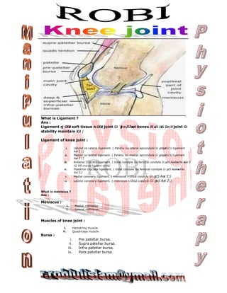
  • 1. What is Ligament ? Ans : Ligament nj GKwU soft tissue hv GKwU joint Gi `yyB ev Z‡ZvwaK bones ‡K a‡i iv‡L Ges H joint Gi stability maintain K‡i | Ligament of knee joint : i. Lateral co-lateral ligament. ( Patella Ges lateral epicondyle Gi gvSvgvwS†Z G ligament Aew¯’Z | ) ii. Medial co-lateral ligament. ( Patella Ges medial epicondyle Gi gvSvgvwS†Z G ligament Aew¯’Z | ) iii. Anterior cruciate ligament. ( tibial condyle Ges femoral condyle Gi gv‡S AvovAvwofv‡e &Aew¯’Z e‡j G‡K cruciate ligament ejv nq| iv. Posterior cruciate ligament. ( tibial condyle Ges femoral condyle Gi gv‡S AvovAvwofv‡e Aew¯’Z|) v. Medial coronary ligament. ( meniscus ও tibial condyle Gi gv‡S Aew¯’ Z|) vi. Lateral coronary ligament. ( meniscus ও tibial condyle Gi gv‡S Aew¯’Z|) What is meniscus ? Ans : Meniscus : i. Medial meniscus. ii. Lateral meniscus. Muscles of knee joint : i. Hamstring muscle. ii. Quadriceps muscle. Bursa : i. Pre patellar bursa. ii. Supra patellar bursa. iii. Infra patellar bursa. iv. Para patellar bursa.
  • 2. Muscle injury : Muscle injury n‡j H muscle Gi resisted test positive or painful Ges opposite passive movement positive n‡e| Ligament test : Lateral co-lateral ligament test : ( Varus test ) Knee flex n‡e 5- 10º . Medial co-lateral ligament test : ( Valgus test ) Knee flex n‡e 5- 10º. Anterior Cruciate Ligament : ( Anterior Dawer Test ). Posterior Cruciate Ligament: (Posterior Dawer Test ). Miniscus Test : Medial & Lateral Miniscus : ( Mckmarry Test ). Medial Gi ‡ÿ‡Î tibiaÕi lateral rotation Ges Lateral Gi ‡ÿ‡Î medial rotation কের “D” Shape Ki‡Z n‡e. Sign & Symptoms of Injury ( Knee Joint ) Miniscus injury : Sudden traumatic cause. Sign :  Immediate loss of function.  Swelling. Symptoms :  Twings – Sharp pain during knee flexion & extension.  Locking –flexion I extension G AvuU‡K hvIqv .  Giving way – tibia mvg‡bi w`‡K P‡j hvIqv. Ligament Injury : Main injury nq MCL, M.miniscus & M.coronary ligament G.  Pain – started after 1-2 hrs. Healing stage of Soft tissue : 0-5 days – Inflammatory stage. ( Leukocyte,Lymphocyte & macrophage– blood cell for healing soft tissue.) 5-21 days – Repair stage. 3 complication are founds in this stage –  Cross linking.  Excessive scar.  Adhesion.
  • 3. 21 day -6 month+ – Remodeling stage. Treatment for reduce this complication Inflammatory stage : PRICE – 0-5 days. P = Protection – by Creps bandage, Plaster of parries, Back slab. R = Rest. I = Ice. C = Compression. E = Elevation. Repair stage : 05-21 days. PRICE- continue.  GTFM – Gentle Transverse Friction Massage.  Pain free mobilization. ( Pain limitation )  Electrical modalities – UST/Wax, & LASER.  Tapping.  Gentle stretching. Remodeling stage :  DTFM – Deep Transverse Friction Massage.  Strong stretching.  ROM mobilization.  Manipulation.  UST.  Balance, co-ordination & proprioception ex’s. Proprioception : Joint position sense. Stage of Injury : 1. Acute – 2 wk. 2. Sub-acute – 2-5 wk. 3. Chronic – 6+ wk. Treatment of Knee Joint Friction. Mulligan.− Caudal (non weight) & lateral glide-1st wk. Caudal (MWM) after 1st wk. Mobilization. Manipulation. Manipulation of Knee Joint :  Miniscus manipulation.  Loose body flexion manipulation.  Loose body flexion rotation manipulation.  Loose body extension varus manipulation.  MCL manipulation towards flexion.  MCL manipulation towards medial rotation.  MCL manipulation towards lateal rotation. Miniscus Injury :
  • 4. Loss of function. ( Immediately )  Swelling.  Haematoma. Adhesion – MCL (test) :  Passive extension + lateral rotation.(Slight pain)  Valgus strain – Markedly painful.  Flexion – ROM (5˚-10˚) limited.  Pain is felt medially. Examination of patient (
  • 5. Shoulder Joint Shoulder Capsulitis : 3 question – symptoms :  Can you lie on that side at night.  Is there any pain at rest.  Has the pain spread below elbow. Sign : LAM L = Lateral rotation. A = Abduction. M = Medial rotation.
  • 6. Causes of Frozen shoulder :  Capsulitis.  O.A .  Traumatic arthritis.  # of humerus.  Immobilize – prolong time by POP. Stage : 3 unfavorable ans – stage ІІ 3 favorable ans – stage І 2 favorable+1 unfavorable ans – stage ІІ- 2 unfavorable+1 favorable ans – stage ІІ+ Stage-I – 15 days below ─ no physiotherapy. Only pain killer, heat therapy, SWD, UST/IFT. Management – after 15 days- o Mulligan’s mobilization. (continue) o Home stretching – after 70%-80% Functional Examination : Active abduction/elevation : Diagnosis :  Capsular pattern.  Shoulder fold.  ROM.  Tendonitis / Muscle injury.  Hard end feel. ( Osteo Arthrosis ). Passive elevation : Diagnosis :  Capsular pattern.  Shoulder fold.  ROM.  Tendonitis / Muscle injury.  Hard end feel. ( Osteo Arthrosis ). Painful arc : Painful arc is a painful section between two pain free section. Generally 90˚-120˚. Causes of painful arc –  Subacromion bursitis.  Acromio-clavicular jt. ligament lesion.  Supraspinatus tendonitis.
  • 7. Passive abduction ( Glenohumoral jt.) : GH(90˚)+ ST(150˚)+ GH(180˚). Diagnosis :  Capsular pattern.  Shoulder fold.  ROM.  Tendonitis / Muscle injury. Passive Lateral Rotation : Diagnosis :  Anterior capsule shortening.  Medial rotator muscle lesion.  Capsular pattern.  AC joint ligament lesion. Passive Medial Rotation : Diagnosis :  posterior capsule shortening.  lateral rotator muscle lesion.  Capsular pattern.  AC joint ligament lesion. Resisted Adduction : Diagnosis :  Adductor muscle lesion / tendonitis. Resisted Abduction : Diagnosis :  Abductor muscle lesion / tendonitis. Resisted Medial Rotation : Diagnosis :  Medial rotator muscle lesion / tendonitis. Resisted Lateral Rotation : Diagnosis :  Lateral rotator muscle lesion / tendonitis.
  • 8. Resisted Elbow Flexion : Diagnosis :  Flexor muscle lesion / tendonitis. Resisted Elbow Extension : Diagnosis :  Extensor muscle lesion / tendonitis. Passive Horizontal Adduction : Diagnosis :  AC lesion.  Subscapularis muscle injury / tendonitis.  Sub coracoid bursitis. Muscle – for movt. Of shoulder joint : Adduction :  Pectoralis major.(Anterior)  Latissimus dorsi.(Posterior)  Teris major- Posterior side pain + resisted medial rotation positive/painful.  Teris minor- Posterior side pain + resisted lateral rotation positive/painful. Subscapularis muscle : Action – Medial rotation. (Pain on anterior side). - Initial abduction.(0˚-30˚) Abduction :  Subscapularis muscle.  Deltoid muscle fiber. Elbow flexion :  Biceps.  Brachialis. Elbow extension :  Triceps. Sub deltoid bursitis :