SlideShare a Scribd company logo
1 of 51
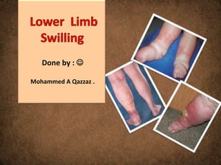
Done by : 

Mohammed A Qazzaz .
Basic anatomy

   The primary function of the lower limbs is
   to support the weight of the body and to
   provide a stable foundation in
   standing, walking, and running; they have
   become specialized for locomotion.

   The lower limbs are divided into the
   gluteal region, the thigh, the knee, the
   leg, the ankle, and the foot.
Swelling

Swelling is the enlargement of a body part
or organ beyond it normal size and usually
causing a distortion of the shape and
structure of the affected area.

Fluid

Gas

Mass
The swollen leg

    Swelling is a sign for many conditions
    affect the human body .

    These conditions could be vascular or non
    vascular .

    It could be unilateral or bilateral .
Non vascular or lymphatic

     General disease states :

     Cardiac failure from any cause .
     Liver failure .
     Hypoproteinaemia due to nephrotic
     syndrome, malabsorption, protein-losing
     enteropathy
     Hypothyroidism (myxoedema)
     Allergic disorders, including angioedema and
     idiopathic cyclic oedema
     Prolonged immobility and lower limb dependency
Non vascular or lymphatic

      Local disease processes :


      Ruptured Baker’s cyst
      Myositis ossificans
      Bony or soft-tissue tumours
      Arthritis
      Haemarthrosis
      Calf muscle haematoma
      Achilles tendon rupture
      Cellulitis
      Athlete’s foot
Non vascular or lymphatic
   Retroperitoneal fibrosis May lead to arterial, venous and lymphatic
   abnormalities

   Gigantism: Rare

              All tissues are uniformly enlarged

   Drugs : Corticosteroids, oestrogens, progestagens

   Monoamine                                                   oxidase
   inhibitors, phenylbutazone, methyldopa, hydralazine, nifedipine

   Trauma : Painful swelling due to reflex sympathetic dystrophy

   Obesity : Lipodystrophy

            Lipoidosis
Venous

Deep venous thrombosis

Phlebitis

Post-thrombotic syndrome

Varicose veins

Klippel–Trenaunay’s syndrome

External venous compression
Arterial and lymphatic


      Aneurysm

      Inflammation in the lymph nodes

      Lymphedema
Common Causes of Leg Edema in the United States

  Table 1
                 Unilateral                                Bilateral

    Acute (<72 hours)      Chronic           Acute (<72 hours)          Chronic
  Deep vein           Venous insufficiency                       Venous
  thrombosis                                                     insufficiency
                                                                 Pulmonary
                                                                 hypertension
                                                                 Heart failure
                                                                 Idiopathic edema

                                                                 Lymphedema
                                                                 Drugs
                                                                 Premenstrual
                                                                 edema
                                                                 Pregnancy
                                                                 Obesity
Less Common Causes of Leg Edema in the United
States
 Table 2
                         Unilateral                                                Bilateral
     Acute (<72 hours)                    Chronic                Acute (<72 hours)                  Chronic
Ruptured Baker’s cyst         Secondary lymphedema         Bilateral deep vein           Renal disease (nephrotic
                              (tumor, radiation, surgery,  thrombosis                    syndrome,
                              bacterial infection)                                       glomerulonephritis)
Ruptured medial head of       Pelvic tumor or lymphoma Acute worsening of systemic       Liver disease
gastrocnemius                 causing external pressure on cause (heart failure, renal
                              veins                        disease)
Compartment syndrome          Reflex sympathetic dystrophy                               Secondary lymphedema
                                                                                         (secondary to tumor,
                                                                                         radiation, bacterial infection,
                                                                                         filariasis)
                                                                                         Pelvic tumor or lymphoma
                                                                                         causing external pressure
                                                                                         Dependent edema
                                                                                         Diuretic-induced edema
                                                                                         Dependent edema
                                                                                         Preeclampsia
                                                                                                  8
                                                                                         Lipidema
                                                                                         Anemia
Rare Causes of Leg Edema in the United States

Table 3
                  Unilateral                                         Bilateral
  Acute (<72 hours)             Chronic          Acute (<72 hours)                 Chronic
                       Primary lymphedema                                Primary lymphedema
                       (congenital                                       (congenital
                       lymphedema,                                       lymphedema, lymphede
                       lymphedema praecox,                               ma
                       lymphedema tarda)                                 praecox, lymphedema
                                                                         tarda)
                       Congenital venous                                 Protein losing
                       malformations                                     enteropathy,
                                                                         malnutrition,
                                                                         malabsorption
                       May-Thurner syndrome                              Restrictive pericarditis
                       (iliac-vein compression
                                   51
                       syndrome)
                                                                         Restrictive
                                                                         cardiomyopathy
                                                                         Beri Beri
                                                                         Myxedema
How can we start ?   Figure 1
How can we start ?   Figure 2
Idiopathic edema

 diopathic edema is a pitting edema of unknown cause that
 occurs primarily in pre-menopausal women who do not have
 evidence of heart, liver, or kidney disease. In this condition, the
 fluid retention at first may be seen primarily pre-menstrually
 (just prior to menstruation), which is why it sometimes is called
 "cyclical" edema. However, it can become a more constant and
 severe problem. Obesity and depression can be associated with
 this syndrome, and diuretic abuse is common

 Spironolactone is considered the drug of choice for idiopathic
 edema
 avoiding environmental heat, low salt diet, avoiding excessive
 fluid intake, and weight loss for obese patients. It may be
 helpful to ask about depression, eating disorder
Venous insufficiency

 Venous insufficiency is characterized by chronic pitting edema,
 often associated with brown hemosiderin skin deposits on the
 lower legs. The skin changes can progress to dermatitis and
 ulceration, which usually occur over the medial maleoli. Other
 common findings include varicose veins and obesity. Most patients
 are asymptomatic but a sensation of aching or heaviness can
 occur. The diagnosis is usually made clinically but can be
 confirmed with a Doppler study. Although chronic venous
 insufficiency is thought to result from previous deep vein
 thrombosis, only one third of patients will give that history


 compression stockings
How can we start ?   Figure 1
How can we start ?   Figure 3
How can we start ?   Figure 4


                                DVT
How can we start ?   Figure 5
History

Key elements of the history include

 What is the duration of the edema (acute [<72 hours] vs. chronic)? If the onset is acute,
deep vein thrombosis should be strongly considered.


Is the edema painful ? Deep vein thrombosis and reflex sympathetic dystrophy are usually
painful. Chronic venous insufficiency can cause low-grade aching. Lymphedema is usually
painless.

What drugs are being taken? Calcium channel blockers, prednisone, and anti-inflammatory
drugs are common causes of leg edema


Is there a history of systemic disease (heart, liver, or kidney disease)?
Antihypertensive drugs
        Calcium channel blockers
        Beta blockers
        Clonidine
        Hydralazine
        Minoxidil
        Methyldopa
Hormones
        Corticosteroids
        Estrogen
        Progesterone
        Testosterone
Other
        Nonsteroidal anti-inflammatory drugs
        Pioglitazone, Rosiglitazone
        Monoamine oxidase inhibitors
Physical Examination

   Body mass index. Obesity
   Distribution of edema
   Tenderness
   Pitting

   Varicose veins

   Kaposi-Stemmer sign
   Skin changes

   Signs of systemic disease: findings of heart failure (especially jugular
   venous distension and lung crackles) and liver disease (ascites, spider
   hemangiomas, and jaundice) may be helpful in detecting a systemic cause.
Kaposi-Stemmer sign: inability to pinch a fold of skin at base of
second toe because of thickened skin indicates lymphoedema
cellulitis
Varicose veins


Clinical presentation :

    Local pain and edema

    Local inflammation

    Local hemorrhage into
    the surrounding tissue

    Dilated superficial veins
Definition


  Lymphedema may be defined as abnormal limb swelling caused
  by the accumulation of increased amounts of high protein ISF
  secondary to defective lymphatic drainage in the presence of
  (near) normal net capillary filtration.

  At birth, 1 in 6000 persons will develop lymphoedema
Classification


  Two main types of lymphoedema are recognised:

  1 primary lymphoedema, in which the cause is unknown (or at
  least uncertain and unproven); it is thought to be caused by
  congenital lymphatic dysplasia

  2 secondary or acquired lymphoedema, in which there is a clear
  underlying cause.
primary lymphoedema
     Three types of primary lymphedema are distinguished by
     age of onset.
        Congenital lymphedema is present at birth or occurs
        early in infancy.
            It accounts for fewer than 10% of primary
            lymphedema cases.
            Lymphedema that is both congenital and
            hereditary is known as Milroy's disease.
        Lymphedema praecox occurs at any time from puberty
        until the end of the third decade.
            Most cases of primary lymphedema are of this
            type.
            It is three times more common in women than in
            men.
        Lymphedema tarda occurs after age 30.
secondary lymphoedema


   Secondary lymphedema is due to obstruction from a variety of causes,
   including infection, parasites, mechanical injury (including surgery),
   postphlebitic syndrome, and neoplasms.

       In developed countries, the most common causes are obstruction by
       malignancies, postsurgical lymphedema (e.g., after mastectomy), and
       lymphatic destruction from therapeutic radiation.

       In less well-developed countries, parasitic obstruction (elephantiasis)
       is a common cause. Wuchereria bancrofti is the most common
       offending parasite.
Filariasis

 This is the most common cause of lymphoedema worldwide,
 affecting up to 100 million individuals. It is particularly prevalent
 in Africa, India and South America where 5–10% of the
 population
 may be affected. The viviparous nematode Wucheria bancrofti,
 whose only host is man, is responsible for 90% of cases and is
 spread by the mosquito.
  The disease is associated with poor sanitation.
 The parasite enters lymphatics from the blood and lodges
 in lymph nodes, where it causes fibrosis and obstruction
elephantiasis
management

Relief of pain

Control of swelling

Skin care

Manual lymphatic drainage

Exercise
Lower  limb swilling

More Related Content

What's hot (20)

Acute cholecystitis.pptx
Acute cholecystitis.pptxAcute cholecystitis.pptx
Acute cholecystitis.pptx
 
Varicose Veins
Varicose VeinsVaricose Veins
Varicose Veins
 
ascites
 ascites ascites
ascites
 
Haematuria
HaematuriaHaematuria
Haematuria
 
Leg ulcers
Leg ulcers Leg ulcers
Leg ulcers
 
Acute cholecystitis..
Acute cholecystitis..Acute cholecystitis..
Acute cholecystitis..
 
Intussusception
IntussusceptionIntussusception
Intussusception
 
Lower GI - Bleed
Lower GI - Bleed Lower GI - Bleed
Lower GI - Bleed
 
Vasculitis
VasculitisVasculitis
Vasculitis
 
Obstructive jaundice.
Obstructive jaundice.Obstructive jaundice.
Obstructive jaundice.
 
Obstructive jaundice
Obstructive jaundiceObstructive jaundice
Obstructive jaundice
 
Venous ulcer:A pedal misery!
Venous ulcer:A pedal misery!Venous ulcer:A pedal misery!
Venous ulcer:A pedal misery!
 
varicose vein surgery
 varicose vein surgery varicose vein surgery
varicose vein surgery
 
Chronic limb ischemia
Chronic limb ischemia Chronic limb ischemia
Chronic limb ischemia
 
Ascites
AscitesAscites
Ascites
 
Approach to child with generalized body swelling
Approach to child with generalized body swellingApproach to child with generalized body swelling
Approach to child with generalized body swelling
 
Cholangitis
CholangitisCholangitis
Cholangitis
 
NEPHRITIC SYNDROME / APSGN IN CHILDREN
NEPHRITIC SYNDROME / APSGN IN CHILDREN NEPHRITIC SYNDROME / APSGN IN CHILDREN
NEPHRITIC SYNDROME / APSGN IN CHILDREN
 
Obstructive jaundice
Obstructive jaundiceObstructive jaundice
Obstructive jaundice
 
Hepatospleenomegaly in children
Hepatospleenomegaly in childrenHepatospleenomegaly in children
Hepatospleenomegaly in children
 

Viewers also liked

Edema
EdemaEdema
Edemajecty
 
Deep Vein Thrombosis
Deep Vein ThrombosisDeep Vein Thrombosis
Deep Vein Thrombosisdbridley
 
Doppler ultrasound in deep vein thrombosis
Doppler ultrasound in deep vein thrombosisDoppler ultrasound in deep vein thrombosis
Doppler ultrasound in deep vein thrombosisSamir Haffar
 
Intermittent pneumatic compression pump therapy for lymphedema •
Intermittent pneumatic compression pump therapy for lymphedema • Intermittent pneumatic compression pump therapy for lymphedema •
Intermittent pneumatic compression pump therapy for lymphedema • MaxiMedRx
 
20131020 03 黃睦升_淋巴水腫
20131020 03 黃睦升_淋巴水腫20131020 03 黃睦升_淋巴水腫
20131020 03 黃睦升_淋巴水腫Kit Leong
 
DEEP VEIN THROMBOSIS
DEEP VEIN THROMBOSISDEEP VEIN THROMBOSIS
DEEP VEIN THROMBOSISVinay Teradal
 
淋巴水腫之物理治療 楊靜蘭
淋巴水腫之物理治療 楊靜蘭淋巴水腫之物理治療 楊靜蘭
淋巴水腫之物理治療 楊靜蘭Kit Leong
 
Lymphoscintigraphy As an Imaging Modality in Lymphatic System
Lymphoscintigraphy As an Imaging Modality in Lymphatic SystemLymphoscintigraphy As an Imaging Modality in Lymphatic System
Lymphoscintigraphy As an Imaging Modality in Lymphatic SystemApollo Hospitals
 
Determining a vascular cause for leg pain and referrals
Determining a vascular cause for leg pain and referralsDetermining a vascular cause for leg pain and referrals
Determining a vascular cause for leg pain and referralsSpecialistVeinHealth
 
Measuring for Lower Extremity Compression Garments
Measuring for Lower Extremity Compression GarmentsMeasuring for Lower Extremity Compression Garments
Measuring for Lower Extremity Compression GarmentsOSUCCC - James
 
Non-Thrombotic Iliac Vein Lesions: Permissive Role in CVD Pathogenicity
Non-Thrombotic Iliac Vein Lesions: Permissive Role in CVD PathogenicityNon-Thrombotic Iliac Vein Lesions: Permissive Role in CVD Pathogenicity
Non-Thrombotic Iliac Vein Lesions: Permissive Role in CVD PathogenicityVein Global
 
N 295 lecture 10 peripheral vasc student copy
N 295 lecture 10 peripheral vasc student copyN 295 lecture 10 peripheral vasc student copy
N 295 lecture 10 peripheral vasc student copyElly Hb
 

Viewers also liked (20)

Edema
EdemaEdema
Edema
 
Deep Vein Thrombosis
Deep Vein ThrombosisDeep Vein Thrombosis
Deep Vein Thrombosis
 
DVT
DVTDVT
DVT
 
Dvt
DvtDvt
Dvt
 
Doppler ultrasound in deep vein thrombosis
Doppler ultrasound in deep vein thrombosisDoppler ultrasound in deep vein thrombosis
Doppler ultrasound in deep vein thrombosis
 
Intermittent pneumatic compression pump therapy for lymphedema •
Intermittent pneumatic compression pump therapy for lymphedema • Intermittent pneumatic compression pump therapy for lymphedema •
Intermittent pneumatic compression pump therapy for lymphedema •
 
20131020 03 黃睦升_淋巴水腫
20131020 03 黃睦升_淋巴水腫20131020 03 黃睦升_淋巴水腫
20131020 03 黃睦升_淋巴水腫
 
DEEP VEIN THROMBOSIS
DEEP VEIN THROMBOSISDEEP VEIN THROMBOSIS
DEEP VEIN THROMBOSIS
 
D V T
D V TD V T
D V T
 
淋巴水腫之物理治療 楊靜蘭
淋巴水腫之物理治療 楊靜蘭淋巴水腫之物理治療 楊靜蘭
淋巴水腫之物理治療 楊靜蘭
 
Lymphoscintigraphy As an Imaging Modality in Lymphatic System
Lymphoscintigraphy As an Imaging Modality in Lymphatic SystemLymphoscintigraphy As an Imaging Modality in Lymphatic System
Lymphoscintigraphy As an Imaging Modality in Lymphatic System
 
A3 - Symptom Management
A3 - Symptom ManagementA3 - Symptom Management
A3 - Symptom Management
 
Determining a vascular cause for leg pain and referrals
Determining a vascular cause for leg pain and referralsDetermining a vascular cause for leg pain and referrals
Determining a vascular cause for leg pain and referrals
 
Dvt
DvtDvt
Dvt
 
Measuring for Lower Extremity Compression Garments
Measuring for Lower Extremity Compression GarmentsMeasuring for Lower Extremity Compression Garments
Measuring for Lower Extremity Compression Garments
 
Vascular disorders
Vascular disordersVascular disorders
Vascular disorders
 
Leg Ulcer
Leg UlcerLeg Ulcer
Leg Ulcer
 
Non-Thrombotic Iliac Vein Lesions: Permissive Role in CVD Pathogenicity
Non-Thrombotic Iliac Vein Lesions: Permissive Role in CVD PathogenicityNon-Thrombotic Iliac Vein Lesions: Permissive Role in CVD Pathogenicity
Non-Thrombotic Iliac Vein Lesions: Permissive Role in CVD Pathogenicity
 
N 295 lecture 10 peripheral vasc student copy
N 295 lecture 10 peripheral vasc student copyN 295 lecture 10 peripheral vasc student copy
N 295 lecture 10 peripheral vasc student copy
 
Dvt
DvtDvt
Dvt
 

Similar to Lower limb swilling

Scleroderma long case summary
Scleroderma long case  summaryScleroderma long case  summary
Scleroderma long case summaryDr. Rubz
 
Deep Vein Thrombosis and Pulmonary Embolism, by Prof. Minnu M. Panditrao
Deep Vein Thrombosis and Pulmonary Embolism, by Prof. Minnu M. PanditraoDeep Vein Thrombosis and Pulmonary Embolism, by Prof. Minnu M. Panditrao
Deep Vein Thrombosis and Pulmonary Embolism, by Prof. Minnu M. PanditraoMinnu Panditrao
 
Etiology –lower gi bleed
Etiology –lower gi bleedEtiology –lower gi bleed
Etiology –lower gi bleedAbino David
 
Approach to a patient with body swelling-1 new.pptx
Approach to a patient with body swelling-1 new.pptxApproach to a patient with body swelling-1 new.pptx
Approach to a patient with body swelling-1 new.pptxtowinb34
 
Cardiomyopathy,myocarditis,pericarditi,tumor
Cardiomyopathy,myocarditis,pericarditi,tumorCardiomyopathy,myocarditis,pericarditi,tumor
Cardiomyopathy,myocarditis,pericarditi,tumorpathologydept
 

Similar to Lower limb swilling (7)

clinical aspects of vein
clinical aspects of veinclinical aspects of vein
clinical aspects of vein
 
Scleroderma long case summary
Scleroderma long case  summaryScleroderma long case  summary
Scleroderma long case summary
 
Deep Vein Thrombosis and Pulmonary Embolism, by Prof. Minnu M. Panditrao
Deep Vein Thrombosis and Pulmonary Embolism, by Prof. Minnu M. PanditraoDeep Vein Thrombosis and Pulmonary Embolism, by Prof. Minnu M. Panditrao
Deep Vein Thrombosis and Pulmonary Embolism, by Prof. Minnu M. Panditrao
 
Etiology –lower gi bleed
Etiology –lower gi bleedEtiology –lower gi bleed
Etiology –lower gi bleed
 
Approach to a patient with body swelling-1 new.pptx
Approach to a patient with body swelling-1 new.pptxApproach to a patient with body swelling-1 new.pptx
Approach to a patient with body swelling-1 new.pptx
 
Cardiomyopathy,myocarditis,pericarditi,tumor
Cardiomyopathy,myocarditis,pericarditi,tumorCardiomyopathy,myocarditis,pericarditi,tumor
Cardiomyopathy,myocarditis,pericarditi,tumor
 
2012 cardiac lecture 2 pathologies
2012 cardiac lecture 2 pathologies2012 cardiac lecture 2 pathologies
2012 cardiac lecture 2 pathologies
 

More from mohammed Qazzaz (20)

Nocturnal Enuresis
Nocturnal EnuresisNocturnal Enuresis
Nocturnal Enuresis
 
‫أَيَّاً يَكُن
‫أَيَّاً يَكُن ‫أَيَّاً يَكُن
‫أَيَّاً يَكُن
 
DVT.. Deep vein thrombosis.
DVT.. Deep vein thrombosis.DVT.. Deep vein thrombosis.
DVT.. Deep vein thrombosis.
 
Croup
CroupCroup
Croup
 
Blood film
Blood filmBlood film
Blood film
 
Iron deficiency in children
Iron deficiency in childrenIron deficiency in children
Iron deficiency in children
 
Poliomyelitis
PoliomyelitisPoliomyelitis
Poliomyelitis
 
Epstein barr virus
Epstein barr virus Epstein barr virus
Epstein barr virus
 
Migraine
MigraineMigraine
Migraine
 
Breastfeeding
Breastfeeding Breastfeeding
Breastfeeding
 
Rickettsial diseases
Rickettsial diseasesRickettsial diseases
Rickettsial diseases
 
Herbal medicine
Herbal medicineHerbal medicine
Herbal medicine
 
Conjuctival diseases
Conjuctival diseasesConjuctival diseases
Conjuctival diseases
 
الاجهاض
الاجهاضالاجهاض
الاجهاض
 
Diabetic Ketoacidosis
Diabetic KetoacidosisDiabetic Ketoacidosis
Diabetic Ketoacidosis
 
lacrimal system anatomy and clinical
lacrimal system anatomy and clinicallacrimal system anatomy and clinical
lacrimal system anatomy and clinical
 
Enegry drink hazards
Enegry drink hazardsEnegry drink hazards
Enegry drink hazards
 
Herpes simplex virus
Herpes simplex virusHerpes simplex virus
Herpes simplex virus
 
Autism
AutismAutism
Autism
 
Vomiting
VomitingVomiting
Vomiting
 

Lower limb swilling

  • 1. Done by :  Mohammed A Qazzaz .
  • 2. Basic anatomy The primary function of the lower limbs is to support the weight of the body and to provide a stable foundation in standing, walking, and running; they have become specialized for locomotion. The lower limbs are divided into the gluteal region, the thigh, the knee, the leg, the ankle, and the foot.
  • 3. Swelling Swelling is the enlargement of a body part or organ beyond it normal size and usually causing a distortion of the shape and structure of the affected area. Fluid Gas Mass
  • 4. The swollen leg Swelling is a sign for many conditions affect the human body . These conditions could be vascular or non vascular . It could be unilateral or bilateral .
  • 5. Non vascular or lymphatic General disease states : Cardiac failure from any cause . Liver failure . Hypoproteinaemia due to nephrotic syndrome, malabsorption, protein-losing enteropathy Hypothyroidism (myxoedema) Allergic disorders, including angioedema and idiopathic cyclic oedema Prolonged immobility and lower limb dependency
  • 6. Non vascular or lymphatic Local disease processes : Ruptured Baker’s cyst Myositis ossificans Bony or soft-tissue tumours Arthritis Haemarthrosis Calf muscle haematoma Achilles tendon rupture Cellulitis Athlete’s foot
  • 7. Non vascular or lymphatic Retroperitoneal fibrosis May lead to arterial, venous and lymphatic abnormalities Gigantism: Rare All tissues are uniformly enlarged Drugs : Corticosteroids, oestrogens, progestagens Monoamine oxidase inhibitors, phenylbutazone, methyldopa, hydralazine, nifedipine Trauma : Painful swelling due to reflex sympathetic dystrophy Obesity : Lipodystrophy Lipoidosis
  • 8. Venous Deep venous thrombosis Phlebitis Post-thrombotic syndrome Varicose veins Klippel–Trenaunay’s syndrome External venous compression
  • 9. Arterial and lymphatic Aneurysm Inflammation in the lymph nodes Lymphedema
  • 10.
  • 11. Common Causes of Leg Edema in the United States Table 1 Unilateral Bilateral Acute (<72 hours) Chronic Acute (<72 hours) Chronic Deep vein Venous insufficiency Venous thrombosis insufficiency Pulmonary hypertension Heart failure Idiopathic edema Lymphedema Drugs Premenstrual edema Pregnancy Obesity
  • 12. Less Common Causes of Leg Edema in the United States Table 2 Unilateral Bilateral Acute (<72 hours) Chronic Acute (<72 hours) Chronic Ruptured Baker’s cyst Secondary lymphedema Bilateral deep vein Renal disease (nephrotic (tumor, radiation, surgery, thrombosis syndrome, bacterial infection) glomerulonephritis) Ruptured medial head of Pelvic tumor or lymphoma Acute worsening of systemic Liver disease gastrocnemius causing external pressure on cause (heart failure, renal veins disease) Compartment syndrome Reflex sympathetic dystrophy Secondary lymphedema (secondary to tumor, radiation, bacterial infection, filariasis) Pelvic tumor or lymphoma causing external pressure Dependent edema Diuretic-induced edema Dependent edema Preeclampsia 8 Lipidema Anemia
  • 13. Rare Causes of Leg Edema in the United States Table 3 Unilateral Bilateral Acute (<72 hours) Chronic Acute (<72 hours) Chronic Primary lymphedema Primary lymphedema (congenital (congenital lymphedema, lymphedema, lymphede lymphedema praecox, ma lymphedema tarda) praecox, lymphedema tarda) Congenital venous Protein losing malformations enteropathy, malnutrition, malabsorption May-Thurner syndrome Restrictive pericarditis (iliac-vein compression 51 syndrome) Restrictive cardiomyopathy Beri Beri Myxedema
  • 14. How can we start ? Figure 1
  • 15. How can we start ? Figure 2
  • 16. Idiopathic edema diopathic edema is a pitting edema of unknown cause that occurs primarily in pre-menopausal women who do not have evidence of heart, liver, or kidney disease. In this condition, the fluid retention at first may be seen primarily pre-menstrually (just prior to menstruation), which is why it sometimes is called "cyclical" edema. However, it can become a more constant and severe problem. Obesity and depression can be associated with this syndrome, and diuretic abuse is common Spironolactone is considered the drug of choice for idiopathic edema avoiding environmental heat, low salt diet, avoiding excessive fluid intake, and weight loss for obese patients. It may be helpful to ask about depression, eating disorder
  • 17. Venous insufficiency Venous insufficiency is characterized by chronic pitting edema, often associated with brown hemosiderin skin deposits on the lower legs. The skin changes can progress to dermatitis and ulceration, which usually occur over the medial maleoli. Other common findings include varicose veins and obesity. Most patients are asymptomatic but a sensation of aching or heaviness can occur. The diagnosis is usually made clinically but can be confirmed with a Doppler study. Although chronic venous insufficiency is thought to result from previous deep vein thrombosis, only one third of patients will give that history compression stockings
  • 18.
  • 19.
  • 20.
  • 21. How can we start ? Figure 1
  • 22. How can we start ? Figure 3
  • 23. How can we start ? Figure 4 DVT
  • 24. How can we start ? Figure 5
  • 25. History Key elements of the history include What is the duration of the edema (acute [<72 hours] vs. chronic)? If the onset is acute, deep vein thrombosis should be strongly considered. Is the edema painful ? Deep vein thrombosis and reflex sympathetic dystrophy are usually painful. Chronic venous insufficiency can cause low-grade aching. Lymphedema is usually painless. What drugs are being taken? Calcium channel blockers, prednisone, and anti-inflammatory drugs are common causes of leg edema Is there a history of systemic disease (heart, liver, or kidney disease)?
  • 26. Antihypertensive drugs Calcium channel blockers Beta blockers Clonidine Hydralazine Minoxidil Methyldopa Hormones Corticosteroids Estrogen Progesterone Testosterone Other Nonsteroidal anti-inflammatory drugs Pioglitazone, Rosiglitazone Monoamine oxidase inhibitors
  • 27. Physical Examination Body mass index. Obesity Distribution of edema Tenderness Pitting Varicose veins Kaposi-Stemmer sign Skin changes Signs of systemic disease: findings of heart failure (especially jugular venous distension and lung crackles) and liver disease (ascites, spider hemangiomas, and jaundice) may be helpful in detecting a systemic cause.
  • 28. Kaposi-Stemmer sign: inability to pinch a fold of skin at base of second toe because of thickened skin indicates lymphoedema
  • 30.
  • 31. Varicose veins Clinical presentation : Local pain and edema Local inflammation Local hemorrhage into the surrounding tissue Dilated superficial veins
  • 32.
  • 33.
  • 34.
  • 35.
  • 36. Definition Lymphedema may be defined as abnormal limb swelling caused by the accumulation of increased amounts of high protein ISF secondary to defective lymphatic drainage in the presence of (near) normal net capillary filtration. At birth, 1 in 6000 persons will develop lymphoedema
  • 37. Classification Two main types of lymphoedema are recognised: 1 primary lymphoedema, in which the cause is unknown (or at least uncertain and unproven); it is thought to be caused by congenital lymphatic dysplasia 2 secondary or acquired lymphoedema, in which there is a clear underlying cause.
  • 38. primary lymphoedema Three types of primary lymphedema are distinguished by age of onset. Congenital lymphedema is present at birth or occurs early in infancy. It accounts for fewer than 10% of primary lymphedema cases. Lymphedema that is both congenital and hereditary is known as Milroy's disease. Lymphedema praecox occurs at any time from puberty until the end of the third decade. Most cases of primary lymphedema are of this type. It is three times more common in women than in men. Lymphedema tarda occurs after age 30.
  • 39. secondary lymphoedema Secondary lymphedema is due to obstruction from a variety of causes, including infection, parasites, mechanical injury (including surgery), postphlebitic syndrome, and neoplasms. In developed countries, the most common causes are obstruction by malignancies, postsurgical lymphedema (e.g., after mastectomy), and lymphatic destruction from therapeutic radiation. In less well-developed countries, parasitic obstruction (elephantiasis) is a common cause. Wuchereria bancrofti is the most common offending parasite.
  • 40.
  • 41.
  • 42.
  • 43.
  • 44. Filariasis This is the most common cause of lymphoedema worldwide, affecting up to 100 million individuals. It is particularly prevalent in Africa, India and South America where 5–10% of the population may be affected. The viviparous nematode Wucheria bancrofti, whose only host is man, is responsible for 90% of cases and is spread by the mosquito. The disease is associated with poor sanitation. The parasite enters lymphatics from the blood and lodges in lymph nodes, where it causes fibrosis and obstruction
  • 45.
  • 46.
  • 48.
  • 49.
  • 50. management Relief of pain Control of swelling Skin care Manual lymphatic drainage Exercise