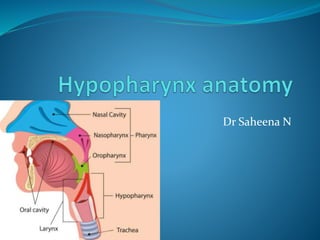
2. PHARYNX
It is a funnel shaped fibro-muscular tube of about 13 cm . length
It extends form the base of the skull to the level of the 6th cervical vertebrae.
Its anterior wall is deficient and lies behind the cavities of the nose, mouth and larynx
Accordingly it is divided into 3 parts: Nasopharynx , oropharynx & laryngeopharynx .
a)Base of skull
(basioociput and basi
sphenoid) soft palate
b)Plane of hard palate
hyoid bone
c) Hyoid bone lower
border of cricoid
cartilage
Nasopharynx
Oropharynxx
Larygeopharynx
5. Pyriform fossa
Lies on either side of larynx
Extends from pharyngo epiglottic
fold to the upper end of oesophagus
Boundaries
Superiorly- pharyngo epiglottic fold
Laterally – thyrohyoid membrane & thyroid cartilage
Medially – aryepiglottic fold ,posterolateral suface of
arytenoid & cricoid cartilages
Inferiorly – continues as esophagus
7. ..pyriform fossa
Also known as smuggler’s fossa
Forms the lateral channel for food
Foreign bodies may lodge in the PF
Internal laryngeal nerve –runs submucosally in the
lateral wall of the sinus- accessible for
local anesthasia
Referred otalgia in pyriform
fossa malignancy
9. Post cricoid region
Part of anterior wall of laryngopharynx between the
upper and lower border of cricoid cartilage
It is the common site for ca in females with
plummer –vinson syndrome
10. Posterior pharyngeal wall
Extends from the level of hyoid bone to the level
of cricoarytenoid joint
The posterior hypo pharyngeal
wall continuates the posterior
oropharynx wall;
It is composed of mucosa and
the constrictor muscle.
Smooth bulge of posterior pharyngeal wall
with intact mucosa
due to retropharyngeal malignancy.
11. WALLS OF THE PHARYNX
Consists of the following from within outwards:
1. Mucous coat lined by stratified squamous epithelium.
2. Inner fibrous coat (pharyngo-basilar fascia).
3. Muscular coat.
4. Outer fibrous coat (buccopharyngeal fascia).
Muscles of the pharynx
Sup.constrictor
mid.constrictor
inf.constrictor
Outer circular layer:
3 constrictors of the pharynx.
Inner longitudinal layer:
3 longitudinal muscles.
Constrictors of the pharynx.
1- Sup.constrictor
2- mid.constrictor
N.B. All muscles of pharynx take nerve supply from pharyngeal plexus except stylopharyngeus
from glossopharyngeal nerve
3- inf.constrictor
longitudinal muscles.
1 Stylo-pharyngeus
2Salpingo – pharyngeus
3-Palato-pharyngeus:
12. Hypopharyngeal diverticulum
Zenker’s diverticulum
Failure of cricopharyngeal sphincter relaxation when
pharyngeal muscles are contracting
Pharyngeal mucosa herniates through killian’s
dehiscence- week area between two parts of inferior
constrictor muscle
Gateway of tears- perforation can occur at this site
during oesophagostomy.
14. Lymphatic drainage
Pyriform sinus- richly supplied by lymphatics
(so nodal mets are common in ca pyriform fossa)
- Lymphatics exit through the thyrohyoid membrane
and drain into the upper jugular chain
Post cricoid region- drain into para pharyngeal
lymph nodes
-Also drain into supraclavicular and para tracheal
nodes
Posterior pharyngeal wall- lateral or para pharyngeal
nodes and then to deep cervical nodes