Downloaded 20 times
![JOURNAL OF WOUND CARE Vol 22. No 1. EWMA Documen t 2013 S 13
T
his chapter describes various debridement
techniques with autolytic, enzymatic and/
or absorptive properties. Many different
products are currently available on the market,
offering different combinations of components
and suitable for different wound characteristics and
stages. We will provide an overview of the various
product types, defined in four overall categories:
autolytic dressings, enzymatic dressings, absorptive
dressings and honey.
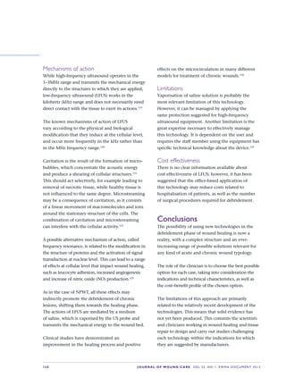
Autolytic dressings
Background
The term ‘autolytic debridement’ describes a
natural process in all kinds of wounds, which
can be supported by a moist wound-mangament
strategy. Autolytic debridement products can
be found in many different varieties, including
different properties, benefits and limitations. They
can be defined in the following groups:
a Hydrogels, or hydrogel-based dressings, are
three-dimensional, cross-linked homopolymers
Autolytic dressings,
enzymatic dressings,
absorbtive dressings
and honey
or copolymers, saturated with water. The
proportion of water in hydrogel dressings can
vary from 30% to 90%. Different gel-forming
agents, such as carboxymethylcellulose, are
incorporated into most hydrogels.34
b Hydrocolloids are composed of carbomethyl
cellulose, gelatin, pectin, elastomers and
adhesives that turn in to a gel when exudate
is absorbed.35
c Highly absorptive dressings with autolytic
and occlusive properties, such as dressings
with a multifunctional polymeric membrane
formulation and hydrations techniques
(e.g. hydration response technology [HRT]).
These dressings are designed for exudation
management, aiming to create a moist and
physiological environment for autolytic
debridement.36
Some dressings comprise a hydrophilic polymer-
modified starch in its three-dimensional network
with physically-bound iodine.](https://image.slidesharecdn.com/ewmadebridementdocumentjwcfinal-170325085136/85/Ewma-debridement-document_jw_cfinal-13-320.jpg)

![S42 JOURNAL OF WOUND CARE Vol 22. No 1. EWMA Docu men t 2013
References
1 Hinchliffe, R.J.,Valk, G.D.,Apelqvist,
J. et al.A systematic review of the
effectiveness of interventions to
enhance the healing of chronic
ulcers of the foot in diabetes.
Diabetes Metab Res Rev. 2008; 24
(Suppl. 1): 119–144.
2 O’Brien, M. Exploring methods of
wound debridement. Br J Comm
Nurs. 2002; 7: 10–18.
3 Oxford English Dictionary.
Available from: http://
oxforddictionaries.com/
4 Gethin, G., Cowman, S., Kolbach,
D.N. Debridement for venous leg
ulcers. Cochrane Database Sys Rev.
2010; 7: CD008599.
5 Vowden, K.R.,Vowden, P.Wound
debridement. Part 2: sharp
techniques. J Wound Care. 1999; 8:
291–294.
6 Department of Health (DH).
Reference guide to consent for
examination or treatment. DH, 2001.
7 Fisher-Jeffes, L.F., Barton, C., Finlay,
F. Clinicians’ knowledge of informed
consent. J Med Ethics. 2007; 33:
181–184.
8 Department of Health (DH).
Seeking consent: working with
children. DH, 2001.
9 Pape ,T. Legal and ethical
considerations of informed consent.
AORN J. 1997; 65: 122–127.
10 Kanerva,A., Suominen,T., Leino-
Kilpi, H. Informed consent for short
stay surgery. Nurs Ethics. 1999; 6:
483–493.
11 Fisher, F., McDonald, N.J.,Weston,
R. Medical EthicsToday: Its practice
and Philosophy. LatimerTrend, 1995.
12 Victorian Healthcare Association
(VHA). Informed Consent for
Treatment/InterventionVHA
Clinical Governance in Community
Health Discussion Paper, 2009.
13 Leclercq,W.K., Keulers, B.J.,
Scheltinga, M.R. et al.A review of
surgical informed consent: past,
present, and future.A quest to help
patients make better decisions.
World J Surg. 2010; 34: 1406–1415.
14 Rodeheaver, G.T. Pressure
ulcer debridement and cleansing:
a review of current literature.
Ostomy Wound Manage. 1999; 45
(Suppl. 1A): 80S–85S.
15 Eneroth, M., van Houtum,W.H.
The value of debridement and
vacuum-assisted closure (V.A.C.)
therapy in diabetic foot ulcers.
Diabetes Metab Res Rev. 2008; 24
(Suppl. 1): S76–80.
16 Armstrong, M., Price, P.Wet-to-
dry gauze dressings: fact and fiction
Wounds. 2004; 16: 56–62.
17 Kammerlander, G.,Andriessen,
A.,Asmussen, P. et al. Role of the
wet-to-dry phase of cleansing in
preparing the chronic wound bed
for dressing application. J Wound
Care. 2005; 14: 349–353.
18 Ovington, L.G. Hanging wet-
to-dry dressings out to dry. Home
Healthc Nurse. 2001; 19: 477–484.
19 Spoelhof, G.D., Ide, K. Pressure
ulcers in nursing home patients.
Am Fam Physician. 1993;Vol 47:
1207–1215.
20 Nash, M.S., Nash, L.H., Garcia,
R.G., Neimark, P. Nonselective
debridement and antimicrobial
cleansing of a venting ductal breast
carcinoma.Arch Phys Med Rehabil.
1999; 80: 118–121.
21 Donati, L.,Vigano, M. Use of the
hydrocolloidal dressing Duoderm
for skin donor sites for burns. Int J
Tissue React. 1988; 4: 267–272.
22 Barnea,Y.,Amir,A., Leshem, D.
et al. Clinical comparative study of
Aquacel and paraffin gauze dressing
for split-skin donor site treatment,
Ann Plast Surg. 2004; 53: 132–136.
23 Dryburgh ,N., Smith, F., Donaldson,
J., Mitchell, M. Debridement for
surgical wounds. Cochrane Database
Syst Rev. 2008; 3, CD006214.
24 Edwards, J., Stapley, S.
Debridement of diabetic foot
ulcers. Cochrane Database Syst
Rev. 2010; 1, CD003556.
25 Vermeulen, H., Ubbink, D.T.,
Goossens,A.et al. Dressings and
topical agents for surgical wounds
healing by secondary intention.
Cochrane Database Syst Rev. 2004;
1, CD003554.
26 RagnarsonTennvall, G.,Apelqvist,
J. Cost-effective management
of diabetic foot ulcers: a review.
Pharmacoeconomics. 1997; 12:
42–53.
27 Lewis, R.,Whiting, P., ter Riet, G.,
et al.A rapid and systematic review
of the clinical effectiveness and cost
effectiveness of debriding agents in
treating surgical wounds healing by
secondary intention. HealthTechnol
Assess. 2001; 5: 1–141.
28 Benbow, M. Using debrisoft for
wound debridement. J Comm Nurs.
2001; 25: 5, 17–18.
29 Haemmerle, G., Duelli, H.,
Abel, M., Strohal, R.The wound
debrider: a new monofilament fibre
technology. Br J Nurs. 2011; 20: 6
(Suppl.), S35–42.
30 Dam,W.,Winther, C.,
Rasmussen, G.S. Methods for
cleaning and debridement of
wounds — experiences with
Debrisoft. SAR. 2011; 19: 182–184.
31 Vowden, K.,Vowden, P. Debrisoft:
Revolutionising debridement. Br J
Nurs. 2011; 20: 20 (Suppl.), S1–S16.
32 Bahr, S., Mustafi, N., Hättig, P.
et al. Clinical efficacy of a new
monofilament fibre-containing
wound debridement product. J
Wound Care. 2011; 20: 242–248.
33 Gray, D.,Acton, C., Chadwick, P.
et al. Consensus guidance for the
use of debridement techniques in
the UK.Wounds UK. 2011; 7: 77–84.
34 Dissemond, J. Modern wound
dressings for the therapy of chronic
wounds [in German]. Hautarzt.
2006; 10: 881–887.
35 Bouza, C., Munoz,A.,Amate, J.M.
Efficacy of modern dressings in the
treatment of leg ulcers: a systematic
review.Wound Repair Regen. 2005;
13: 218–229.
36 König, M.,Vanscheidt,W.,Augustin,
M., Kapp, H. Enzymatic versus
autolytic debridement of chronic leg
ulcers:A prospective randomised trial.
JWound Care. 2005; 14: 320–323.
37 Caruso, D.M., Foster, K.N., Blome-
Eberwein, S.A. et al. Randomized
clinical study of Hydrofiber dressing
with silver or silver sulfadiazine in
the management of partial-thickness
burns. J Burn Care Res. 2006; 27:
298–309.
38 Vandenbulcke, K., Horvat, L.I.,
De Mil, M. et al. Evaluation of the
antibacterial activity and toxicity of 2
new hydrogels: a pilot study. Int J Low
ExtremWounds. 2006; 5: 109–114.
39 Jull,A.B., Rodgers,A.,Walker, N.
Honey as a topical treatment for
wounds. Cochrane Database Syst
Rev. 2008; 4, CD005083.
40 Wiegand, C.,Abel, M., Ruth,
P., Hipler, U.C. Comparison of
the antimicrobial effect of two
superabsorbent polymer-containing
wound dressings in vitro. 4th
International Workshop on Wound
Technology (IWWT), Paris, 2012.
41 Apelqvist, J., RagnarsonTennvall,
G. Cavity foot ulcers in diabetic
patients: a comparative study of
cadexomer iodine ointment and
standard treatment.An economic
analysis alongside a clinical trial.Acta
DermVenereol. 1996; 76: 231–235.](https://image.slidesharecdn.com/ewmadebridementdocumentjwcfinal-170325085136/85/Ewma-debridement-document_jw_cfinal-42-320.jpg)
![JOURNAL OF WOUND CARE Vol 22. No 1. EWMA Documen t 2013 S 43
42 Moss, C.,Taylor,A.E., Shuster, S.
Comparison of cadexomer iodine
and dextranomer for chronic
venous ulcers. Clin Exp Dermatol.
1987; 12: 413–418.
43 SIGN (Scottish Intercollegiate
Guidelines Network), Part of NHS
Quality Improvement Scotland.
Management of ChronicVenous
Leg Ulcers — A National Clinical
Guideline. SIGN, 2010.
44 Hansson, C., Persson, L.M.,
Stenquist, B. et al.The effects of
Cadexomer iodine paste in the
treatment of veneous leg ulcers
compared with hydrocollid dressing
and paraffin gauze dressing. Int
Dermatol. 1998; 37: 390–396.
45 National Pressure Ulcer
Advisory Panel (NPUAP) and
European Pressure Ulcer Advisory
Panel (EPUAP). Prevention and
Treatment of Pressure Ulcers:
Clinical Practice Guideline. NPUAP/
EPUAP, 2009.
46 Yastrub, D.J. Relationship
between type of treatment and
degree of wound healing among
institutionalized geriatric patients
with stage II pressure ulcers. Care
Manage J. 2004; 5: 213–218.
47 Freise, J., Kohaus, S., Körber,A. et
al. Contact sensitization in patients
with chronic wounds: Results of
a prospective investigation. J Eur
Acad DermatolVenereol. 2008; 22:
1203–1207.
48 Bowler, P.G., Jones, S.A., Davies,
B.J., Coyle, E. Infection control
properties of some wound
dressings. J Wound Care. 1999; 8:
499–502.
49 de la Brassinne, M.,Thirion, L.,
Horvat, L.I.A novel method of
comparing the healing properties of
two hydrogels in chronic leg ulcers.
J Eur Acad DermatolVenereol.
2006; 20: 131–135.
50 Best Practice Statement:The Use
ofTopical Antiseptic/Antimicrobial
Agents in Wound Management (2nd
edn).Wounds UK, 2011.
51 Sundberg, J., Meller, R.A
retrospective review of the use of
cadexomer iodine in the treatment
of chronic wounds.Wounds. 1997;
9: 68–86.
52 Falabella,A.F. Debridement and
wound bed preparation. Dermatol
Ther. 2006; 19: 317–325.
53 Pullen, R., Popp, R.,Volkers, P.,
Fusgen, I. Prospective randomized
double-blind study of the wound-
debriding effects of collagenase and
fibrinolysin/deoxyribonuclease in
pressure ulcers.Age Ageing. 2002;
31: 126–130.
54 Ramundo, J., Gray, M. Collagenase
for enzymatic debridement: a
systematic review. J Wound Ostomy
Continence Nurs. 2009; 36: 4–11.
55 Shapira, E., Giladi,A., Neiman, Z.
Use of water insoluble papain for
debridement of burn eschar and
necrotic tissue. Plast Reconstr Surg.
2005; 52: 279.
56 Hellgren, L., Mohr,V.,Vincent,
J. Proteases of Antarctic krill — a
new system for effective enzymatic
debridement of necrotic ulcerations.
Experientia. 1986; 42: 403–404.
57 Mekkes,J.R., Le Poole,I.C.,Das,P.K.
et al.Efficient debridement of necrotic
wounds using proteolytic enzymes
derived from Antarctic krill:a double-
blind, placebo-controlled study in a
standardized animal wound model.
Wound Repair Regen. 1998;6: 50–57.
58 Dissemond, J., Goos, M.
Conditioning of chronic wounds with
proteolytic enzymes [in German].
Hautarzt. 2003; 54: 1073–1079
59 Smith, R.G. Enzymatic debriding
agents: an evaluation of the medical
literature. Ostomy Wound Manage.
2008; 54: 8, 16–34.
60 Goode,A.W., Glazer, G., Ellis,
B.W.The cost effectiveness of
Dextranomer and Eusol in the
treatment of infected surgical
wounds. Br J Clin Pract. 1979; 33:
325–328.
61 Heel, R.C., Marton, P., Brogden,
R.N. et al. Dextranomer: a review of
its general properties and therapeutic
efficacy. Drugs. 1979; 18: 89–102.
62 Dissemond, J., Goos, M. Options
for debridement in the therapy
of chronic wounds [in German].
J Dtsch Dermatol Ges. 2004; 2:
743–751.
63 Greenwood, D. Honey for
superficial wounds and ulcers.
Lancet. 1993; 341: 8837, 90–91.
64 Schneider, L.A., Körber,A.,
Grabbe, S., Dissemond, J. Influence
of pH on wound healing: a new
perspective for wound therapy?
Arch Dermatol Res. 2007; 298:
418–420.
65 Gethin, G., Cowman, S.
Manuka honey vs. hydrogel — a
prospective, open label, multicentre,
randomised controlled trial to
compare desloughing efficacy and
healing outcomes in venous ulcers. J
Clin Nurs. 2009; 18: 466–474.
66 Tartibian, B., Maleki, B.H.The
effects of honey supplementation
on seminal plasma cytokines,
oxiditive stress biomarkers and
antioxidants during 8 weeks of
intensive cyclying training. J Androl.
2012; 33: 449–461.
67 Bauer, L., Kohlich,A.,
Hirschwehr, R. et al. Food allergy
to honey: pollen or bee products?
Characterization of allergenic
proteins in honey by means of
immunoblotting. J Allergy Clin
Immunol. 1996; 97 (1 Pt 1): 65–73.
68 Horobin,A., Shakeesheff, K.,
Pritchard, D. Maggots and wound
healing: an investigation of the
effects of secretions from Lucilla
sericata larvae upon the migration
of human dermal fibroblasts over a
fibronectin-coated surface.Wound
Repair Regen. 2005; 13: 422–433.
69 Baer,W.S.The treatment of
chronic osteomyelitis with the
maggot (larvae or blowfly) J Bone
Joint Surg. 1931; 13: 428–475
70 Margolin, L., Gialanella, P.
Assessment of the antimicrobial
properties of maggots. Int Wound J.
2010; 7: 202–204.
71 Sherman, R.A., Shimoda, K.J.
Presurgical maggot debridement
of soft tissue wounds is associated
with decreased rates of
postoperative infection. Clin Infect
Dis. 2004; 39: 1067–1070.
72 Chambers, L.,Woodrow, S.,
Brown A.P. et al. Degradation of
extracellular matrix components
by defined proteinases from the
greenbottle larva Lucilia ssericata
used for clinical debridement of
non healing wounds. Br J Dermatol.
2003; 148: 14–23.
73 Sherman, R.A. Maggot versus
conservative debridement therapy
for the treatment of pressure
ulcers.Wound Repair Regen. 2002;
10: 208–214.
74 Mumcuoglu, KY., Miller, J.,
Mumcuoglu, M. et al. Destruction
of bacteria in the digestive tract of
the maggot of Lucilia sericata. J Med
Entomol. 2001; 38: 161–166.
75 Steenvoorde, P., Jukema, G.N.
The antimicrobial activity of
maggots in vivo results. JTissue
Viability. 2004; 14: 97–101.
76 Van der Plas, M.J.A., Jukema, G.N.,
Wai, S.W. et al. Maggot excretions/
secretions are differentially effective
against biofilms of Staphylococcus
aureus and Pseudomonas aeruginosa.
J Antimicrob Chemother. 2008; 61:
377–379.](https://image.slidesharecdn.com/ewmadebridementdocumentjwcfinal-170325085136/85/Ewma-debridement-document_jw_cfinal-43-320.jpg)

![JOURNAL OF WOUND CARE Vol 22. No 1. EWMA Documen t 2013 S 49
Appendix 4. Mechanical debridement, excluded articles
1 Edstrom, L.E., Robson, M.C., Macchiaverna, J.R., Scala,A.D. Prospective
randomized treatments for burned hands: nonoperative vs operative.
Preliminary report. Scand J Plast Reconstr Surg. 1979; 13: 131–135.
2 Xakellis, G.C., Chrischilles, E.A. Hydrocolloid versus saline-gauze
dressings in treating pressure ulcers: a cost-effectiveness analysis.Arch Phys
Med Rehabil. 1992; 73: 463–469.
3 Brown, G.S. Reporting outcomes for stage IV pressure ulcer healing: a
proposal.Adv Skin Wound Care. 2000; 13: 277–283.
4 Piaggesi,A., Baccetti, F., Rizzo, L. et al. Sodium carboxyl-methyl-cellulose
dressings in the management of deep ulcerations of diabetic foot. Diabet
Med. 2001; 18: 320–324.
5 Caravaggi, C., De Giglio, R., Pritelli, C. et al. HYAFF 11-based autologous
dermal and epidermal grafts in the treatment of noninfected diabetic
plantar and dorsal foot ulcers: a prospective, multicenter, controlled,
randomized clinical trial. Diabetes Care. 2003; 26: 2853–2859.
6 Eginton, M.T., Brown, K.R., Seabrook, G.R. et al.A prospective
randomized evaluation of negative-pressure wound dressings for diabetic
foot wounds.AnnVasc Surg. 2003; 17: 645–649.
7 Wanner, M.B., Schwarzl, F., Strub, B. et al.Vacuum-assisted wound
closure for cheaper and more comfortable healing of pressure sores: a
prospective study. Scand J Plast Reconstr Surg Hand Surg. 2003; 37: 28–33.
8 Allie, D.E., Hebert, C.J., Lirtzman, M.D. et al. Novel treatment strategy for
leg and sternal wound complications after coronary artery bypass graft
surgery: bioengineered Apligraf.AnnThorac Surg. 2004; 78: 673–678.
9 Cohn, S.M., Lopez, P.P., Brown, M. et al. Open surgical wounds: how does
Aquacel compare with wet-to-dry gauze? J Wound Care. 2004; 13: 10–12.
10 Mouës, C.M.,Vos, M.C., van den Bemd, G.J. et al. Bacterial load in
relation to vacuum-assisted closure wound therapy: a prospective
randomized trial.Wound Repair Regen. 2004; 12: 11–17.
11 Brigido, S.A.The use of an acellular dermal regenerative tissue matrix
in the treatment of lower extremity wounds: a prospective 16-week pilot
study. Int Wound J. 2006; 3: 181–187.
12 Huang,W.S., Hsieh, S.C., Hsieh, C.S. et al. Use of vacuum-assisted
wound closure to manage limb wounds in patients suffering from acute
necrotizing fasciitis.Asian J Surg. 2006; 29: 135–139.
13 Yao, C.,Yao, P.,Wu, H., Zha, Z.Acceleration of wound healing in
traumatic ulcers by absorbable collagen sponge containing recombinant
basic fibroblast growth factor. Biomed Mater. 2006; 1: 33–37.
14 Mouës, C.M., van den Bemd, G.J., Heule, F., Hovius, S.E. Comparing
conventional gauze therapy to vacuum-assisted closure wound therapy: a
prospective randomised trial. J Plast Reconstr Aesthet Surg. 2007; 60: 672–681
15 Koller, J., Bukovcan, P., Orság, M. et al. Enzymatic necrolysis of acute
deep burns—report of preliminary results with 22 patients.Acta Chir
Plast. 2008; 50: 4, 109–114.
16 Wang, J.W.,Teng,Y.J. Efficacy of ionic silver dressing and gel in local
treatment of dog bite wounds: a randomised control study [in Chinese]. J
Clin RehabilTissue Eng Res. 2008; 12: 2659–2662.
17 El-Nahas, M., Gawish, H.,Tarshoby, M., State, O.The impact of topical
phenytoin on recalcitrant neuropathic diabetic foot ulceration. J Wound
Care. 2009; 18: 33–37.
18 Saba, S.C.,Tsai, R., Glat, P. Clinical evaluation comparing the efficacy of
aquacel ag hydrofiber dressing versus petrolatum gauze with antibiotic
ointment in partial-thickness burns in a pediatric burn center. J Burn Care
Res. 2009; 30: 380–385.
19 Martin, F.T., O’Sullivan, J.B., Regan, P.J. et al. Hydrocolloid dressing in
pediatric burns may decrease operative intervention rates. J Pediatr Surg.
2010; 45: 600–605.
20 Perez, D., Bramkamp, M., Exe, C. et al. Modern wound care for the
poor: a randomized clinical trial comparing the vacuum system with
conventional saline-soaked gauze dressings.Am J Surg. 2010; 199: 14–20.
21 Solway, D.R., Clark,W.A., Levinson, D.J.A parallel open-label trial to
evaluate microbial cellulose wound dressing in the treatment of diabetic
foot ulcers. Int Wound J. 2011; 8: 69–73.
22 Brenes, R.A., Sobotka, L.,Ajemian, M.S. et al. Hyaluronate-iodine
complex: a new adjunct for the management of complex sternal wounds
after a cardiac operation.Arch Surg. 2011; 146: 1323–1325.
23 Uccioli, L., Giurato, L., Ruotolo,V. et al.Two-step autologous grafting
using HYAFF scaffolds in treating difficult diabetic foot ulcers: results of a
multicenter, randomized controlled clinical trial with long-term follow-up.
Int J Low Extrem Wounds. 2011; 10: 80–85.
24 Warriner, R.A. 3rd, Cardinal, M.,TIDE Investigators. Human fibroblast-
derived dermal substitute: results from a treatment investigational device
exemption (TIDE) study in diabetic foot ulcers.Adv Skin Wound Care.
2011; 24: 306–311.
25 Zhen, Z.J., Lai, E.C., Lee, Q.H. et al. Conventional wound management
versus a closed suction irrigation method for infected laparotomy wound-
-a comparative study. Int J Surg. 2011; 9: 378–381.](https://image.slidesharecdn.com/ewmadebridementdocumentjwcfinal-170325085136/85/Ewma-debridement-document_jw_cfinal-49-320.jpg)


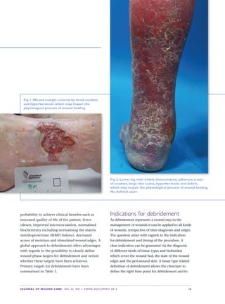
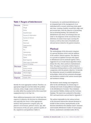
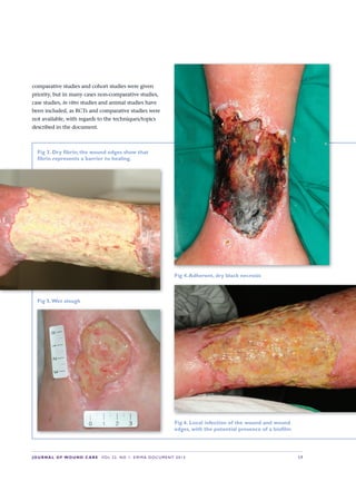
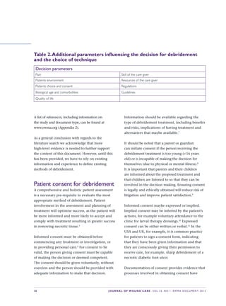
This document provides an overview of debridement, which is defined as removing necrotic material, eschar, devitalized tissue, infected tissue, hyperkeratosis, slough, pus, hematomas, foreign bodies, debris, bone fragments or any other type of bioburden from a wound to promote healing. Debridement is a central part of wound management and can be applied to all wound types. Indications for debridement include the diagnosis of different tissue types covering the wound bed, state of the wound edges and periwound skin. The document discusses various debridement methods including mechanical, autolytic, enzymatic, larval therapy, direct and indirect technologies, and surgical deb


![Hypothalamus short ppt by Dr. Neha [PT].pptx](https://cdn.slidesharecdn.com/ss_thumbnails/hypothalamusbydr-260124145759-b9f94a93-thumbnail.jpg?width=640&height=640&fit=bounds)


