Download to read offline


![Pan Pacific Clinical Practice Guideline for the Prevention and Management of Pressure Injury
page 29
• medication use (e.g. sedatives, hypnotics and analgesics), and
• surgery.
5.2.2 Reduction in tissue tolerance
Tissue tolerance is the ability of the skin and its supporting structures to tolerate the effects of pressure
by acting as a cushioning and transferring pressure loads from the skin surface to the skeleton. In the
presence of pressure, both extrinsic and intrinsic factors influence tissue tolerance.1
Extrinsic factors
Shear, friction and moisture all impact on the ability of skin to tolerate pressure. Shear is a mechanical
force created from a parallel (tangential) load that causes the body to slide against resistance between
the skin and a contact surface. The outer layers of the skin (the epidermis and dermis) remain stationary
while deep fascia moves with the skeleton, creating distortion in the blood vessels and lymphatic
system between the dermis and deep fascia. This leads to thrombosis and capillary occlusion. Shear is a
mechanical force created from parallel loads that cause the body to slide against resistance between
the skin and a contact surface. The outer layers of the skin (the epidermis and dermis) remain stationary
while deep fascia moves with the skeleton, creating distortion in the blood vessels between the dermis
and deep fascia. This leads to thrombosis and capillary occlusion.1, 6, 7
Friction is a mechanical force that occurs when two surfaces move across one another, creating
resistance between the skin and contact surface that leads to shear.1, 4, 6
Moisture alters resilience of the epidermis to external forces by causing maceration, particularly when
the skin is exposed for prolonged periods. Moisture can occur due to incontinence, wound exudate and
perspiration. Some forms of moisture, particularly faecal incontinence, create added risks by exposing
the skin to bacteria and enzymes that raise the skin pH.1, 4, 6
Intrinsic factors
Intrinsic factors reduce the skin’s tolerance through impacting its supporting structures, vascular and
lymphatic system.
Advancing age is the demographic characteristic most associated with an increased risk of PI. Patients
aged over 65 years are at a greater risk, and the risk increases in those aged over 75 years.1, 4, 6
While
some studies have identified men and Caucasians at increased risk,1
there is no consensus on the role of
these demographic characteristics.4, 25
In a SR on risk factors in patients with SCI, males were more likely
to develop PI in the chronic phase of SCI (eight studies, odds ratio [OR] 1.3, 95% CI 1.1 to 1.7). Age and
ethnicity appeared unrelated to PI risk in this population.26, 27
Chronic illnesses that influence tissue perfusion, the lymphatic system and sensation also increase PI risk.
In addition, illness and conditions that impair oxygen delivery to the tissues are also associated with an
increased PI risk. In patients in the chronic phase of SCI, past history of deep vein thrombosis, lower limb
fracture and pneumonia were all identified as risk factors for PI.27
Chronic illnesses and conditions that
impair oxygen delivery, tissue perfusion, sensation and/or lymphatic function identified as increasing PI
risk include, but are not limited to:1, 6
• diabetes mellitus,
• carcinoma,
• peripheral arterial disease,
• cardiopulmonary disease,
• lymphoedema,
• renal impairment or failure,
• low blood pressure,
• circulatory abnormalities,
• anaemia and
• smoking.](https://image.slidesharecdn.com/2012awmapanpacificguidelines-180309101051/85/2012-awma-pan_pacific_guidelines-31-320.jpg)


![page 40
Pan Pacific Clinical Practice Guideline for the Prevention and Management of Pressure Injury
baseline incomparability (e.g. level of dependence, PI risk using Norton Scale).38
The other three trials were too
small to detect significant differences.38, 39
A pooling of the studies was not heterogeneous and the results showed
significantly lower incidence of PIs associated with ONS (odds ratio [OR] 0.75, 95% CI 0.62 to 0.89, p=not reported).40
An additional trial investigated using ETF (standard formula delivering 1500 k/cal daily) in addition to a standard
hospital diet. This was compared to a routine hospital diet. Adding this trial to the meta-analysis for prevention of PI
using ONS maintained the homogeneity. Pooled results from the five studies (n=1325) showed an OR of 0.74 (95%
CI 0.62 to 0.88, p=not reported), which translated to a numbers needed to treat (NNT) of 19.25 (i.e. 19.25 patients
need to be treated with ONS to prevent one PI).40
However, acceptability of the ETF intervention was questionable,
as only 40% of the intervention population agreed to have ETF in the first week of the trial, and by the second week
this had reduced to 16%.38
The results from these low quality trials indicate that high protein ONS given twice daily
may reduce the risk of developing PIs in adults at risk of PIs.38, 40
(Level I evidence)
The NPUAP/EPUAP evidence based guideline4
included a grade A recommendation supporting the use of high
protein oral nutrition support on the same trials reported above. (Level I evidence)
The WOCNS guideline6
included a consensus based recommendation supporting the use of high protein oral
nutrition support based on the recommendation included in the NPUAP/EPUAP guideline. (Consensus)
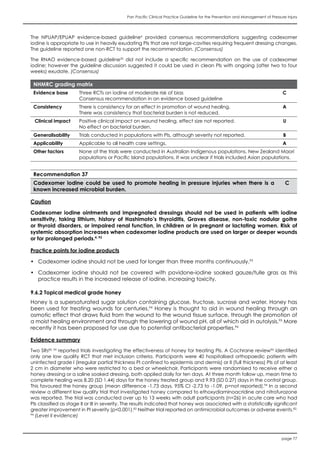
NHMRC grading matrix
Evidence base Two SRs and one MA A
Consistency Small trials did not find any significant effects B
Clinical impact Small clinical impact C
Generalisability Trials in populations with high risk of PI A
Applicability Applicable to all health care settings. A
Other factors None of the trials were conducted in Australian Indigenous populations, New Zealand
Maori populations, Pacific Island populations or Asian populations.
Recommendation 10
Provide high protein oral nutritional supplements in addition to a regular diet for patients at a
high risk of pressure injury.
B
Practice points for nutrition
• To reduce the risk of PI, patients who have been identified as being malnourished or at nutritional risk
require:4
• a minimum of 30 to 35 kcal per kg body weight per day
• 1.25 to 1.5 g per kg body weight daily of protein
• 1 ml of fluid intake per kcal per day
• Patients with SCI have reduced energy needs due to decreased activity and muscle atrophy. These
patients require:41
• Paraplegic patients: 29.8 ±1.2 kcal/kg body weight per day
• Tetraplegic patients: 24.3 ± 1.1 kcal/kg body weight per day
• When determining dietary intake requirements, consider concurrent diagnoses.41
• Refer to appropriate national clinical guidelines for strategies to improve oral dietary intake.
• When the decision to use enteral feeding in a person at risk of PIs has been made, practice should
be guided by relevant evidence based guidelines.
• Consider referring patients with identified nutritional deficits or high risk of PI to a dietitian.6
6.4 Support surfaces
A support surface is a surface on which the patient is placed to manage pressure load, shear, friction
and microclimate. This includes bed, trolley and operating table mattresses; integrated bed systems;
and seat cushions. Support surfaces are designed to reduce interface pressure through increasing the
body surface area or alternating the area of the body in contact with the support surface (i.e. pressure
reduction and pressure relief).4](https://image.slidesharecdn.com/2012awmapanpacificguidelines-180309101051/85/2012-awma-pan_pacific_guidelines-42-320.jpg)


![page 64
Pan Pacific Clinical Practice Guideline for the Prevention and Management of Pressure Injury
The following pain assessment tools have been validated for use in adult populations with PIs:4, 62, 69
• Visual analogue scale (VAS)
• Wong-Baker FACES Pain Rating Scale (FRS)73
• McGill Pain Questionnaire (MPQ)74
The following pain assessment tools have been validated in child populations (not with PIs) and could
be considered appropriate for assessing PI associated pain in children:4, 72
• 0 to 10 pain rating scale
• Wong-Baker FRS73
• Face, Legs, Activity, Cry, Consolability (FLACC) scale75
• Revised-FLACC76
• Crying; Requires O2 for Saturation >95%; Increasing vital signs; Expression; Sleepless (CRIES) scale77
Evidence summary
Three SRs62, 69, 72
reported on pain scales that have been used in PI associated pain assessment. The review by
Girouard et al.69
was based on 26 studies of various design that addressed pain assessment, prevalence, incidence
and management. Pieper et al72
identified three pain assessment scales that have been used in PI assessment in
adults, as well as numerous tools appropriate for assessing PI pain in children. The third SR62
focused on assessment
and management of pain, odour and exudate associated with PIs and included 13 studies of different design
(Level II, III and IV evidence).62
Girourard et al.69
reported on pain assessment from 26 studies and concluded that prevalence of PI associated pain
was higher in studies that used a validated assessment tool compared to those using non-validated assessment
tools. Findings from the evidence suggested that patients may have preferences for types of pain assessment tools
(e.g. numerical versus pictorial scales) and accuracy of pain assessment may be increased if the patient is offered
a choice.69, 70
(Descriptive studies)
Visual analogue scale
A standard visual analogue scale (VAS) was reported as a suitable tool for measuring pain intensity associated
with PIs. One trial reported a moderate correlation between VAS and wound stage (r=0.37, p<0.01), a moderate
correlation between VAS and generalised pain (r=0.59) and a strong correlation between the VAS and Wong-Baker
FRS (r=0.90).4, 62, 69
Girouard et al.69
reported a second trial found that there is significant variability in VAS ratings when
pain is rated at the end of the FRS scale.73
(Level III evidence)
Faces Pain Rating Scales
One cross-sectional trial conducted in 44 participants (not cognitively impaired) with PIs in an acute care setting
found a strong correlation (r=0.92, p<0.01) between the Wong-Baker FRS and VAS, and that was confirmed in a
secondary analysis of the trial.62, 69
The Wong-Baker FRS was also reported to be reliable in cognitively impaired
adults.69
Girouard et al.69
included a trial that reported on the validity of the Revised Faces Ratings Scale, a pain
scale developed by the International Association for the Study of Pain and generally referred to as the Faces Pain
Scale-Revised (FPS-R).78
The trial reported statistically significant and high correlation (r=0.90) between pain intensity
and FPS-R rating.69
(Level III evidence)
McGill Pain Questionnaire
Trials on the McGill Pain Questionnaire (MPQ)74
have focused on the different descriptors and ratings of pain given
by patients with PIs. Reliability and validity of the scale for measuring PI associated pain was established in a trial of
47 patients with stage II to IV PIs. There was a strong correlation between MPQ and FRD (r=0.90).4
Studies on general
pain have also provided evidence of the tool’s internal consistency, construct validity and sensitivity.62
One trial
found the MPQ to be a time consuming tool for clinical use.69
Use of the Present Pain Intensity subscale of the MPQ
by cognitively impaired adults was suggested.69
(Level III evidence)
Pain assessment in children
A number of pain assessment tools for use in children were reported; however, none have been specifically validated
for assessing PI associated pain. The FLACC scale75
was reported to have high inter-rater reliability [r=0.9] when used
to assess post-operative pain in children.4, 72
The CRIES scale77
was reported to be highly reliable in children up to
6 months in age.4, 72
The revised-FLACC76
was reported to have medium to high intra-class correlation coefficients
(range 0.76 to 0.90).72
(Level III evidence)](https://image.slidesharecdn.com/2012awmapanpacificguidelines-180309101051/85/2012-awma-pan_pacific_guidelines-66-320.jpg)


![Pan Pacific Clinical Practice Guideline for the Prevention and Management of Pressure Injury
page 93
11. INTERVENTIONS NOT CURRENTLY RECOMMENDED
11.1 Therapeutic ultrasound
Ultrasound therapy delivers acoustic vibrations at either low (20 to 50 kilohertz [kHz]) or high (0.5 to
3.0 megahertz [MHz]) frequencies4
in either a continuous or a pulsed manner to the area under
treatment.125, 131, 132
Usually a water or gel based coupling agent is used between the wound area and
the ultrasound applicator. The benefits of ultrasound are achieved from both thermal effects and non-
thermal effects. Thermal effects, generally achieved through continuous ultrasound, are hypothesised
to increase blood flow to the area. Non-thermal effects, such as acoustic streaming and cavitation are
achieved through pulsed ultrasound.131, 132
These are variously theorised to contribute to wound healing
through enzymatic fibrinolysis; stimulation of protein synthesis; and an increase in cell proliferation that
stimulates inflammation and promotes angiogenesis. However, there is insufficient research in this area
to determine the validity of these theories. These non-thermal effects are distinguished from the use of
ultrasound for debridement.
Evidence summary
One Cochrane SR125
investigated the effectiveness of therapeutic ultrasound for healing PIs. The review updated
a previous SR53
conducted by the same research team. The SR reported three RCTs meeting the inclusion criteria,
only one of which was described as being of good quality. Two of the trials compared ultrasound therapy at a
frequency of 3 MHz to sham ultrasound. In the first, described as being of good quality, 88 patients with at least
partial thickness PIs were treated with ultrasound at a frequency of 3.28 MHz with pulse duration of 2ms and pulse
repetition of 100Hz five times per week for 12 weeks or until complete healing. There was no significant difference
in complete healing compared to patients receiving sham ultrasound (40% versus 44% of PIs, p=0.61). The relative
risk (RR)of healing at 12 weeks was not significant (RR 0.91, 95% CI 0.55 to 1.48, p=0.69). The second RCT included
40 participants with superficial PIs. Ultrasound at a frequency of 3 MHz was applied for a minimum of five minutes
to PIs up to 3 cm2
in size, with an additional one minute for each additional 0.5cm2
to a maximum of ten minutes.
The therapy was delivered three times per week and the control group received placebo ultrasound on the same
regimen. The number of PIs healed was not significantly different between groups (48% compared to 42%, p=not
reported). Relative risk of healing (mean treatment time of 34 days) was not significant (RR 1.13, 955 CI 0.57 to 2.26,
p=0.73). The findings from these trials were pooled in meta-analysis and the RR for number of PIs healed was not
significant (RR 0.97, 95% CI 0.65 to 1.45, p=0.89). In the third RCT 18 participants with SCI and PIs described as ‘skin
wounds’ received either laser (820nm laser diode), ultrasound and ultraviolet light treatment alternating for five
days per week or standard wound care (cleansing, Jelonet™ dressing and pressure care). At 12 weeks there was
no significant difference in number of PIs healed between the therapy groups and the standard wound care group.
Relative risk of healing with ultrasound and ultraviolet light therapy compared to standard therapy was 1.18 (95%
CI 0.76 to 1.83, p=0.43). Findings from this review suggested there is no benefit of using ultrasound therapy to heal
PIs.125
(Level I evidence)
The NPUAP/EPUAP4
guideline provided consensus based recommendations on the use of therapeutic ultrasound.
The guideline suggests that low frequency (40 kHz) ultrasound be considered for treating stage III and IV PIs, providing
support for the consensus opinion from a small RCT of unknown quality and two small, non-randomised trials. Use of
ultrasound is suggested for treating infected PIs,4
based on findings of one of the moderate quality trials reported in
the Cochrane review.125
(Level II evidence)
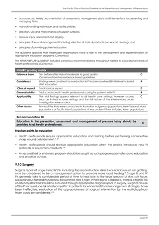
NHMRC grading matrix
Evidence base One meta-analysis at low risk of bias including 3 RCTs at low risk of bias A
Consistency Findings were consistent for no effect A
Clinical impact Low D
Generalisability Trials conducted in populations with stage I and II PIs B
Applicability The trial findings appear relevant to all health care settings; however access may
be limited.
B
Other factors None of the trials were conducted in Australian Indigenous populations, New Zealand Maori
populations or Pacific Island populations. It was unclear if trials included Asian populations.
Recommendation 52
Therapeutic ultrasound does not improve healing in stage I or II pressure injuries. A
Recommendation 53
The effectiveness of therapeutic ultrasound in treating stage III or IV pressure injuries is unknown. CBR](https://image.slidesharecdn.com/2012awmapanpacificguidelines-180309101051/85/2012-awma-pan_pacific_guidelines-95-320.jpg)


![page 98
Pan Pacific Clinical Practice Guideline for the Prevention and Management of Pressure Injury
15. REFERENCES
1. Australian Wound Management Association Inc, Clinical Practice Guidelines for the Prediction and Prevention of Pressure
Ulcers. 2001, West Leederville: Cambridge Publishing.
2. Australian Wound Management Association Inc, Standards for wound management. 2nd ed. 2010: AWMA.
3. Australian Wound Management Association Inc (AWMA), Position Document of the Australian Wound Management
Association: Bacterial impact on wound healing: From contamination to infection. 2011
4. National Pressure Ulcer Advisory Panel (NPUAP) and European Pressure Ulcer Advisory Panel (EPUAP), Prevention and
Treatment of Pressure Ulcers: Clinical Practice Guideline. 2009, Washington DC: NPUAP.
5. The Trans Tasman Dietetic Wound Care Group, Evidence based practice guidelines for the nutritional management of adults
with pressure injuries. 2011, http://daa.asn.au/wp-content/uploads/2011/09/Trans-Tasman-Dietetic-Wound-Care-Group-
Pressure-Injury-Guidelines-2011.pdf.
6. Wound Ostomy and Continence Nurses Society (WOCNS), Guideline for Prevention and Management of Pressure Ulcers.
2010, Mount Laurel (NJ): WOCNS.
7. Reger, S.; Ranganathan, V.; Orsted, H.; Ohura, T. and Gefen, A., Shear and friction in context, in Pressure Ulcer Prevention:
pressure, shear, friction and microclimate in context, L. MacGregor, Editor. 2010, Wounds International: London.
8. Merlin, T.; Weston, A.; Tooher, R.; Middleton, P.; Salisbury, J.; Coleman, K.; Norris, S.; Grimmer-Somers, K. and Hillier, S., NHMRC
levels of evidence and grades for recommendations for developers of guidelines. 2009, NHMRC: Canberra.
9. Victorian Quality Council, The Pressure Ulcer Point Prevalence Survey (PUPPS) Report 2003. 2003, Victorian Quality Council,:
Melbourne.
10. Prentice, J. and Stacey, M., Pressure ulcers: the case for improving prevention and management in Australian health care
settings. Primary Intention, 2001. 9: p. 111-120.
11. Prentice, J. and Stacey, M., European Wound Management Association Journal Evaluating Australian clinical practice
guidelines for pressure ulcer prevention., 2002. 2(2): p. 11-15.
12. Prentice, J., An Evaluation of Clinical Practice Guidelines for the Prediction and Prevention of Pressure Ulcers, in School of
Surgery and Pathology, Faculty of Medicine, Dentistry and Health Science. 2007, The University of Western Australia: Perth. p.
331.
13. Australian Government Department of Health and Ageing (DOHA), Clinical IT in aged care product trial. Trails of a system
for prevention and management of pressure ulcers. 2006, Canberra: DOHA. http://www.health.gov.au/internet/main/
publishing.nsf/content/AB2946CC847A1D16CA2573B5007ACD9C/$File/PRIME%20Product.pdf.
14. Graves, N.; Birrell, F. and Whitby, M., Modeling the economic losses from pressure ulcers among hospitalised Australians.
Wound Repair & Regeneration, 2005. 13(5): p. 462-467.
15. Gorecki, C.; Brown, J.; Nelson, E.; Briggs, M.; Schoonhoven, L.; Dealey, C.; Defloor, T.; Nixon, J. and European Quality of Life
Pressure Ulcer Project group, Impact of pressure ulcers on quality of life in older patients: a systematic review. Journal of the
American Geriatrics Society, 2009 57(7): p. 1175-83.
16. Mucous Membrane Task Force National Pressure Ulcer Advisory Panel (NPUAP). Mucosal Pressure Ulcers: An NPUAP Position
Statement. online [cited 2011 August]; NPUAP.
17. Queensland Health, Pressure Ulcer Prevention and Management Resource Guideline 2009 2008, Queensland: Queensland
Health, . http://www.health.qld.gov.au/patientsafety
18. Whitney, J.; Phillips, L.; Aslam, R.; Barbul, A.; Gottrup, F.; Lisa Gould, L.; Robson, M.; Rodeheaver, G.; Thomas, D. and Stotts, N.,
Guidelines for the treatment of pressure ulcers. Wound Rep Reg., 2006. 14: p. 663-679.
19. Institute for Clinical Systems Improvement (ICSI), Pressure ulcer prevention and treatment. Health care protocol. 2010,
Bloomington (MN): ICSI.
20. Registered Nurses’ Association of Ontario (RNAO), Assessment and management of stage I to IV pressure ulcers. 2007,
Toronto, Ontario RNAO.
21. Stechmiller, J.; Cowan, L.; Whitney, J.; Phillips, L.; Aslam, R.; Barbul, A. and et al., Guidelines for the prevention of pressure
ulcers. Wound Rep Reg., 2008. 16(2): p. 151-68.
22. Stockton, L.; Gebhardt, K. and Clark, M., Seating and pressure ulcers: Clinical practice guidelines. J Tissue Viability, 2009. 18:
p. 98-108.
23. Pace, K.; Little, B.; Styles, M. and van Lill, S., Dietetic Interventions in the Management of Adults with Pressure Ulcers 2007
Wellington: New Zealand Dietetic Association,.
24. Braden, B. and Bergstrom, N., A conceptual scheme for the study of the etiology of pressure sores. Rehabilitation Nursing,
1987. 12: p. 8-16.
25. World Union of Wound Healing Societies, Principles of best practice: minimising pain at wound dressing-related procedures.
A consensus document. 2004, London, UK: MEP Ltd.
26. Gelis, A.; Dupeyron, A.; Legros, P.; Benaim, C.; Pelissier, J. and Fattal, C., Pressure ulcer risk factors in persons with SCI: Part I:
Acute and rehabilitation stages. Spinal Cord, 2009. 47(2): p. 99-107.
27. Gelis, A.; Dupeyron, A.; Legros, P.; Benaim, C.; Pelissier, J. and Fattal, C., Pressure ulcer risk factors in persons with spinal cord
injury part 2: the chronic stage. Spinal Cord, 2009. 47(9): p. 651-61.
28. Australian Commission on Safety and Quality in Health Care (ACSQHC), National Safety and Quality Health Service Standards.
2011, Sydney: ACSQHC.
29. Australian Wound Management Association Inc (AWMA) and New Zealand Wound Care Society Inc (NZWCS), Australia and
New Zealand Clinical Practice Guideline for Prevention and Management of Venous Leg Ulcers. 2011, Australia: AWMA.
30. Braden, B. and Bergstrom, N., Braden Scale for Predicting Pressure Sore Risk. 1988. http://www2.kumc.edu/coa/Education/
GeriatricSkillsFair/Station4/BradenInstructionSheet.pdf.
31. Doreen Norton, D.; McLaren, R. and Exton-Smith, A., An Investigation of Geriatric Nursing Problems in the Hospital. . 1962,
London: National Corporation for the Care of Old People (now the Centre for Policy on Ageing). http://www.leika.ca/
filesNVIAdmin/571.pdf.
32. Waterlow, J. Waterlow Score Card. 1985, revised 2005 [cited 2011 September]. [Available from: http://www.judy-waterlow.
co.uk/downloads/Waterlow%20Score%20Card-front.pdf.
33. Pancorbo-Hidalgo, P.; Garcia-Fernandez, F.; Lopez-Medina, I. and Alvarez-Nieto, C., Risk assessment scales for pressure ulcer
prevention: a systematic review. Journal of Advanced Nursing., 2006. 54(1): p. 94-110.
34. Kottner, J.; Hauss, A.; Schluer, A. and Dassen, T., Validation and clinical impact of paediatric pressure ulcer risk assessment
scales: A systematic review. International Journal of Nursing Studies, 2011. in press.](https://image.slidesharecdn.com/2012awmapanpacificguidelines-180309101051/85/2012-awma-pan_pacific_guidelines-100-320.jpg)
![Pan Pacific Clinical Practice Guideline for the Prevention and Management of Pressure Injury
page 99
35. Moore, Z. and Cowman, S., Risk assessment tools for the prevention of pressure ulcers. Cochrane Database of Systematic
Reviews, 2008. Issue 3(CD006471).
36. Kottner, J.; Dassen, T. and Tannen, A., Inter- and intrarater reliability of the Waterlow pressure sore risk scale: a systematic
review. International Journal of Nursing Studies, 2009. 46(3): p. 369-79.
37. Watterson, C.; Fraser, A.; Banks, M.; Isenring, E.; Miller, M.; Silvester, C.; Hoevenaars, R.; Bauer, J.; Vivanti, A. and Ferguson,
M., DAA Evidence Based Practice Guidelines for the Nutritional Management of Malnutrition in Adult Patients across the
Continuum of Care. Nutrition and Dietetics 2009. 66(Suppl. 3): p. S1-S34.
38. Langer, G.; Schloemer, G.; Knerr, A.; Kuss, O. and Behrens, J., Nutritional interventions for preventing and treating pressure
ulcers. Cochrane Database of Systematic Reviews, 2003. Issue 4(CD003216).
39. Reddy, M.; Gill, S. and Rochon, P., Preventing pressure ulcers: a systematic review. JAMA, 2006. 296(8): p. 974-84.
40. Stratton, R.; Ek, A.; Engfer, M.; Moore, Z.; Rigby, P.; Wolfe, R. and Elia, M., Enteral nutritional support in prevention and treatment
of pressure ulcers: a systematic review and meta-analysis. Ageing Research Reviews, 2005. 4(3): p. 422-50.
41. Trans Tasman Dietetic Wound Care Group, Evidence Based Practice Guidelines for the Dietetic Management of Adults with
Pressure Injuries. Review 1. 2011: www.ttdwcg.org
42. Reenalda, J.; Jannink, M.; Nederhand, M. and Izerman, M., Clinical use of interface pressure to predict pressure ulcer
development: a systematic review. Assistive Technology, 2009. 21(2): p. 76-85.
43. McInnes, E.; Jammali-Blasi, A.; Bell-Syer, S.; Dumville, J. and Cullum, N., Support surfaces for pressure ulcer prevention.
Cochrane Database of Systematic Reviews, 2011. Issue 4(CD001735).
44. Wounds International, International Review. Pressure Ulcer Prevention: Pressure, Shear, Friction and Microclimate in Context.
2010, London: Wounds International.
45. Dean, S. and Young, C. Pressure reduction foam mattress replacements Part 1 What are you buying ? The Product . . in 5th
National Australian Wound Management Asssociation Conference. 2004. [Available from: http://www.health.qld.gov.au/
patientsafety/documents/whatis.pdf].
46. Committee PL/36 on Flexible Polyurethane, Australian Standard® AS2281-1993 Flexible cellular polyurethane for seat
cushioning and bedding. 1993, Homebush, NSW.: Standards Australia.
47. British Standard, Hospital bedding - Part 2: Combustion modified, flexible polyurethane, general purpose foam mattress cores
- specification, BS 5223-2. 1999.
48. Brienza, D. and Geyer, M., Understanding Support Surface Technologies. Advances in Skin & Wound Care, 2000. 13(5): p. 237-
243.
49. Thomas, S., Surgical Dressings and Wound Management. 2010, Great Britain: Medetec Publications.
50. Rithalia, S. and Kenny, L., Review Hospital bed mattresses: an overview of technical aspects. Journal of Medical Engineering
& Technology, 2003. 24(1): p. 32-39.
51. Clubb, M., Water vapour permeable materials for mattress coverings. Journal of Tissue Viability, 1998. 8(1): p. 12-14.
52. Rithalia, S., Pressure sores: which foam mattress and why? Journal of Tissue Viability, 1996. 6(11): p. 115-119.
53. Cullum, N.; Nelson, E.; Flemming, K. and Sheldon, T., Systematic reviews of wound care management: (5) beds; (6)
compression; (7) laser therapy, therapeutic ultrasound, electrotherapy and electromagnetic therapy. Health Technology
Assessment (Winchester, England), 2001. 5(9): p. 1-221.
54. Cullum, N.; Deeks, J.; Sheldon, T.; Song, F. and Fletcher, A., Beds, mattresses and cushions for pressure sore prevention and
treatment. Cochrane Database of Systematic Reviews, 2000. Issue 2(CD001735).
55. CSIRO Division of Wool Technology Leather Research Centre, Australian Medical Sheepskin. 1997, Melbourne, Vic: CSIRO.
http://www.csiro.au/files/files/p8u0.pdf
56. Junkin, J. and Gray, M., Are pressure redistribution surfaces or heel protection devices effective for preventing heel pressure
ulcers? Journal of Wound, Ostomy and Continence Nursing, 2009. 36(6): p. 602-8.
57. Tissue Viability Society (TVS), Seating and Pressure Ulcers: Clinical Practice Guideline. 2009, London: TVS.
58. Fife, C.; Yankowsky, K.; Ayello, E.; Capitulo, K.; Krasner, D.; Mulder, G. and Sibbald, R., Legal issues in the care of pressure ulcer
patients: Key concepts for healthcare providers—A consensus paper from the International Expert Wound Care Advisory
Panel. Advances in Skin & Wound Care, 2011. Nov: p. 493-507.
59. Krapfl, L. and Gray, M., Does regular repositioning prevent pressure ulcers? Journal of Wound, Ostomy and Continence
Nursing, 2008. 35(6): p. 571-7.
60. Michael, S.; Porter, D. and Pountney, T., Tilted seat position for non-ambulant individuals with neurological and neuromuscular
impairment: a systematic review. Clinical Rehabilitation, 2007. 21(12): p. 1063-74.
61. van Lis, M.; van Asbeck, F. and Post, M., Monitoring healing of pressure ulcers: a review of assessment instruments for use in
the spinal cord unit. . Spinal Cord, 2010. 48(2): p. 92-9.
62. de Laat, E.; Scholte op Reimer, W. and van Achterberg, T., Pressure ulcers: diagnostics and interventions aimed at wound-
related complaints: a review of the literature. Journal of Clinical Nursing, 2005. 14(4): p. 464-72.
63. Shea, J., Pressure sores: classification and management. Clinical Orthopaedics and Related Research, 1975. 112: p. 89-100.
64. National Pressure Ulcer Advisory Panel (NPUAP), Pressure ulcers prevalence, cost and risk assessment: consensus development
conference statement. Decubitus, 1989. 2(2): p. 24-28.
65. European Pressure Ulcer Advisory Panel (EPUAP), Pressure Ulcer Treatment Guidelines. . 1998: EPUAP.
66. Kottner, J.; Raeder, K.; Halfens, R. and Dassen, T., A systematic review of interrater reliability of pressure ulcer classification
systems. Journal of Clinical Nursing, 2009. 18(3): p. 315-36.
67. Ankrom, M.; Bennett, R.; Sprigle, S.; Langemo, D.; Black, J.; Berlowitz, D.; Lyder, C. and National Pressure Ulcer Advisory Panel
(NPUAP), Pressure-related deep tissue injury under intact skin and the current pressure ulcer staging systems. Advances in Skin
& Wound Care, 2005. 18(1): p. 35-42.
68. Panel on the Prediction and Prevention of Pressure Ulcers in Adults, Pressure Ulcers in Adults: Prediction and Prevention.
Clinical Practice Guideline, No. 3. AHCPR Publication No. 92-0047. 1992, Rockville, MD: Agency for Health Care Policy and
Research (AHCPR).
69. Girouard, K.; Harrison, M. and Van Den Kerkof, E., The symptom of pain with pressure ulcers: a review of the literature. Ostomy
Wound Management, 2008. 54(5): p. 30-40.
70. Gorecki, C.; Closs, J.; Nixon, J. and Briggs, M., Patient-reported pressure ulcer pain: A mixed-methods systematic review.
Journal of Pain and Symptom Management, 2011. 42(3): p. 443-59.
71. Dallam, L.; Smyth, C.; Jackson, B.; Krinsky, R.; O’Dell, C.; Rooney, J.; Badillo, C.; Amella, E.; Ferrara, L. and Freeman, K., Pressure
ulcer pain: assessment and quantification. J WOCN, 1995. 22(5): p. 211-218.
72. Pieper, B.; Langemo, D. and Cuddigan, J., Pressure ulcer pain: a systematic literature review and national pressure ulcer](https://image.slidesharecdn.com/2012awmapanpacificguidelines-180309101051/85/2012-awma-pan_pacific_guidelines-101-320.jpg)
![page 100
Pan Pacific Clinical Practice Guideline for the Prevention and Management of Pressure Injury
advisory panel white paper. Ostomy Wound Management, 2009. 55(2): p. 16-31.
73. Wong, D.; Hockenberry-Eaton, M.; Wilson, D.; Winkelstein, M. and Schwartz, P., Wong’s Essentials of Pediatric Nursing. 6 ed.
2001, St. Louis: Mosby Inc.
74. Melzack, R., The McGill Pain Questionnaire: Major properties and scoring methods. Pain, 1975. 1: p. 277-299.
75. Merkel, S. and et al, The FLACC: A behavioral scale for scoring postoperative pain in young children. Pediatr Nurse, 1997.
23(3): p. 293-297.
76. Malviva, S.; Voepel-Lewis, T.; Burke, C.; Merkel, S. and Tait, A., The revised FLACC observational pain tool: improved reliability
and validity for pain assessment in children with cognitive impairment. . Paediatr Anaesth, 2006. 16(3): p. 258-65
77. Krechel, S. and Bildner, J., CRIES: a new neonatal postoperative pain measurement score - initial testing of validity and
reliability. Paediatric Anaesthesia, 1995. 5: p. 53-61.
78. International Association for the Study of Pain. The Faces Pain Scale - Revised. 2001 [cited 2011 May ]; University of
Saskatachewan,. [Available from: http://www.usask.ca/childpain/fpsr/.
79. World Health Organization (WHO), Cancer pain relief and palliative care. Report of a WHO expert committee (World Health
Organization Technical Report Series, 804). 1990, Geneva, Switzerland: WHO.
80. World Health Organization (WHO), Cancer Pain Relief. With a Guide to Opioid Availability. 2 ed. 1996, Geneva: WHO. http://
whqlibdoc.who.int/publications/9241544821.pdf
81. Jacobsen, J. Topical Opioids for Pain. Fast Facts and Concepts #185. 2007 [cited 2001 May]; Medical College of Wisconsin
End of Life/Palliative Research Centre. [Available from: http://www.eperc.mcw.edu/fastFact/ff_185.htm.
82. Reddy, M.; Gill, S.; Kalkar, S.; Wu, W.; Anderson, P. and Rochon, P., Treatment of pressure ulcers: a systematic review. JAMA,
2008. 300(22): p. 2647-62.
83. Gray, M., Does oral supplementation with vitamins A or E promote healing of chronic wounds? Journal of Wound Ostomy
and Continence Nursing 2003. 30(6): p. 290-294.
84. Gray, M.; Ratliff, C. and Whitney, J., Is hyperbaric oxygen therapy effective for the management of chronic wounds? Journal
of Wound, Ostomy and Continence Nursing, 2006. 33(1): p. 21-5.
85. Gray, M. and Whitney, J., Does vitamin C supplementation promote pressure ulcer healing? J. Wound Ostomy Continence
Nurs., 2003. 30(5): p. 245-249.
86. Gray, M., Does oral zinc supplementation promote healing of chronic wounds? J. Wound Ostomy Continence Nurs., 2003.
30(6): p. 295-299.
87. Moore, Z. and Cowman, S., Repositioning for treating pressure ulcers. Cochrane Database of Systematic Reviews, 2009. Issue
2(CD006898).
88. Konig, M.; Vanscheidt, W.; Augustin, M. and Kapp, H., Enzymatic versus autolytic debridement of chronic leg ulcers: a
prospective randomised trial. Journal of Wound Care, 2005. 14(7): p. 320-3.
89. Enoch, S. and Harding, K., Wound Bed Preparation: Wound Debridement Wounds (available: http://www.medscape.com/
viewarticle/459733_6), 2003. 15(7).
90. Falabella, A.; Carson, P.; Eaglstein, W. and Falanga, V., The safety and efficacy of a proteolytic ointment in the treatment of
chronic ulcers of the lower extremity. Journal of the American Academy of Dermatology, 1998. 39(5 Pt 1): p. 737-40.
91. Moore, Z. and Cowman, S., A systematic review of wound cleansing for pressure ulcers. Journal of Clinical Nursing, 2008.
17(15): p. 1963-72.
92. Vermeulen, H.; Westerbos, S. and Ubbink, D., Benefit and harm of iodine in wound care: A systematic review. J. Hosp. Infect.,
2010. 76(3): p. 191-199.
93. Smith & Nephew. Cadexomer Iodine Dressings. 2010 [cited 2010 August]; Smith & Nephew,. [Available from: http://wound.
smith-nephew.com/au/Standard.asp?NodeId=3821.
94. Jull, A.; Rodgers, A. and Walker, N., Honey as a topical treatment for wounds Cochrane Database of Systematic Reviews,
2008. Issue 4.
95. Gethin, G. and Cowman, S., Manuka honey vs. hydrogel--a prospective, open label, multicentre, randomised controlled trial
to compare desloughing efficacy and healing outcomes in venous ulcers. Journal of Clinical Nursing., 2009. 18(3): p. 466-74.
96. Jull, A.; Walker, N.; Parag, V.; Molan, P.; Rodgers, A. and on behalf of the Honey as Adjuvant Leg Ulcer Therapy trial
collaborators, Randomized clinical trial of honey-impregnated dressings for venous leg ulcers. British Journal of Surgery, 2008.
95(2): p. 175-82.
97. White, R. and Cutting, K., Exploring the Effects of Silver in Wound Management - What is Optimal? Wounds, 2006. 18(11): p.
307-314.
98. O’Meara, S.; Al-Kurdi, D. and Ovington, L., Antibiotics and antiseptics for venous leg ulcers. Cochrane Database of Systematic
Reviews, 2010. Issue 1.
99. Haller, G.; Faltin-Traub, E.; Faltin, D. and Kern, C., Oxygen embolism after hydrogen peroxide irrigation of a vulvar abscess. Br
J Anaesth, 2002. 88: p. 597-9
100. Henley, N.; A., D.; Kaehr, D. and Clements, B., Air Embolism associated with irrigation of external fixator pin sites with hydrogen
peroxide. The Journal of Bone and Joint Surgery, 2004. 86: p. 821-822.
101. Brennan, S. and Leaper, D., The effect of antiseptics on the healing wound: a study using the rabbit ear chamber. British
Journal of Surgery, 1985. 72(10): p. 780-782.
102. Lineaweaver, W.; Howard, R.; Soucy, D.; McMorris, S.; Freeman, J.; Crain, C.; Robertson, J. and T., R., Topical antimicrobial
toxicity. Archives of Surgery., 1985. 120(3): p. 267-270.
103. Sleigh, J. and Linter, S., Hazards of hydrogen peroxide. British Medical Journal, 1985. 291(6510): p. 1706.
104. Ward, R. and Saffle, J., Topical agents in bum and wound care. Physical Therapy, 1995. 75: p. 526-5381.
105. NSW Health. NSW Health factshhet: Antibiotic use. 2005 [cited 2010 August]; NSW Health. [Available from: http://www.health.
nsw.gov.au/factsheets/general/antibiotic_use.html.
106. Meaume, S.; Ourabah, Z.; Cartier, H.; Granel-Brocard, F.; Combemale , P.; Bressieux, J. and Bohbot, S., Evaluation of a
lipidocolloid wound dressing in the local management of leg ulcers. Journal of Wound Care, 2005. 14(7): p. 329-34.
107. Bouza, C.; Saz, Z.; Munoz, A. and Amate, J., Efficacy of advanced dressings in the treatment of pressure ulcers: a systematic
review. Journal of Wound Care, 2005. 14(5): p. 193-9.
108. Bradley, M.; Cullum, N.; Nelson, E.; Petticrew, M.; Sheldon, T. and Torgerson, D., Systematic reviews of wound care
management: (2). Dressings and topical agents used in the healing of chronic wounds. Health Technology Assessment
(Winchester, England), 1999. 3(17 Pt 2): p. 1-35.
109. Heyneman, A.; Beele, H.; Vanderwee, K. and Defloor, T., A systematic review of the use of hydrocolloids in the treatment of
pressure ulcers. Journal of Clinical Nursing, 2008. 17(9): p. 1164-73.](https://image.slidesharecdn.com/2012awmapanpacificguidelines-180309101051/85/2012-awma-pan_pacific_guidelines-102-320.jpg)
![Pan Pacific Clinical Practice Guideline for the Prevention and Management of Pressure Injury
page 101
110. Reid, J. and Morison, M., Towards a consensus: classification of pressure sores. Journal of Wound Care, 1994. 3: p. 157-159.
111. Bergstrom, N.; Allman, R.; Alvarez, O.; Bennett, M.; Carlson, C.; Frantz, R.; Garber, S.; Jackson, B.; Kaminski, M.; Kemp, M.;
Krouskop, T.; Lewis, V.; Maklebust, J.; Margolis, D.; Marvel, E.; Reger, S.; Rodeheaver, G.; Salcido, R.; Xakellis, G. and Yarkony,
G., Treatment of Pressure Ulcers. Clinical Practice Guideline, No. 15. AHCPR Publication No. 95-0652. . 1994, Rockville, MD:
U.S. Department of Health and Human Services, Public Health Service, Agency for Health Care Policy and Research,. http://
http://www.ahcpr.gov.
112. Ubbink, D.; Wester bos, S.; Nelson, E. and Vermeulen, H., A systematic review of topical negative pressure therapy for acute
and chronic wounds. British Journal of Surgery, 2008. 95(6): p. 685-92.
113. Vikatmaa, P.; Juutilainen, V.; Kuukasjarvi, P. and Malmivaara, A., Negative pressure wound therapy: a systematic review on
effectiveness and safety. European Journal of Vascular and Endovascular Surgery, 2008. 36(4): p. 438-48.
114. Xie, X.; McGregor, M. and Dendukuri, N., The clinical effectiveness of negative pressure wound therapy: a systematic review.
Journal of Wound Care, 2010. 19(11): p. 490-5.
115. Van Den Boogaard, M.; De Laat, E.; Spauwen, P. and Schoonhoven, L., The effectiveness of topical negative pressure in the
treatment of pressure ulcers: A literature review. European Journal of Plastic Surgery, 2008. 31(1): p. 1-7.
116. Sullivan, N.; Snyder, D.; Tipton, K.; Uhl, S. and Schoelles, K., Negative Pressure Wound Therapy Devices Technology Assessment
Report. 2009, Rockville, MD: Agency for Healthcare Research and Quality (AHRQ),. http://www.ahrq.gov/clinic/ta/
negpresswtd/npwtd02.htm.
117. Junger, M.; Arnold, A.; Zuder, D.; Stahl, H. and Heising, S., Local therapy and treatment costs of chronic, venous leg ulcers with
electrical stimulation (Dermapulse): a prospective, placebo controlled, double blind trial. Wound Repair & Regeneration.,
2008. 16(4): p. 480-7.
118. Gardner, S.; Frantz, R. and Schmidt, F., Effect of electrical stimulation on chronic wound healing: a meta-analysis. Wound
Repair and Regeneration, 1999. 7(6): p. 495-503.
119. Watson, T. Electrical therapy on the web: an educational resource: contraindications. 2010 [cited 2011 August]; Physio Med.
[Available from: http://www.electrotherapy.org/modalities/contragrid.htm.
120. McGaughey, H.; Dhamija, S.; Oliver, L.; Porter-Armstrong, A. and McDonough, S., Pulsed electromagnetic energy in
management of chronic wounds: a systematic review. Physical Therapy Reviews, 2009. 14(2): p. 132-46.
121. Ravaghi, H.; Flemming, K.; Cullum, N. and Olyaee Manesh, A., Electromagnetic therapy for treating venous leg ulcers.
Cochrane Database of Systematic Reviews, 2006. Issue 2(CD002933).
122. Aziz, Z.; Flemming, K.; Cullum, N. and Olyaee Manesh, A., Electromagnetic therapy for treating pressure ulcers. Cochrane
Database of Systematic Reviews, 2010. Issue 11(CD002930).
123. Dbaly, J., Pulsed electromagnetic field therapy: The best option for many patients. . Swiss Medical Tribune, 2005.
124. Energy Medicine Developments, The EnerMed therapy. 2004, EMD.
125. Akbari Sari, A.; Flemming, K.; Cullum, N. and Wollina, U., Therapeutic ultrasound for pressure ulcers. . Cochrane Database of
Systematic Reviews., 2006. Issue 3(CD001275).
126. Soban, L.; Hempel, S.; Munjas, B.; Miles, J. and Rubenstein, L., Preventing pressure uclers in hospitals: A systematic review of
nurse-focussed quality improvement interventions. Joint Commission Journal on Quality and Patient Safety, 2011. 37(6): p.
245-16AP(-228).
127. Dunk, A. and Arbon, P., Is it time for a new descriptor ‘pressure injury’: a bibliometric analysis. Wound Practice and Research,
2009. 17(4): p. 201-207.
128. Fleurence, R., Cost-effectiveness of pressure-relieving devices for the prevention and treatment of pressure ulcers. Int. J.
Technol. Assess. Health Care, 2005. 21(3): p. 334-341.
129. Legood, R. and McInnes, E., Pressure ulcers: guideline development and economic modelling. Journal of Advanced Nursing.,
2005. 50(3): p. 307-14.
130. Clark, M., Pressure ulcers and quality of life. Nurs Stand., 2002. 16: p. 74-80.
131. Al-Kurdi, D.; Bell-Syer, S.E. and Flemming, K., Therapeutic ultrasound for venous leg ulcers. Cochrane Database of Systematic
Reviews, 2008. Issue 1(CD001180).
132. Robinson, V.; Brosseau, L.; Peterson, J. and et al, Therapeutic ultrasound for osteoarthritis of the knee. Cochrane Database of
Systematic Reviews, 2001. Issue 3(CD003132).
133. Kopera, D.; Kokol, R.; Berger, C. and Haas, J., Does the use of low-level laser influence wound healing in chronic venous leg
ulcers? Journal of Wound Care, 2005. 14(8): p. 391-4.
134. Robson, M.; Phillips, T.; Falanga, V.; Odenheimer, D.; Parish, L.; Jensen, J. and Steed, D., Randomized trial of topically applied
repifermin (recombinant human keratinocyte growth factor-2) to accelerate wound healing in venous ulcers. Wound Repair
& Regeneration., 2001. 9(5): p. 347-52.
135. da Costa, R.; F., R.J.; Aniceto, C. and Mendes, M., Double-blind randomized placebo-controlled trial of the use of granulocyte-
macrophage colony-stimulating factor in chronic leg ulcers. American Journal of Surgery, 1997. 173(3): p. 165-8.
136. da Costa, R.; F., R.J.; Aniceto, C. and Mendes, M., Randomized, double-blind, placebo-controlled, dose- ranging study of
granulocyte-macrophage colony stimulating factor in patients with chronic venous leg ulcers. Wound Repair & Regeneration,
1999. 7(1): p. 17-25.
137. Mölnlycke Health Care. Xelma What is the extracellular matrix? 2009 [cited 2010 October]; Mölnlycke Health Care, . [Available
from: http://www.xelma.com/en/What-is-Xelma/What-is-the-extracellular-matrix/.
138. Falanga, V.; Carson, P.; Greenberg, A.; Hasan, A.; Nichols, E. and McPherson, J., Topically applied recombinant tissue
plasminogen activator for the treatment of venous ulcers. Preliminary report. Dermatologic Surgery., 1996 22(7): p. 643-4.
139. Black, J.; Edsberg, L.; Baharestani, M.; Langemo, D.; Goldberg, M.; McNichol, L.; Cuddigan, J. and National Pressure Ulcer
Advisory Panel (NPUAP), Pressure Ulcers: Avoidable or Unavoidable? Results of the National Pressure Ulcer Advisory Panel
Consensus Conference. Ostomy Wound Management, 2011. 57(2): p. 24-37.
140. Prentice, J.; Stacey, M. and Lewin, G., An Australian model for conducting pressure ulcer prevalence surveys. . Prim Intention,
2003. 11(2): p. 87-88,90-91,93-96,98-100,102-109.
141. Wilson, J.; O’Donnell, M.; McAuliffe, L.; Nay, R. and Pitcher, A., Assessment of Pain in Older Adultis with Dementia in Acute, Sub
Acute and Residential Care. 2008, Canberra: Australian Centre of Evidence Based Aged Care (ACEBAC) and Royal College
of Nursing Australia (RCNA).](https://image.slidesharecdn.com/2012awmapanpacificguidelines-180309101051/85/2012-awma-pan_pacific_guidelines-103-320.jpg)


This document provides a summary of the Pan Pacific Clinical Practice Guideline for the Prevention and Management of Pressure Injury. The guideline was developed by an international team to provide evidence-based recommendations for assessing and preventing pressure injuries across various healthcare settings. It aims to optimize pressure injury prevention, assessment, and management. The guideline includes recommendations on risk assessment, prevention interventions like support surfaces and positioning, wound assessment and monitoring, pain management, and treatment interventions.


