This document is a handbook on medical emergencies published by the Department of Medicine at Sarawak General Hospital. It was compiled by Dr. Soo Hua Huat with contributions from other doctors and specialists. The handbook provides guidelines on managing various medical emergencies and is intended to be a quick reference for doctors, especially more junior doctors. It covers both common and less common emergency conditions. The handbook aims to give practical treatment and management advice rather than detailed clinical or pathological information.

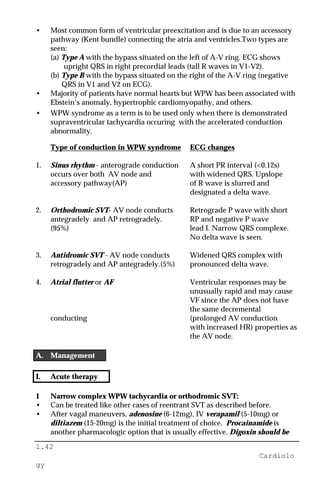
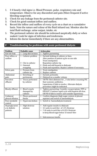
![1.5
Cardiolo
gy
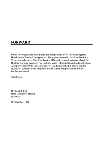
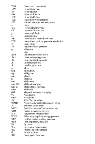
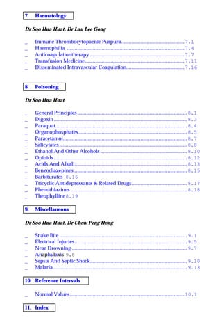
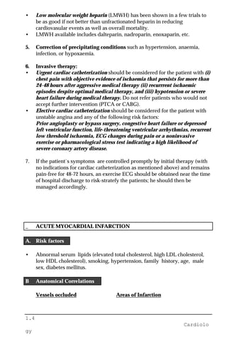
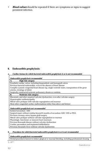
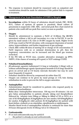
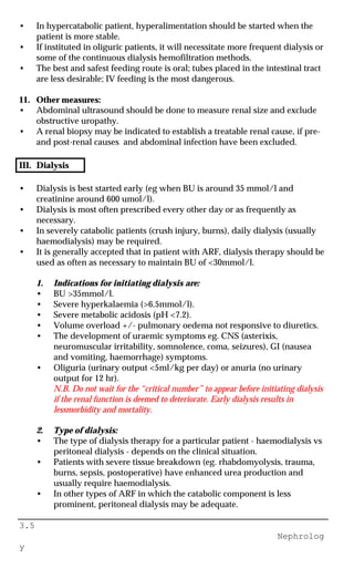
Anterior descending artery Anterior wall of the left ventricle (LV),
anterior part of the septum (affect
intraventricular conduction)
Circumflex artery Lateral or inferior walls of the LV &
posterior wall of LV
Right coronary artery Posterior and inferior surface of the LV,
posterior part of septum, right ventricle
(RV). A-V node in 90% & SA node in
60% of cases (thus inferior and posterior
infaction may be associated with A-V
conduction disturbances)
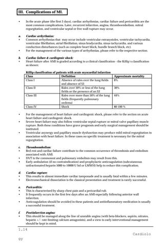
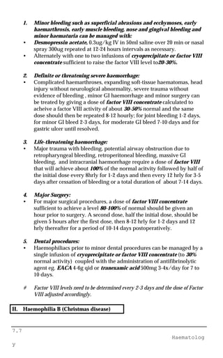
C. Clinical Features
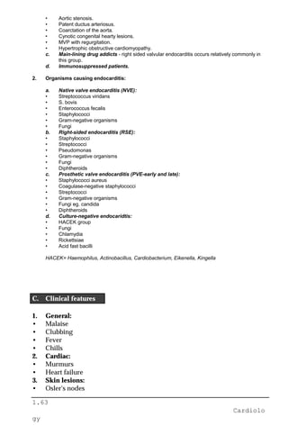
• Pain - characteristically retrosternal, > 20 minutes, radiation to the arms,
neck or jaw. Severe and crushing in nature, not relieved by rest or GTN.
May be epigastric, radiating to the back.
• Symptoms frequently associated with sweating, nausea, vomiting,
dyspnoea, weakness, apprehension, restlessness and anxiety.
• Rarely painless, masquarading as acute left ventricular failure, syncope,
CVA or unexplained shock
• 25% are asymptomatic and detected on routine ECG.
• Non-specific physical signs - pallor, sweating, irregular pulse, hypotension,
and 4th heart sound may be present.
D. Investigations
1. Electrocardiogram (ECG):
• ECG provides valuable information on the size, extent, age of the MI and
some anatomical correlations of vessels occluded.
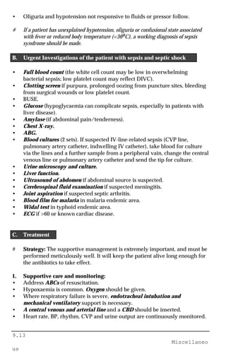
a. Q wave infarction:
ECG changes Onset Duration
ST elevation within minutes days or weeks
or hours
T inversion immediate or hours several months
Q wave hours or days remain indefinitely
(Q wave - _1mm wide[0.04s] and/or _2mm deep [0.2mV] or >25% of the
amplitude of the R wave)
b. Non-Q wave infarction:
• Changes are prolonged ST depression and T inversion.](https://image.slidesharecdn.com/sarawakhandbookcompiledbyllyl-150521205442-lva1-app6891/85/Sarawak-handbook-18-320.jpg)
![1.9
Cardiolo
gy
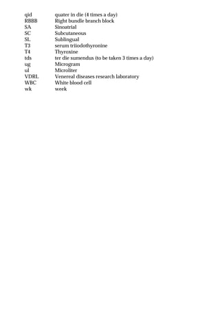
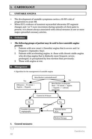
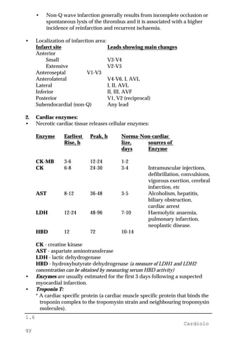
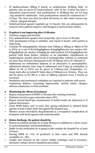
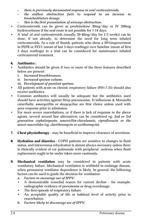
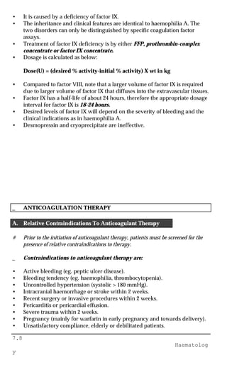
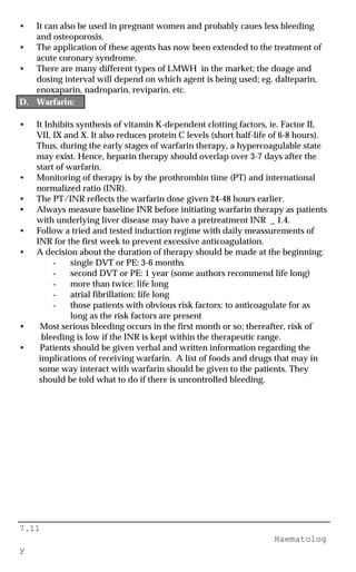
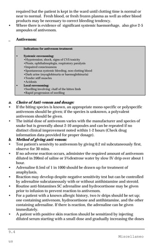
• Sublingual nitroglycerin (0.3-0.5mg) - should be given to patient in the
absence of hypotension repeated every 5 minutes till 3-4 doses have been
given.
• Morphine 5-10mg IV slow bolus with antiemetic.
• Patients who have recurrence of symptoms after given SL GTN and
morphine should be started on IV GTN.
5. Regular leg exercises and low dose heparin (eg. subcutaneous 5,000 units
bd) to prevent deep vein thrombosis.
6. Sedation with small oral doses of diazepam or lorazepam.
7. Diet and bowel care - for the first day after MI, diet should be liquid or soft,
stool softeners or mild laxatives are routinely given.
8. Potassium levels should be maintained at 4-5 mmol/l.
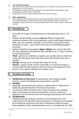
II Specific management to reduce infart size and to improve mortality
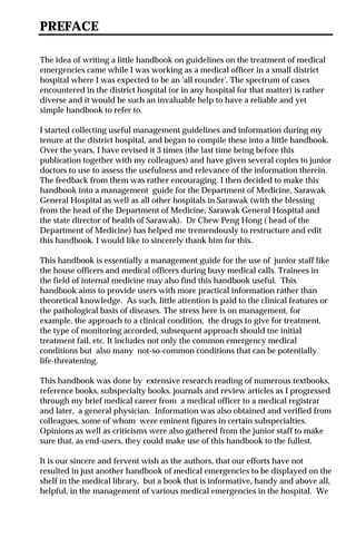
1. Thrombolytic therapy:
• Administration of fibrinolytic agents can achieve early reperfusion in 50-
70% of patients (compared with a spontaneous reperfusion rate of < 30%)
and has been shown to reduce the extent of ventricular damage, and
mortality rates associated with myocardial infarction.
_ All patients fulfilling the following criteria without contraindication
should be given thrombolytic therapy.
a. Clinical:
• Chest pain or chest-pain-equivalent syndrome consistent with acute
myocardial infarction _ 12 hours from symptom onset with:
b. ECG (either one):
• _ 1 mm ST elevation in _ 2 contiguous limb leads.
• _ 2 mm ST elevation in _ 2 contiguous precordial leads.
• New left bundle branch block.
• True posterior MI (Tall R wave in V1 with ST depression; exclude other
causes of tall R wave [eg. RBBB, RVH, WPW syndrome]; right
ventricular infarction[ST elevation V4R]).
# Cardiogenic shock: emergency catheterization and revascularization if
possible; consider thrombolysis if catheterization not immediately available.
# Age is not a limiting factor - can be given up to 80 yo or older if benefit-to-risk
ratio seems favourable.
# Should be avoided in patient with ST segment depression without
concomitant ST segment elevation or with normal ECG.](https://image.slidesharecdn.com/sarawakhandbookcompiledbyllyl-150521205442-lva1-app6891/85/Sarawak-handbook-22-320.jpg)

![1.20
Cardiolo
gy
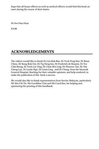
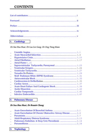
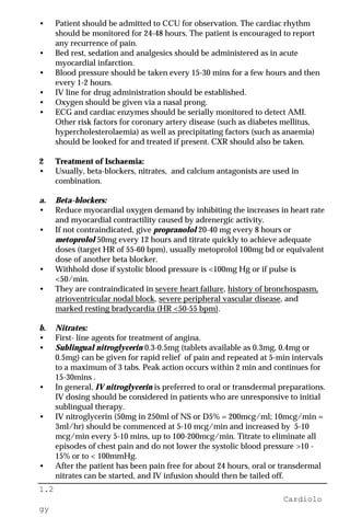
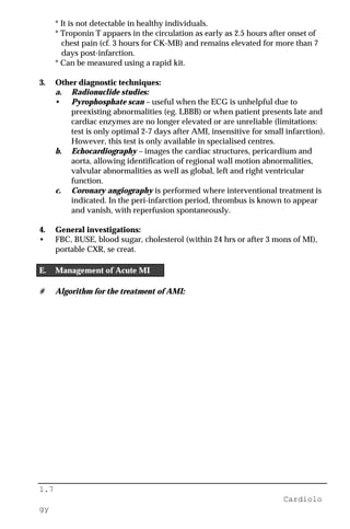
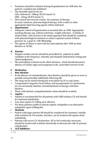
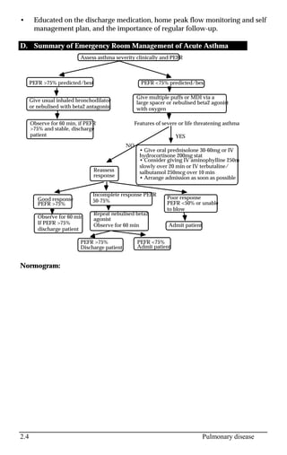
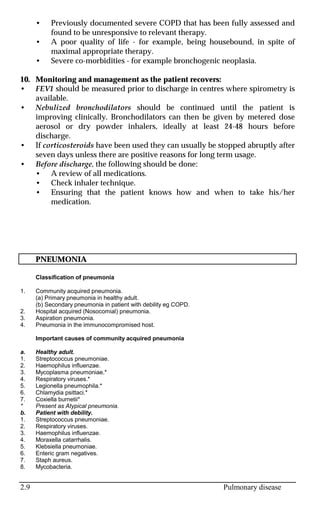
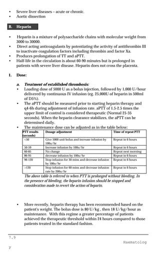
• Hypertensive crisis is defined as a substantial acute increase in blood
pressure, usually with diastolic blood pressure over 120 mmHg with or
without end-organ damage.
• It can be further classified into:
(i) Hypertensive emergencies - increased blood pressure with evidence of
end-organ damage or dysfunction.
• End-organ manifestations include [1] retinal eg. papilloedema [2]
cardiac eg. pulmonary oedema, myocardial ischaemia such as unstable
angina or infarction [3] neurological eg. severe headache, mental
status changes, seizure, coma and [4] renal eg. acute renal failure.
(ii) Hypertensive urgencies - elevation of blood pressure to a level which
may be potentially harmful, but without signs, symptoms or other
evidence of end-organ dysfunction.
A. Management
1. General principles:
• In hypertensive emergencies, blood pressure control should be
accomplished within a few hours to reduce the risk of permanent damage
or death (diastolic of 100-110 mmHg may be adequate for the first 24
hours). IV antihypertensive agents should be used.
• In hypertensive urgencies, blood pressure control can be accomplished
more slowly with oral antihypertensive agents within 24-48 hrs to a
diastolic level of 100-110 mmHg initially. Excessive or rapid decreases in BP
should be avoided to minimize the risk of cerebral hypoperfusion or
coronary insufficiency.
• BUSE, creatinine, urinalysis, CXR and ECG should be performed urgently.
• Drugs of choice for management:
a. Coronary artery disease and heart failure: IV nitroprusside or
nitroglycerin
b. Pheochromocytoma: IV phentolamine or alpha-blocker eg. prazosin
c. Aortic dissection: IV beta-blockers or labetalol +/- nitroprusside
d. Pulmonary oedema: IV frusemide, IV nitroprusside, ACE inhibitors
e. Hypertension in pregnancy: Hydralazine, labetalol and magnesium
sulphate
f. Stroke: Beta-blockers, diuretics or ACE inhibitors.
• Sublingual nifedipine SHOULD BE AVOIDED.
• IV diazoxide should be discouraged.
2. Oral antihypertensive agents:
• Can be used in patients with hypertensive crisis when urgent but not
immediate reduction of BP is indicated. Combination therapy is necessary
for most cases when diastolic blood pressure is > 110 mmHg.
• Beta-blockers eg. atenolol 100mg with or without diuretics, or oral
captopril 12.5mg with or without diuretics may be all that is required. If](https://image.slidesharecdn.com/sarawakhandbookcompiledbyllyl-150521205442-lva1-app6891/85/Sarawak-handbook-33-320.jpg)
![1.24
Cardiolo
gy
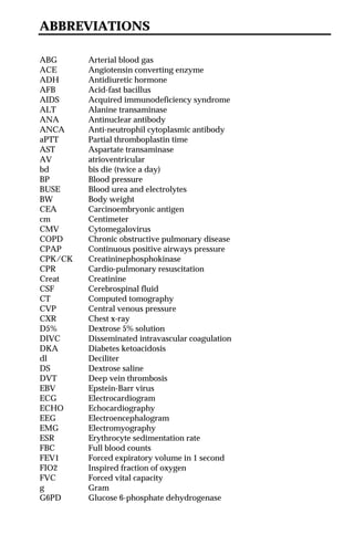
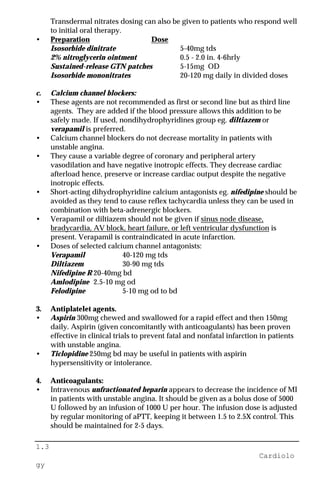
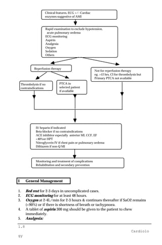
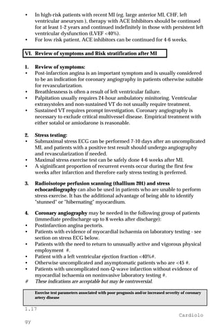
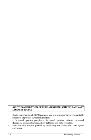
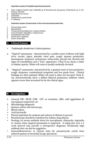
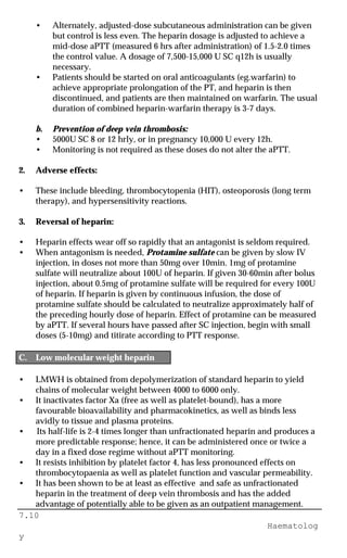
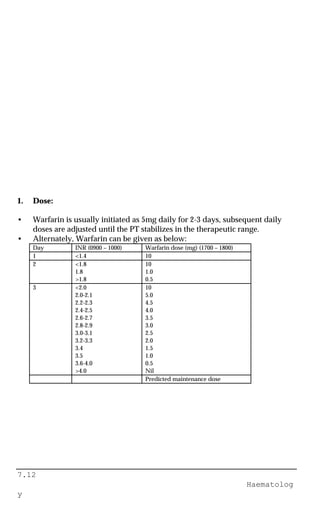
2. Less common - cardiomyopathy, pericarditis, sick sinus syndrome, atrial
myxoma, endocarditis, chronic lung disease, atrial septal defect, acute
alcohol intoxication, WPW syndrome, digoxin toxicity and lone AF.
B. Clinical Features
• May be either chronic or paroxysmal.
• Irregularly irregular pulse, S1 of variable intensity, pulse deficit (rate at
apex is greater than at radial artery).
• ECG: chaotic baseline with no P waves, irregularly irregular but normally
shaped QRS complex (unless concomitant BBB or preexcitation syndrome)
at a rate of 160-200/min. AF with regular ventricular response may suggest
digoxin toxicity.
• Complications of atrial fibrillation are hypotension, cardiac failure,
systemic embolism and troublesome palpitations.
C. Investigations
• BUSE, ECG, CXR, cardiac enzymes, thyroid function, calcium and Echo.
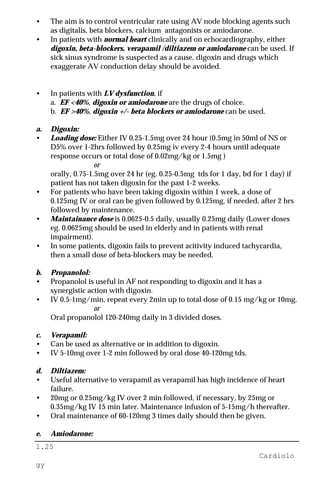
D. Treatment
_ Aims of therapy:
• Control of the ventricular rate using AV node blocking drugs (digitalis, beta
blockers, calcium channel blockers and occasionally amiodarone).
• Reversion to sinus rhythm using membrance stabilizing agents [(class 1a -
quinidine, procainamide, disopyramide), class 1c (propafenone, flecanide)
and class III (sotalol, amiodarone)] or cardioversion.
• Prophylatic therapy to prevent recurrence.
• Anticoagulation to prevent embolization.
1. Emergency synchronised cardioversion is required in patients with rapid
ventricular response (VR >200/min) and acute haemodynamic
deterioration (eg. patients with angina, hypotension, dyspnoea, or heart
failure).
• If initial cardioversion is unsuccessful, procainamide or amiodarone can be
given to facilitate further cardioversion attempts.
• Do not attempt to cardiovert patients with known chronic AF & a known
progressive cause (eg. MS), in which case, rapid rate control with drugs
should be achieved.
• Patients undergoing emergency cardioversion should receive anticoagulant
therapy for 2-3 weeks after conversion unless there is a contraindication
(anticoagulation may not be necessary if AF is <24-48 hr duration).
2. Haemodynamically stable acute AF and Chronic AF:](https://image.slidesharecdn.com/sarawakhandbookcompiledbyllyl-150521205442-lva1-app6891/85/Sarawak-handbook-37-320.jpg)
![1.29
Cardiolo
gy
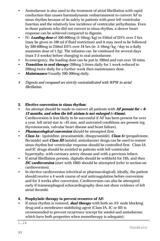
• Atrial flutter rarely occurs without heart disease eg. rheumatic and
ischaemic heart disease, cardiomyopathy, thyrtoxicosis, valvular disease,
septal defect and digoxin toxicity.
B. Clinical Features
• Atrial flutter is a rhythm that originates from a small area within the atria.
• ECG: P waves are replaced by flutter waves resulting in ‘saw-tooth’,or
‘corrugated’ pattern. Flutter may sometimes be obscured by the T wave.
Carotid sinus massage will usually slow the ventricular rate and flutter
waves may then be more obvious. Ventricular rate is often regular but may
vary depending on the degree of A-V block present and is a fraction of
atrial rate 1:2, 1:3, etc. Rarely, 1:1 conduction occurs. Atrial rate is usually
between 250-350/min.
# When ventricular rate is 150/min, suspect atrial flutter with 2:1 block until
proven otherwise.
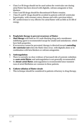
C. Managment
1. DC cardioversion in haemodynamically unstable patients, patients with
symptoms and signs of ischaemia, VR >200/min, known or suspected to
have WPW syndrome. Synchronized DC shock of 25-50J is usually
adequate.
2. Control of the ventricular rate using AV node blocking drugs (digitalis,
beta blockers,calcium antagonists or amiodarone):
• Digoxin often converts atrial flutter to AF and slows the ventricular
response.
• Beta-blockers (eg. IV propranolol or esmolol, oral propranolol or
metoprolol) slows the ventricular response rate by causing AV blockade.
• IV verapamil or diltiazem may reduce the ventricular response but
conversion to sinus rhythm rarely occurs.
• IV amiodarone has also been shown to slow the ventricular rate as
effectively as digoxin.
• Adenosine produces transient AV block and can be used to reveal flutter
waves if the diagnosis of the arrhythmias is in doubt but it generally will
not terminate the atrial flutter and can provoke atrial fibrillation.
# Verapamil and digoxin are contraindicated in patients with WPW syndrome
presenting with atrial flutter or AF.
3. Reversion to sinus rhythm using membrance stabilizing agents [(class 1a -
quinidine, procainamide, disopyramide), class 1c (propafenone, flecanide)
and class III (sotalol, amiodarone)] or DC cardioversion:](https://image.slidesharecdn.com/sarawakhandbookcompiledbyllyl-150521205442-lva1-app6891/85/Sarawak-handbook-42-320.jpg)
![1.59
Cardiolo
gy
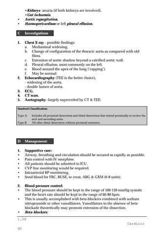
IV propanolol 0.5-1mg q5min until the heart rate is between 60-80 bpm or
maximal dose of 0.15mg/kg followed later by oral medication.
or
Labetolol either as IV infusion or bolus
[Infusion - Mix 200mg in 200 ml of D5% and run at 1-2mg/min (1-
2ml/min). When goal blood pressure is achieved and BP is stable (usually
after 50-200mg total amount infused), the infusion can be stopped and oral
labetolol be started at a dose of 100-200mg 12hrly.
Bolus - IV 50mg over 1-5 min and can be repeated every 5-10min till
maximum dose of 300mg or until desired BP is achieved. Maximum
response occur in 5-10 min and may last for up to 6 hours. BP should be
checked 5-10 mins after each IV boluses. Oral labetolol can then be started.]
or
IV metoprolol 5-10mg q6h.
• Sodium nitroprusside: (mix 50mg in 250ml of D5%= 200mcg/ml;
10mcg/min = 3 ml/hr) start at 0.5mcg/kg/min and titrate until the
desirable blood pressure has been achieved. Average effective dose is
3mcg/kg/min (range from 0.5-10mcg/kg/min).
# If sodium nitroprusside is not available, IV or oral hydralazine, IV GTN or oral
nifedepine can be used instead.
• Hypotension may be due to haemorrhage or cardiac tamponade. When
hypotension occurs, stop all antihypertensive agents & resuscitate patient
with IV fluid.
3. Definitive therapy:
a. Type A: Emergency surgery is indicated.
b. Type B: Medical therapy for stable patients, with surgery reserved for those
with any of the following:
• Appearance of aortic regurgitation.
• Pericardial tamponade.
• Leakage of the aneurysm – rupture or impending rupture.
• Occlusion of a major systemic artery – major organ or limb ischaemia.
• Persistence of severe pain or hypertension.
• Progressive enlargement of the aneurysm.
• Saccular aneurysm formation.
• Dissecting aneurysm in Marfan`s syndrome.
4. Chronic therapy:
• In patients who are treated medically or surgically, chronic therapy with a
beta-blocker is indicated. Those being treated medically require chronic
vasodilator therapy as well. Systolic BP should be aim at 130-140 mmHg.
• Patient will require CT performed once or twice/year.
_ CARDIAC TAMPONADE.
• Cardiac tamponade occurs when a pericardial effusion causes
haemodynamically significant cardiac compression.](https://image.slidesharecdn.com/sarawakhandbookcompiledbyllyl-150521205442-lva1-app6891/85/Sarawak-handbook-72-320.jpg)


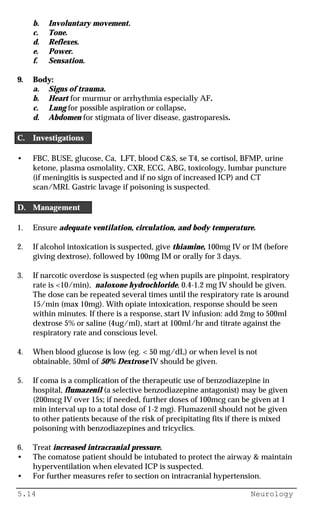
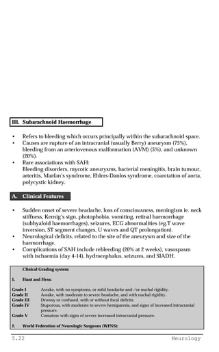
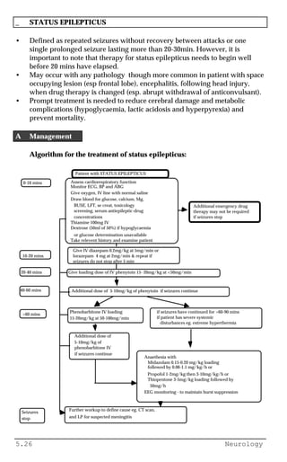

![5.30 Neurology
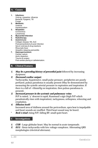
• (i) deterioration of conscious level (ii) dilated, unreactive pupil ipsilateral to
the mass [3rd nerve palsy] (iii) hemiparesis on either side [false localising
sign- produce weakness on the same side].
c. Tonsillar herniation.
• Caused by a subtentorial expanding mass causing herniation of the
cerebellar tonsils through the foramen magnum.
• (i) depression of conscious level (ii) repiratory irregularities or apnoea (iii)
neck stiffness and head tilt.
B. Management
• Neurosurgical intervention is the definitive treatment for some focal causes
and medical treatment is helpful for increased ICP following ischaemic,
anoxic or metabolic brain necrosis.
1. General measures:
• Patient should be placed in bed with the head elevated 30 degrees and in
the midline. Stimulation by family and staff (eg turning the head,
suctioning) should be kept to a minimum.
• Avoid hypotonic IV solutions or fluids that contain large amounts of free
water (eg 5%D/W ).
• Restrict fluid to 1000ml of normal saline/m2 body surface per day and
monitor BP, serum osmolality and urine output.
2. Osmotic agents:
• This can be initiated with mannitol, glycerol or other agents.
• Mannitol given in 0.5-1.0 g/kg (eg. 100-200ml of 20% over 15-30 min) every
6 hourly for 24-72 hours.
• It draws water from the brain tissue compartment into the systemic
vascular compartment. The effect starts within minutes and lasts for several
hours.
• The effect on both urine output and ICP is extended with a loop diuretic. IV
frusemide 40-80 mg 6hourly may be given.
• Indwelling bladder catheter is necessary. Monitor vital signs, electrolytes,
BU and osmolality (aim at 300-320mOsm/liter) frequently.
• Usually used in anticipation of more definitive treatment.
• If the ICP is stabilized with continuous hyperosmolar therapy, this
treatment is best withdrawn slowly and with close neurological
observation.
• Complications included (i) rebound increase in ICP (ii) acute intravascular
volume expansion with pulmonary oedema or congestive cardiac failure
(iii) dehydration and hypernatraemia.
3. Hyperventilation:
• Produces a fall in arterial PCO2 thereby reducing cerebral blood flow.
• Hyperventilation is an excellent acute treatment, but the effect on ICP is not
well-sustained.
• Patient should be sedated, paralysed and ventilated.
• Maintained PaCO2 at 25-30mm Hg.](https://image.slidesharecdn.com/sarawakhandbookcompiledbyllyl-150521205442-lva1-app6891/85/Sarawak-handbook-165-320.jpg)
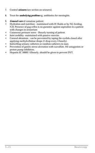
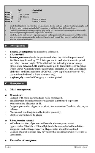
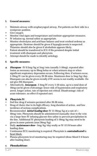
![5.39 Neurology
• In cases associated with withdrawal of dopamine agonists (eg. L-dopa,
bromocriptine) reinstitution of dopaminergic therapy and more gradual
withdrawal should be undertaken.
• In cases following psychotropic therapy, Electro Convulsive therapy might
be indicated in stubborn cases.
3. Re-treatment strategies:
• If neuroleptics are needed, institute treatment as long as possible after an
episode of NMS. Try a neuroleptic from a different family (if possible, a
low-potency agent). Start with a low dose in hospital setting and gradually
increase the dose. The patients should be monitored clinically and also with
CPK levels.
_ GUILLAIN BARRE SYNDROME (ACUTE IDIOPATHIC
DEMYELINATING POLYRADICULONEURITIS [AIDP])
A. Antecedent events / associated diseases
• Acute infectious disease: Campylobacter jejuni, viral (viral exanthems,
CMV, EBV, HIV), myocoplasma
• Surgery.
• Immunization.
• Hodgkin's lymphomas.
• Systemic lupus erythematosus.
• Thrombolytic agents (rare).
B. Clinical features
• Usually preceded by acute illness, as mentioned above.
• Mainly motor polyneuropathy, often progressive and ascending over 3
days to 4 weeks but it may come on rapidly and affect all 4 limbs
simultaneously. Proximal muscle may be more affected.
• Trunk, respiration and cranial nerves (especially VII) may be affected.
• Sensory symptoms are common but signs usually difficult to detect.](https://image.slidesharecdn.com/sarawakhandbookcompiledbyllyl-150521205442-lva1-app6891/85/Sarawak-handbook-174-320.jpg)

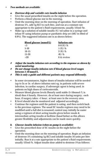

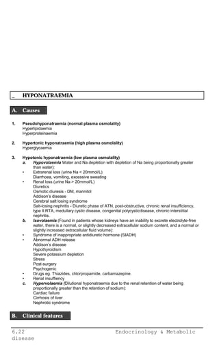
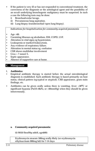
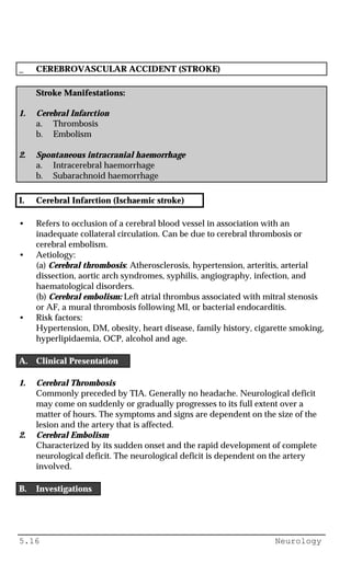
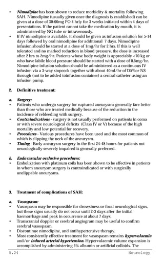
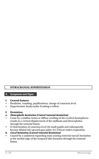
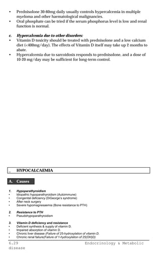
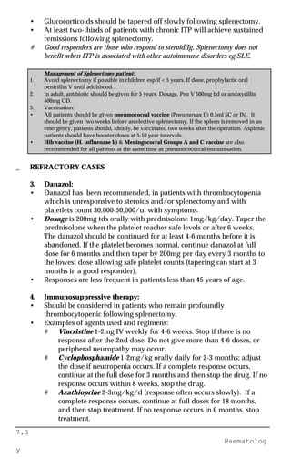
![6.23 Endocrinology & Metabolic
disease
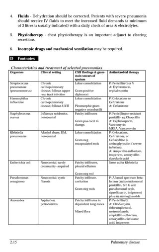
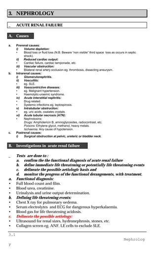
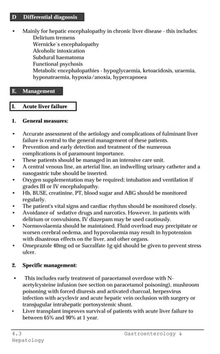
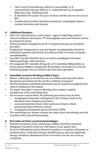
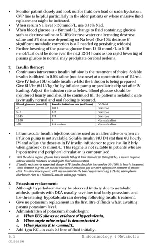
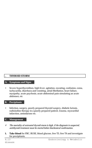
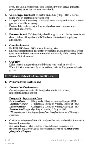
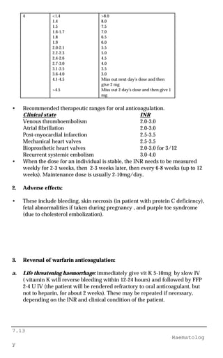
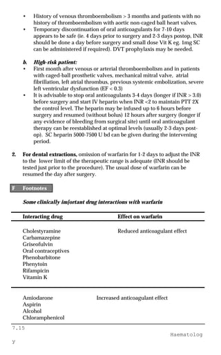
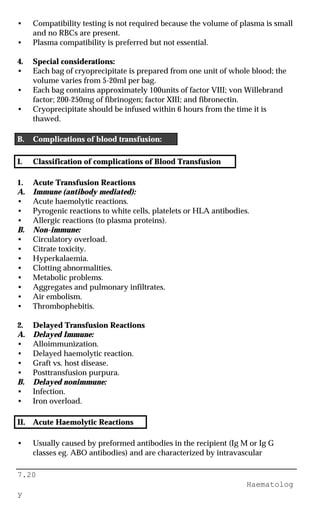
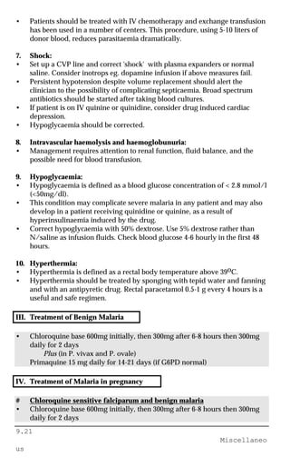
1. The clinical features of acute hyponatraemia are related to osmotic water
shift that leads to increased ICF volume and brain cell swelling.
• Mild hyponatraemia is usually asymptomatic.
• Serum Na of about 120 mmol/L may be associated with disturbed mental
state, restlessness, confusion & irritability.
• As the Na approaches 110 mmol/L, seizures and coma may occur.
2. In chronic hyponatraemia, adaptive mechanisms tend to minimize the
increased in ICF volume & its symptoms.
C. Assessment and Investigations
• Assess the patient's fluid status from skin turgor, tonge moisture, BP, JVP,
presence or absence of pulmonary oedema and fluid chart.
• Measure serum osmolarity and compare it to the calculated osmolarity - an
increased in osmolar gap occurs with substance such as ethylene glycol,
mannitol, hyperglycaemia etc.
• Urine Na combined with clinical assessment of fluid status may help
determine the underlying cause:
- Volume depletion from an extra-renal cause is normally associated
with a low urinary Na (< 20 mmol/L).
- Dehydration with a high urinary Na (>20mmol/L) suggests
inappropriate renal salt-wasting.
- Fluid overload with a low urine Na is seen in conditions such as CCF,
cirrhosis or nephrotic syndrome.
- Isovolaemia with a high urine Na is seen with SIADH.
D. Treatment of hyponatraemia
1. General principles:
• To normalize patient’s extracellular volume and sodium concentration.
• If the plasma Na is >120mmol/L, agressive treatmant is not required.
• If the plasma Na is <120 mmol/L, correct it at a rate of less than 10 mmol/L
per day (0.5-1 mmol/L/hour).
• If the plasma Na is <105 mmol/L, or the patient is symptomatic, urgent
treatment is required with a more rapid rate of increase of 1-2
mmol/L/hour for the first 3-4 hours. However, the total daily increment in
the plasma sodium should still not exceed 10 mmol/L.
• Avoid i) too rapid a correction to normonatraemic or hypernatraemic
levels, and ii) development of hypernatraemia in the period following the
original correction - may cause central pontine myelinolysis.
• Limit correction of serum sodium concentration to no higher than 130 to
135 mmol/l over the first 48 hours.
• Formulas:
1. Osmolarity(mmol/L) = 2(Na + K) + BU + Glucose (all in mmol/L)
[Normal: 270-295]](https://image.slidesharecdn.com/sarawakhandbookcompiledbyllyl-150521205442-lva1-app6891/85/Sarawak-handbook-199-320.jpg)
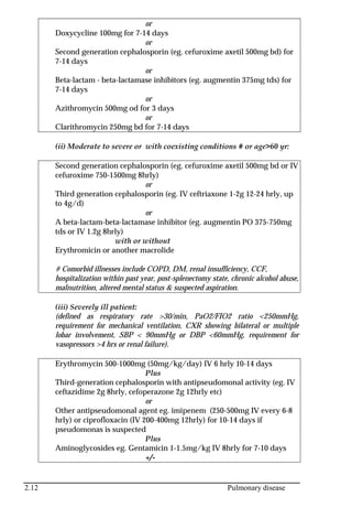
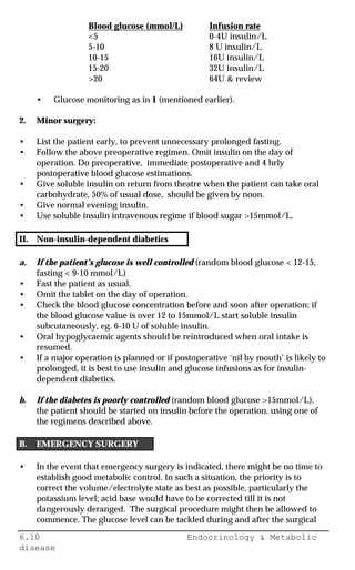
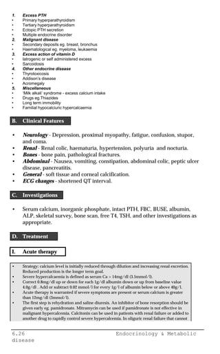
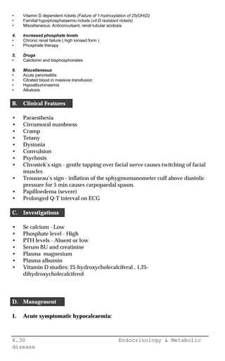
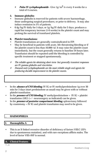
![6.24 Endocrinology & Metabolic
disease
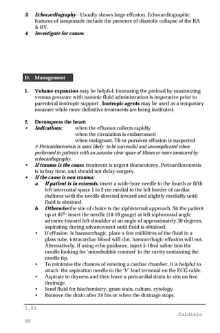
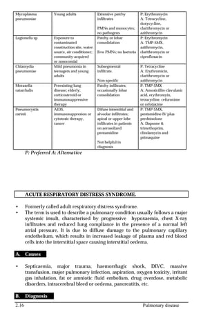
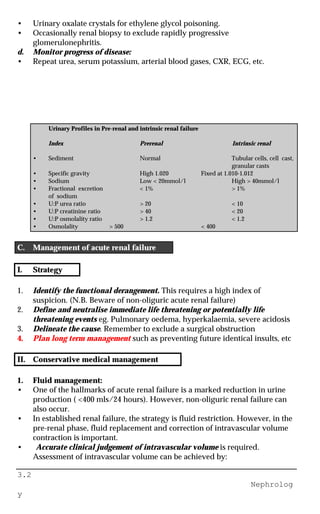
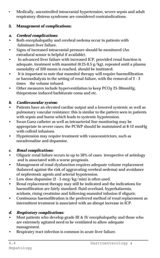
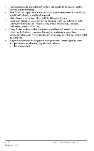
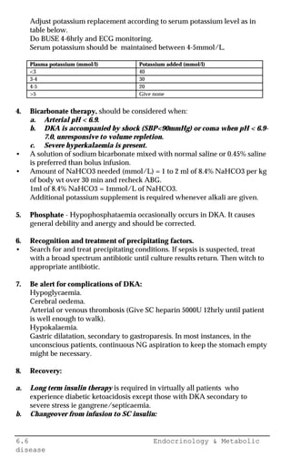
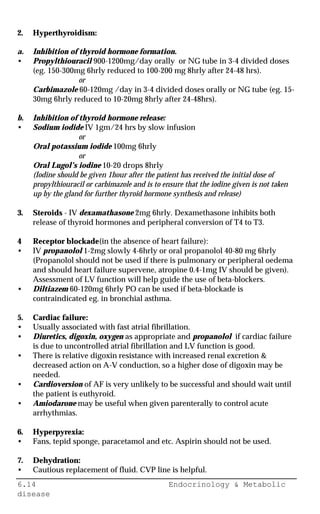
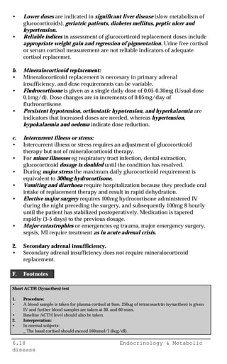
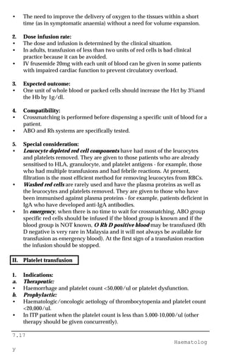
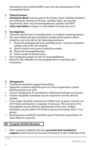
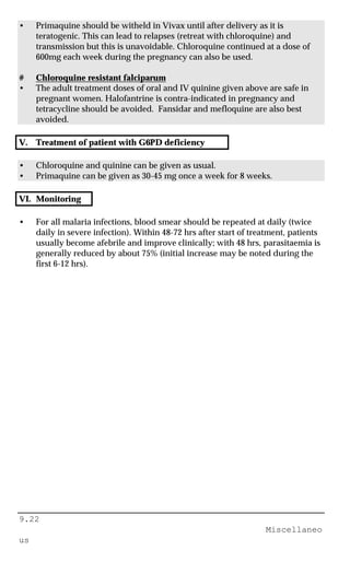
2. Na deficit = (Desired Na-measured serum Na) x0.6x BW(kg)
3. 3% Nacl provides 0.51 mmol/L of Na/ml
4. 0.9% Nacl provides 0.154 mmol/L of Na/ml
5. 1g of Nacl = 17 mmol/L
2. Hypervolaemic hyponatraemia:
• Retriction of Na & water intake, correction of hypokalaemia & promotion of
water loss in excess of Na . This can be done with judicious use of diuretics
with replacement of a proportion of the urinary Na loss to ensure net free
water excretion.
• Treat aetiology.
3. Isovolaemic hyponatraemia:
a. No symptoms
• Strict fluid restriction (e.g. <500mL daily) until serum sodium rises.
• 0.9% saline with frusemide may also be used.
b. Symptomatic
• Rapidly increase the patient’s extracellular tonity to prevent cerebral
oedema and to decrease his extracellular volume (e.g. IV frusemide +
hypertonic 3% saline).
• The principle here is to administer 3% saline at a rate equal to frusemide- or
bumetanide-induced urinary electrolyte losses. The difference in the hourly
rate of urine flow and hypertonic saline infusion will equal the net negative
fluid balance.
• This approach avoids dangerous overexpansion of ECF and corrects
primary cause of the hyponatraemia - excessive total body water.
Example
• To calculate the desired negative water balance:
Weight = 60kg, serum Na = 115mmol/L
Total body water (TBW) = 0.6 x 60 = 36 litres
If desired serum Na = 125mmol/L
then desired TBW = 115/125 X 36 = ˜33 litres (formula as mentioned in hypernatraemia)
Therefore, net negative water balance = (36-33) = 3 litres
• Choose the rate of correction based on clinical circumstances eg. 1 mmol/L. The total change in
plasma Na (125 - serum Na) divided by rate of correction (1 mmol/l) will yield the period of time
over which correction should occur eg. 10/1 = 10 hours.
• The negative water balance divided by the period of time calculated above yields the target rate
of free water removal in liters/hour eg. 3 /10 = 0.3 liter/hour = 300 ml/hour.
• Establish urine output with IV frusemide 40mg and titrate dose to achieve a urine output
(ml/hour) equal to the rate or free water removal ie 300 ml/hour.
• Na deficit (0.6 x [125 - Na] x 60) = 0.6 x[125 -115] x 60 = 360 mmol/L Nacl
• Replace with 3% Nacl = 360/0.51ml = 700 ml of 3% Nacl over 10 hours.](https://image.slidesharecdn.com/sarawakhandbookcompiledbyllyl-150521205442-lva1-app6891/85/Sarawak-handbook-200-320.jpg)
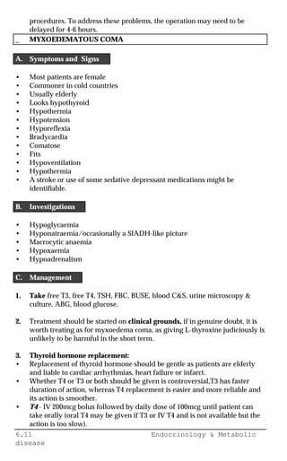
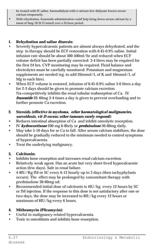
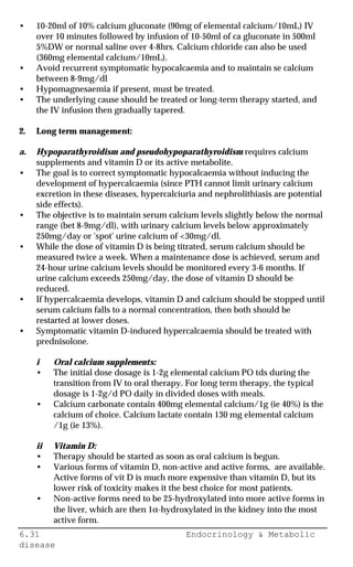
![6.25 Endocrinology & Metabolic
disease
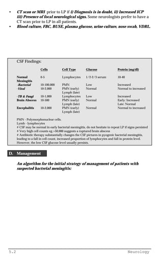
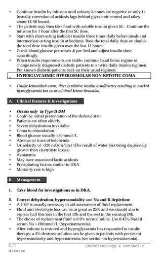
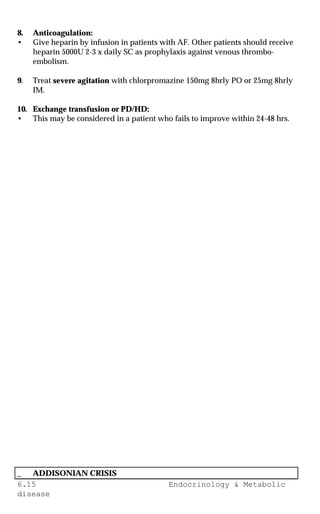
Alternatively
• If we infuse 3% saline at 100ml/hour and hourly urine flow is established at 500ml/hour with
frusemide, a negative balance of 400mls per hour is achieved. We can thus achieve a 3 L negative
water balance over 7.5 hours.
• Hourly serum and urinary electrolytes are mandatory.
3. Hypovolaemic hyponatraemia:
• Rehydration should be accomplished over 2 to 3 days (usually with normal
saline).
• Amount of fluid to be given is calculated as below:
i) Amount depleted: Calculate from degree of dehydration, e.g. mild =
5%, moderate 5-8%, severe = 8-12% = ( %x BW [kg] x 1000) ml
ii) Normal daily fluid requirement: Varies with patient size generally 1 to
2 litres
• Rate: As a rule of thumb,1/2 of calculated deficits + maintenance +
projected losses may be given during the first 24h of treatment. About 1/2
of the total 24h volume is given in the first 8h.
• Type of fluid: usually normal saline
4. Syndrome of inappropriate ADH secretion (SIADH):
• Inclusive criteria for the diagnosis are:
a. Plasma Na <130mmol/l.
Plasma osmolality <275 mosmol/kg.
Urine Na >20mmol/l.
Urine osmolality > plasma osmalality.
b. No oedema or signs of hypovolaemia.
c. Normal renal, thyroid & adrenal fucntion.
d. The patient should not be taking diuretics.
• Treatment - as in isovolaemic hyponatraemia but may also use
demeclocycline or lithium.
_ HYPERCALCAEMIA
A. Causes](https://image.slidesharecdn.com/sarawakhandbookcompiledbyllyl-150521205442-lva1-app6891/85/Sarawak-handbook-201-320.jpg)
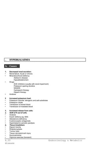
![6.38 Endocrinology & Metabolic
disease
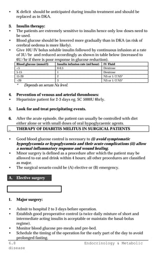
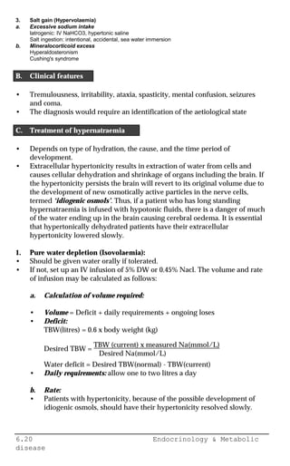
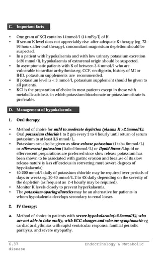
b. In asymptomatic patients without ECG changes, K should be given as
follows:
• At a concentration less than 40mmol/L (<3g KCl in 1L of carrier fluid).
• At a rate of < 20mmol/h (10mmol KCl per hour recommended).
• The plasma K should be monitored regularly, and with ECG monitoring.
c. In emergency eg. cardiac arrhythmias, severe myopathy, K can be given at
rates up to 40mmol/hr and in concentrations of 200-400mmol/l (by mixing
20-40 mmol KCL in 100cc of saline).
• Preferrably the fluid should be dextrose free as fast infusion of dextrose
would result in endogenous insulin secretion, thus simulating an
insuline/glucose infusion.
3. As soon as the ECG changes normalizes , cardiac rhythm returns to normal
or respiratory muscle strength has been restored to normal, IV infusion is
gradually tapered and then discontinued. Oral KCl is then initiated.
_ METABOLIC ACIDOSIS
A. Causes
1. High anion gap metabolic acidosis
a. Renal failure:
• Acute, chronic
b. Ketoacidosis:
• Diabetes mellitus, ethanol, starvation
c. Lactic acidosis:
(i) Type A (tissue hypoxia apparent):
• Severe hypoxia, severe anaemia, shock/haemorrhage, CCF
(ii) Type B (tissue hypoxia not apparent):
• Acquired disease: diabetes mellitus, liver failure, convulsions, tumours.
• Drugs/toxins: biguanides, ethanol, methanol
• Congenital disorders: G6PD deficiency, fructose 1,6 diphosphatase deficiency.
d. Toxin:
• Salicylate, ethanol, methanol, paraldehyde, ethylene glycol
2. Normal anion gap metabolic acidosis
a. Hyperkalaemic:
• Early uraemic acidosis
• Obstructive uropathy
• Renal tubular acidosis Type 4: Mineralocorticoid deficiency, tubule unresponsiveness.
• Ingestions/infusions: HCL, lysine/arginine HCL, ammonium chloride
• Diabetic ketoacidosis: post-therapy
b. Hypokalaemic:
• Renal tubular acidosis: Type 1 & 2
• Carbonic anhydrase inhibitor: acetazolamide
• Urine diversions: ureterosignoidostomy, vesico-colic fistula, obstructed ileal bladder
• Post-hypocapnic acidosis
• Acute diarrhoea.
Anion Gap = [K] + [Na] - [Cl] - [HCO3] (all in mmol/L)
The normal range is 8-16 mmol/L
B. Treatment of acute metabolic acidosis](https://image.slidesharecdn.com/sarawakhandbookcompiledbyllyl-150521205442-lva1-app6891/85/Sarawak-handbook-214-320.jpg)


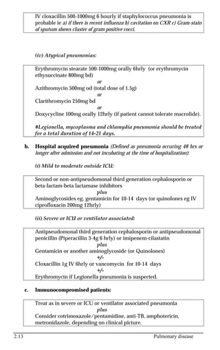
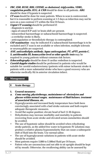
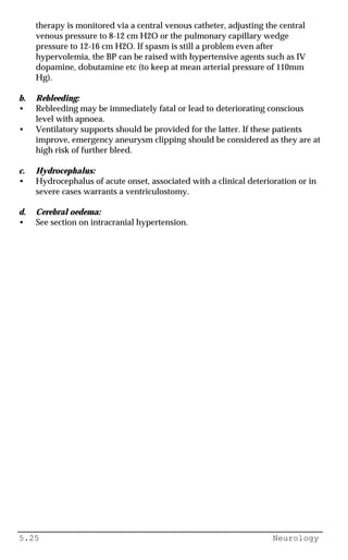
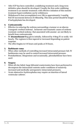
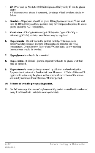
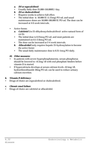
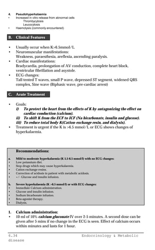
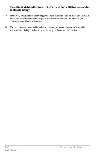
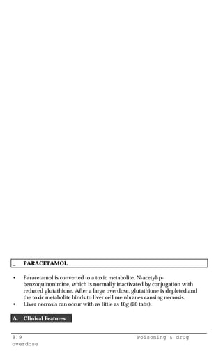
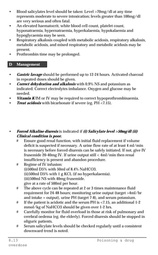
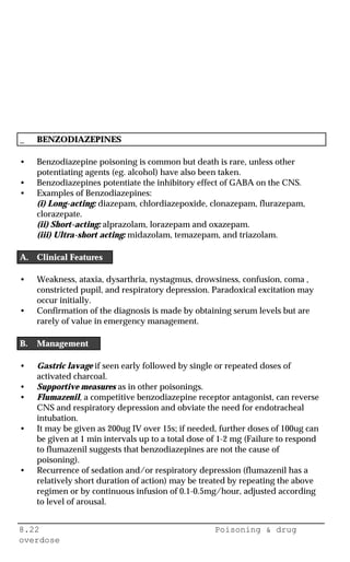
![8.3 Poisoning & drug
overdose
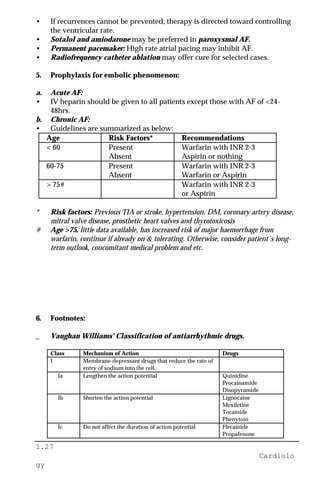
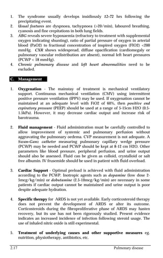
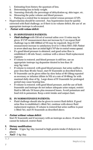
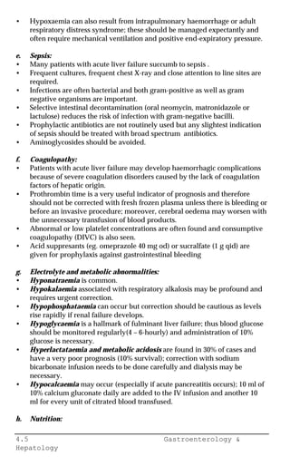
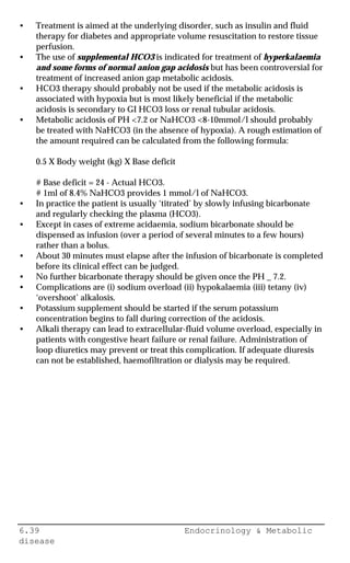
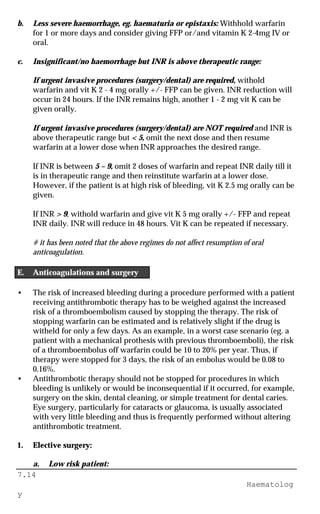
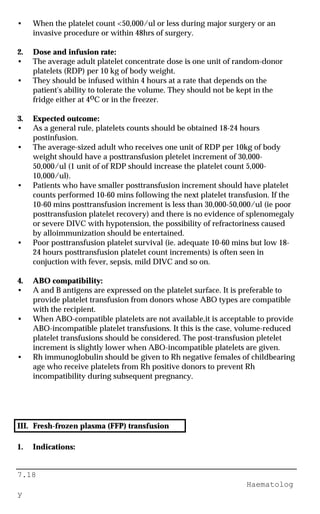
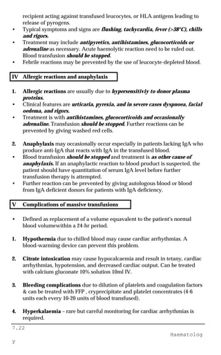
_ DIGOXIN
• Overdosage of digoxin usually arises from its therapeutic usage.
A Symptoms and signs
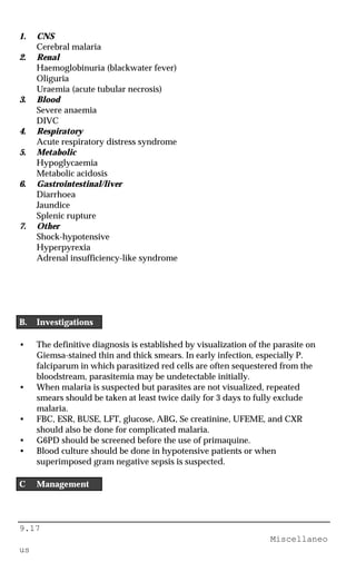
1. GI: Diarrhoea, vomiting, abdominal pain, intestinal ulceration.
2. CNS: Headache, anorexia, confusion, convulsions.
3. Visual: Xanthopsia, haloes, blurring.
4. Cardiac: Arrhythmias (atrial fibrillation with slow ventricular response,
non-paroxysmal AV junctional tachycardia, atrial tachycardia with varying
block[SVT with block], sinus bradycardia, frequent ventricular ectopic
beats, ventricular bigeminy, ventricular tachycardia), worsening of heart
failure. Arrhythmias not seen with digoxin toxicity include: sinus
tachycardia, RBBB, LBBB, hemiblocks and parasystole.
5. Others: Hyperkalaemia in acute overdose, hypokalaemia or
normokalaemia in chronic overdose.
# ECG manifestations of digoxin therapy (not overdose)](https://image.slidesharecdn.com/sarawakhandbookcompiledbyllyl-150521205442-lva1-app6891/85/Sarawak-handbook-246-320.jpg)
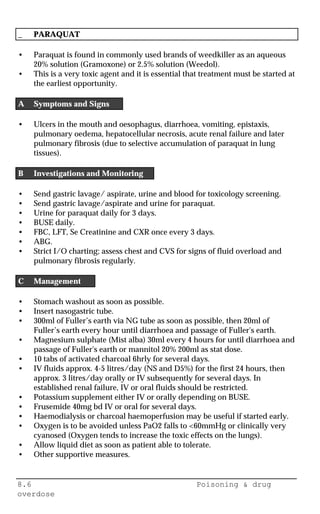
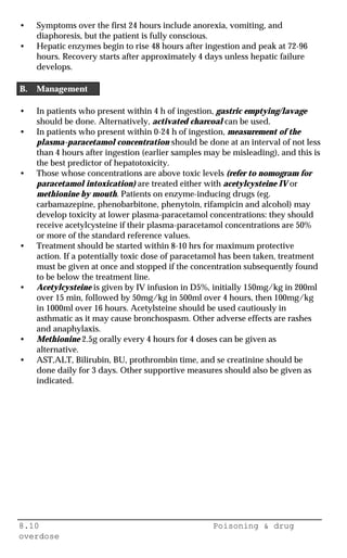
![8.16 Poisoning & drug
overdose
• Methanol produces nausea, vomiting, abdominal pain, headache, vertigo,
and confusion at low overdose. In large overdose, obtundation,
convulsions, and coma.
• Late manifestations include an increased anion gap, metabolic acidosis,
and retinal injury (due to formic acid, lactic acid and ketones).
Opthalmological manifestations (occur 15-19 h later) include clouding and
diminished vision, dancing and flashing spots, dilated or fixed pupils,
hyperaemia of the disc, retinal oedema, and blindness.
• Early diagnosis is suggested by ethanol-like signs of intoxication and an
elevated serum osmolality and is confirmed by measurement of serum
methanol (usually > 20mg/dL) 12-48 hrs following ingestion.
• The diagnosis of methanol-derived formic acidosis is suggested by a large
anion gap, a low serum bicarbonate, an elevated serum formate level, and
an elevated blood methanol.
B. Clinical features of Ethylene Glycol poisoning
• Ethylene glycol poisoning often exhibits three distinct clinical phases after
ingestion due to the toxic metabolites glycolate, glyoxalate and oxalate.
• First, within 12 h, CNS effects predominate. The patient appears intoxicated
without the odor of ethanol in the breath.
• Second, 12-24h after ingestion, cardiopulmonary effects predominate.
Elevated heart and respiratory rate and blood pressure are common. CHF,
ARDS, and circulatory collapse are also noted.
• Third, 24-72h after ingestion, renal effects predominate. Acute tubular
necrosis with acute renal failure occurs if appropriate treatment is not
received.
• Hypocalcaemia may result from precipitation of calcium oxalate into
tissues and may be severe enough to cause tetany and typical ECG changes.
Calcium oxalate crystals are noted on urinalysis. Elevated CPK may be seen
and leukocytosis is common.
• High anion gap metabolic acidosis and a high osmolal gap (Osm measured
- Osm calculated [normal <10 mosm/L]) may be found.
C Management
• Treatment of methanol and ethylene glycol poisoning are similar.
• Gastric lavage if < 4 hrs of ingestion.
• If seizures occur, exclude hypocalcaemia and treat with iv diazepam. If
hypocalcaemic seizures occur, treat with 10 to 20 ml of 10% calcium
gluconate.
• Correct acidosis with sodium bicarbonate (Methanol is metabolised slowly
and the patient may relapse if bicarbonate administration is discontinued
too soon).
• Inhibit metabolism with ethanol: alcohol dehydrogenase (which oxidises
methanol to formaldehyde and then to formic acid; also metabolizes
ethylene glycol) has a much higher affinity for ethanol and hence ethanol is](https://image.slidesharecdn.com/sarawakhandbookcompiledbyllyl-150521205442-lva1-app6891/85/Sarawak-handbook-259-320.jpg)
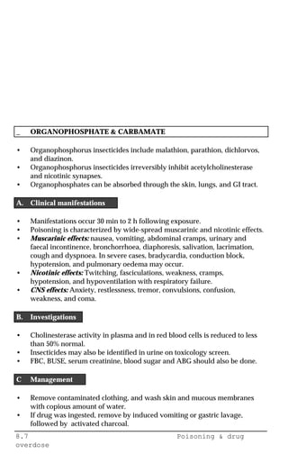
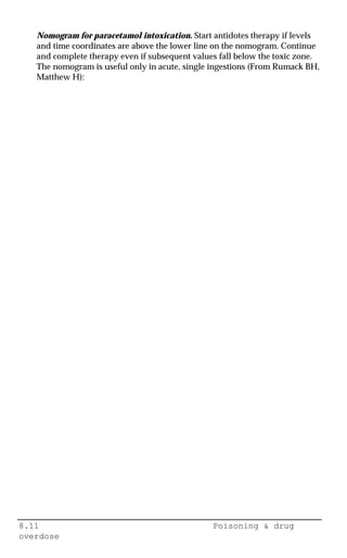
![8.27 Poisoning & drug
overdose
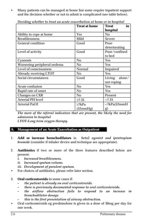
_ THEOPHYLLINE
• Theophylline causes the release of endogenous catecholamines and
prolongs their effects by inhibiting the degradation of cyclic AMP by
phosphodiesterase.
• Aminophylline is 80% theophylline.
A. Clinical Features
• Nausea, vomiting, restlessness, irritability, agitation, tachypnoea,
tachycardia, and muscle tremors. In severe overdose, coma, hypotension,
respiratory depression, generalized tonic-clonic and focal convulsions and
rhabdomyolysis may occur.
• Cardiovascular effects include atrial arrhythmias, multifocal ventricular
ectopics, idioventricular rhythms, ventricular tachycardia, and ventricular
fibrillation.
• Metabolic abnormalities include ketosis, metabolic acidosis, increased
serum amylase, hyperglycaemia, and decreased serum potassium, and
calcium.
• Toxicity occurs at lower theophylline levels with chronic than with acute
poisoning.
• Serial serum levels should be measured to determine the peak
concentration (therapeutic serum level are 55-110 umol/L [10-20mg/L or
mcg/ml]).
B. Management
• Gastric lavage (if within 1-2 hours of ingestion) and multiple doses of
activated charcoal. Activated charcoal given in multiple doses will increase
the elimination of theophylline from the gut.
• Extreme tachycardia is treated with propranolol or esmolol. The possibility
of exacerbating pre-existing obstructive airways disease often precludes the
use of such drugs.
• Hypotension is treated with volume expansion.](https://image.slidesharecdn.com/sarawakhandbookcompiledbyllyl-150521205442-lva1-app6891/85/Sarawak-handbook-270-320.jpg)
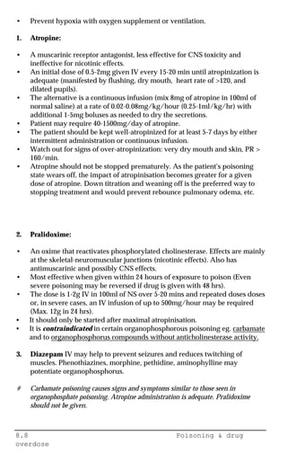
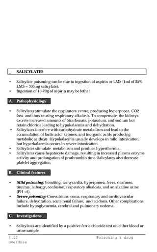
![8.28 Poisoning & drug
overdose
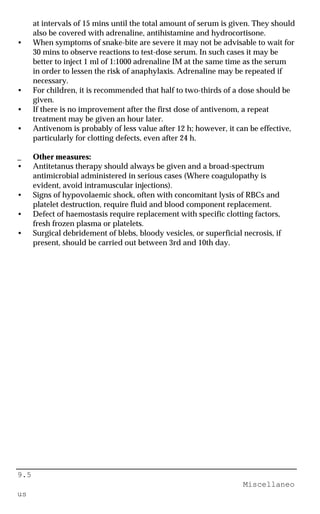
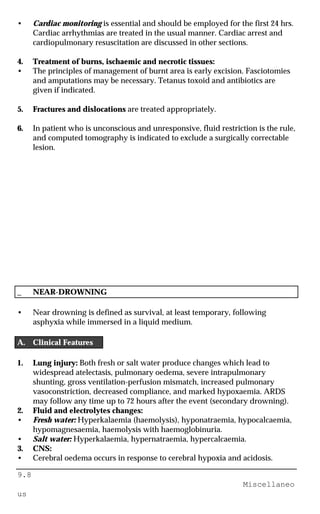
• Serum K level should be measured and monitored regularly. Potassium
supplements are nearly always needed.
• Benzodiazepines and barbiturates are useful for convulsions and
hyperactivity (Phenytoin is ineffective).
• Upper GI haemorrhage may follow theophylline-induced peptic ulceration
and H2-antagonist other than cimetidine should be given since cimetidine
inhibits the metabolism of theophylline.
• Ventricular tachyarrhythmias should be treated with propranolol and
standard antiarrhythmics.
• Haemodialysis, peritoneal dialysis and haemoperfusion are effective in
removing theophylline and are indicated in patients with severe toxicity or
after acute ingestion with serum level > 440 umol/L [80mg/L], and in
chronic ingestion, with serum levels >200-300umol/L [40-60mg/L].](https://image.slidesharecdn.com/sarawakhandbookcompiledbyllyl-150521205442-lva1-app6891/85/Sarawak-handbook-271-320.jpg)


![Differential Diagnosis in Clinical Examination (2012) [PDF] [UnitedVRG].pdf](https://cdn.slidesharecdn.com/ss_thumbnails/differentialdiagnosisinclinicalexamination2012pdfunitedvrg-220407073433-thumbnail.jpg?width=640&height=640&fit=bounds)


