Download to read offline


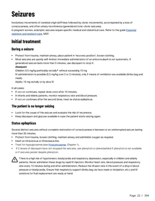
![Page 11 / 394
Shock
Last updated: September 2023
Shock is a condition of widespread reduced tissue perfusion and inadequate oxygen delivery. Prolonged shock can
result in cellular dysfunction and irreversible organ failure. Mortality is high without early diagnosis and treatment.
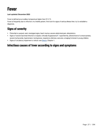
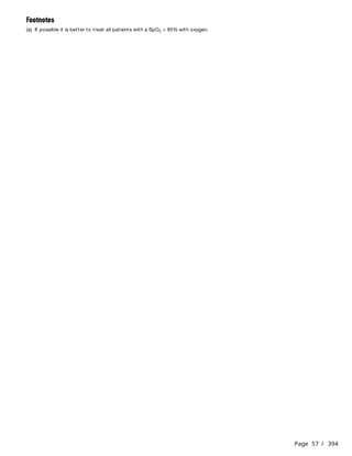
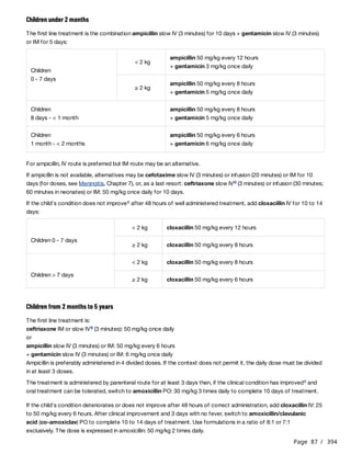
Clinical features
Shock should be suspected in patients with:
In children, accurate BP measurement is difficult, and hypotension is a very late sign of shock. Therefore, critically ill
children should be treated for shock if they present at least one of the following signs: lower limb temperature
gradient , CRT ≥ 3 seconds, weak radial pulse or severe tachycardia .
Clinical features may vary according to the type of shock :
Sign(s) of hypotension: weak pulse, low or declining blood pressure (BP) , narrow pulse pressure
a
Acute onset of signs of tissue hypoperfusion:
Skin: pallor, mottled skin, sweating, cold extremities or lower limb temperature gradient , capillary refill time
(CRT) ≥ 3 seconds
b
Lungs: tachypnea, dyspnoea
Heart: tachycardia, which often occurs before BP decreases
Kidney: oliguria (urine output < 0.5 to 1 ml/kg/hour) or anuria
Brain: thirst, anxiety, agitation, confusional state, apathy, altered mental status
c
b d [1]
[2]](https://image.slidesharecdn.com/guideline-170-en-240313103309-343cfe20/85/guideline-170-en-pdf-11-320.jpg)
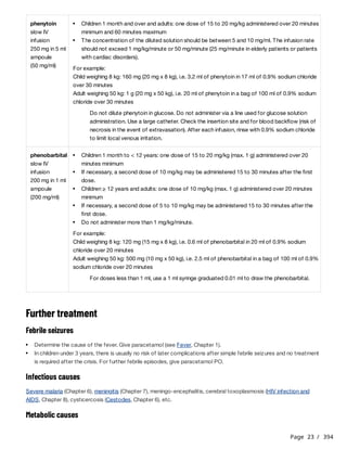
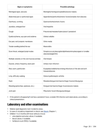
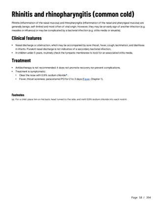
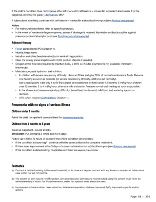
![Page 12 / 394
Type Specific clinical features Risk factors
Distributive
Severe vasodilation
and increased capillary
permeability resulting
in maldistribution of
blood flow
Anaphylaxis: likely when either of the following 2
criteria develop within minutes to hours :
[3]
Involvement of skin and/or mucous membranes (e.g.
generalised urticaria, itching, flushing, swollen
lips/tongue/uvula) AND ≥ 1 of the following:
respiratory symptoms (wheeze, dyspnoea);
low BP or symptoms of end-organ dysfunction
(hypotonia, incontinence);
severe gastrointestinal symptoms (abdominal
pain, repetitive vomiting).
Hypotension, bronchospasm or laryngeal
involvement (stridor, vocal changes, odynophagia)
after exposure to known or probable allergen for
that patient.
Recent exposure to an allergen
(e.g. food, sting, medicine) or
history of anaphylaxis
Septic shock: signs of infection, fever or
hypothermia, altered mental status, dyspnoea,
persisting hypotension despite fluid resuscitation[4]
Infection, recent surgery,
immunodeficiency
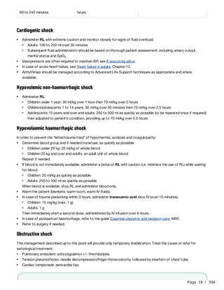
Cardiogenic
Cardiac pump failure
Ischaemia: chest pain, dyspnoea
Arrhythmia
Murmur of valvular heart disease
History of cardiac disease,
advanced age
Acute heart failure: see Heart failure in adults, Chapter
12.
History of cardiac disease, viral
illness, immunodeficiency
Hypovolaemic
Direct blood/fluid loss
or fluid sequestration
into the extravascular
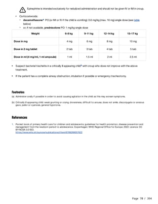
space resulting in
decreased
intravascular volume
Haemorrhagic: external bleeding, signs and symptoms
of internal bleeding, hypotension(a)
Trauma, recent surgery, obstetric
haemorrhage
Non-haemorrhagic: dry mouth, absence of tears,
sunken eyes/fontanelle, low jugular venous pressure
(JVP), altered mental status
Profuse diarrhoea and/or
vomiting, intestinal obstruction
Obstructive
Obstruction to blood
flow to, or from, the
heart or great vessels
Pulmonary embolism (PE): chest pain, tachycardia,
tachypnoea, hypoxia
Deep vein thrombosis (DVT): leg pain, swelling, warmth
Recent surgery or immobilisation,
cancer, history of PE or DVT
Tension pneumothorax: decreased breath sounds,
raised JVP, weak radial pulse, tracheal deviation
Trauma, invasive medical
procedure
Cardiac tamponade: pulsus paradoxus , raised JVP,
narrow pulse pressure, muffled heart sounds
(b) Trauma, immunodeficiency
(a) In children and young adults with hypovolaemic shock, BP may be maintained initially, but subsequently declines rapidly if
fluid loss is not replaced.](https://image.slidesharecdn.com/guideline-170-en-240313103309-343cfe20/85/guideline-170-en-pdf-12-320.jpg)

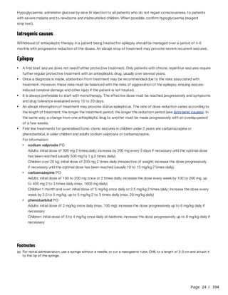
![Page 14 / 394
2) Maintain circulation
Anaphylaxis
Repeat after 5 minutes if no or poor clinical improvement (up to a total of 3 IM injections).
Repeat bolus once if signs of poor perfusion persist after 15 minutes.
Septic shock
Look for source of infection. If possible, take specimens for culture before starting antibiotic treatment.
if needed, insert an oropharyngeal airway.
Auscultate lungs to assess ventilation.
Administer 10 to 15 litres/minute of oxygen with mask to maintain SpO > 94%.
2
If SpO remains ≤ 94% with oxygen, see If resources allow.
2
Control bleeding:
apply direct manual pressure and/or compression/haemostatic dressing to the wound;
in case of massive life-threatening bleeding from an extremity (e.g. leg) not controlled by direct pressure: apply a
windlass tourniquet .
h[5]
Insert 2 peripheral IV lines (catheters 20-22G in children and 14-18G in adults) or an intraosseous (IO) needle.
Administer Ringer lactate (RL) , glucose 5%-Ringer lactate (G5%-RL) , and/or blood, following specific
management described below. Reassess before giving additional fluid therapy. Monitor for fluid overload ,
especially in patients at risk, e.g. severely malnourished children; patients with severe malaria, heart disease, severe
anaemia; older patients.
i j
k
Maintain normal body temperature.
If unable to maintain BP, see If resources allow.
Remove exposure to causal agent.
Administer epinephrine (adrenaline) IM into the mid-anterolateral thigh. Use undiluted solution and a 1 ml syringe
graduated in 0.01 ml :
[6]
Children under 6 months: 0.1 to 0.15 ml
Children 6 months to 5 years: 0.15 ml
Children 6 to 12 years: 0.3 ml
Children over 12 years and adults: 0.5 ml
l
Monitor HR, BP, CRT and clinical response.
In case of stridor, administer nebulized epinephrine: 0.5 mg/kg/dose (max. 5 mg) in 5 ml of 0.9% sodium chloride
over 10 to 15 minutes
If SpO is < 94%, ventilate with bag mask.
2
Administer a bolus of RL:
Children: 10 ml/kg as quickly as possible
Adults: 500 ml as quickly as possible
If shock persists after 3 IM injections of epinephrine, in particular if unable to maintain BP, see If ressources allow.
After initial treatment with epinephrine and IV fluids, some patients (e.g. patients requiring continuing treatment after
2 doses of epinephrine IM or patients with ongoing asthma or shock) may benefit from a short course of
corticosteroid therapy. When the patient is stable, prednisolone PO : 1 to 2 mg/kg (max. 50 mg) once daily in the
morning for 3 to 5 days. Use an IV corticosteroid only if the patient cannot take oral treatment.
Fluid therapy:
Children and adolescents under 15 years: G5%-RL solution as maintenance fluids (see Appendix 1)
Adolescents 15 years and over and adults: one bolus of 250 to 500 ml of RL as quickly as possible
Antibiotic treatment:
Start antibiotics according to the suspected origin of infection within 1 hour of presentation (see tables
below).
[7]](https://image.slidesharecdn.com/guideline-170-en-240313103309-343cfe20/85/guideline-170-en-pdf-14-320.jpg)
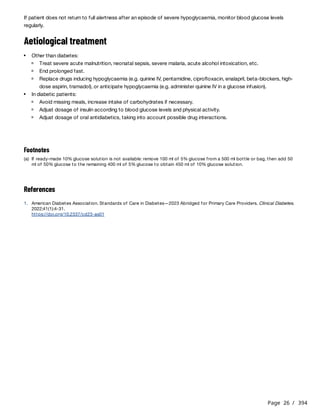
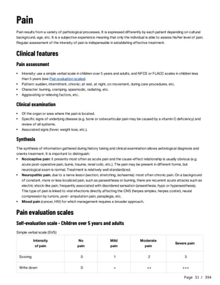
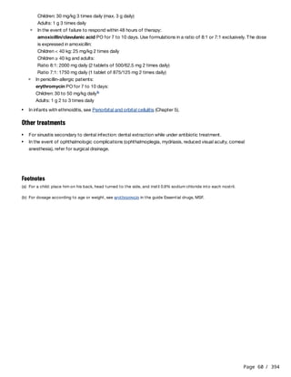
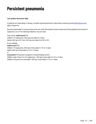
![Page 19 / 394
If ressources allow:
These protocols are for peripheral IV administration. Titrate according to patient's clinical situation. Refer to
management objectives (including BP) under Management.
Manage airways and breathing:
complete airway obstruction: endotracheal intubation or cricothyroidotomy
respiratory failure: non-invasive or invasive mechanical ventilation
Maintain circulation:
If unable to achieve management objectives (in particular BP) using fluid therapy (and no signs of fluid
overload are present) or, in the case of anaphylaxis, if shock persists after 3 IM epinephrine injections,
vasopressors-inotropes (see below) can be used in the following conditions:
close monitoring in a critical care unit;
a large peripheral IV catheter (proximal forearm or above), a central venous catheter or an IO line
dedicated to the infusion;
n
use of an electric syringe or pump to control flow rate ;
o
intensive monitoring of drug administration, particularly during syringe changes.
All infused volumes must be accounted for when recording fluid balance.
Norepinephrine (NEP) tartrate(i) Epinephrine (EPN) (adrenaline)
Indication
Children: 2 choice
Adults: 1 choice
nd
st
Children: 1 choice
Adults: 2 choice
st
nd
Preparation
of
diluted
solution(j)
Children:
Add 1 ml (2 mg) of NEP tartrate to 39 ml
of 0.9% NaCl to obtain a 0.05 mg/ml (50
micrograms/ml) solution.
Children:
Add 2 ml (2 mg) of EPN to 38 ml of 0.9% NaCl
to obtain a 0.05 mg/ml (50 micrograms/ml)
solution.
Adults:
Add 2 ml (4 mg) of NEP tartrate to 38 ml
of 0.9% NaCl to obtain a 0.1 mg/ml (100
micrograms/ml) solution.
Adults:
Add 4 ml (4 mg) of EPN to 36 ml of 0.9% NaCl
to obtain a 0.1 mg/ml (100 micrograms/ml)
solution.
Starting
rate(k)
0.1 microgram/kg/minute
Rate for
increasing(k)
Increase by 0.05 micrograms/kg/minute every 10 minutes for the first hour, then every hour.
Max. 1 microgram/kg/minute.
Rate for
decreasing(k)
Taper down doses when management objectives are attained. Do not stop abruptly.
Decrease by 0.05 micrograms/kg/minute every hour.
(i) 2 mg of NEP tartrate = 1 mg of NEP base.
(j) 0.9% sodium chloride or 5% glucose or RL can be used for dilution.
(k) The infusion rate is calculated as follows: [desired dose (microgram/kg/min) x weight (kg) x 60 min] ÷ concentration
(microgram/ml).
Ongoing care: measure serum potassium, magnesium, calcium and phosphate levels and correct any
abnormalities. Additional investigations (e.g. X-rays, other laboratory tests) may be indicated, depending on
aetiology suspected.](https://image.slidesharecdn.com/guideline-170-en-240313103309-343cfe20/85/guideline-170-en-pdf-19-320.jpg)

![Page 21 / 394
2020;13(10):100472. Published 2020 Oct 30.
https://doi.org/10.1016/j.waojou.2020.100472
4. Singer M, Deutschman CS, Seymour CW, et al. The Third International Consensus Definitions for Sepsis and Septic Shock
(Sepsis-3). JAMA. 2016;315(8):801-810.
https://doi.org/10.1001/jama.2016.0288
5. Richey SL. Tourniquets for the control of traumatic hemorrhage: a review of the literature. World J Emerg Surg. 2007;2:28.
Published 2007 Oct 24.
https://doi.org/10.1186/1749-7922-2-28
6. Emergency treatment of anaphylaxis. Guidelines for healthcare providers. Rescuscitation Council UK. Updated 2021
[Accessed May 31 2023].
https://www.resus.org.uk/sites/default/files/2021-
05/Emergency%20Treatment%20of%20Anaphylaxis%20May%202021_0.pdf
7. Evans L, Rhodes A, Alhazzani W, et al. Surviving sepsis campaign: international guidelines for management of sepsis and
septic shock 2021. Intensive Care Med. 2021;47(11):1181-1247.
https://doi.org/10.1007/s00134-021-06506-y](https://image.slidesharecdn.com/guideline-170-en-240313103309-343cfe20/85/guideline-170-en-pdf-21-320.jpg)
![Page 25 / 394
Hypoglycaemia
Last update: November 2023
Hypoglycaemia is an abnormally low concentration of blood glucose. Severe hypoglycaemia can be fatal or lead to
irreversible neurological damage.
Blood glucose levels should be measured whenever possible in patients presenting symptoms of hypoglycaemia. If
hypoglycaemia is suspected but blood glucose measurement is not available, glucose (or another available sugar)
should be given empirically.
Always consider hypoglycaemia in patients presenting impaired consciousness (lethargy, coma) or seizures.
For diagnosis and treatment of hypoglycaemia in neonates, refer to the guide Essential obstetric and newborn care,
MSF.
Clinical features
Rapid onset of non-specific signs, mild to severe depending on the degree of the hypoglycaemia: sensation of hunger
and fatigue, tremors, tachycardia, pallor, sweats, anxiety, blurred vision, difficulty speaking, confusion, convulsions,
lethargy, coma.
Diagnosis
Capillary blood glucose concentration (reagent strip test):
If blood glucose measurement is not available, diagnosis is confirmed when symptoms resolve after the administration
of sugar or glucose.
Symptomatic treatment
Symptoms improve approximately 15 minutes after taking sugar by oral route.
Neurological symptoms improve a few minutes after the injection.
Check blood glucose after 15 minutes. If it is still low, re-administer glucose by IV route or sugar by oral route according
to the patient’s clinical condition.
If there is no clinical improvement, differential diagnoses should be considered: e.g. serious infection (severe malaria,
meningitis, etc.), epilepsy, unintentional alcohol intoxication or adrenal insufficiency in children.
In all cases, after stabilisation, give a meal or snack rich in complex carbohydrates and monitor the patients for a few
hours.
Non-diabetic patients:
Hypoglycaemia: < 3.3 mmol/litre (< 60 mg/dl)
Severe hypoglycaemia: < 2.2 mmol/litre (< 40 mg/dl)
Diabetic patients on home treatment: < 3.9 mmol/litre (< 70 mg/dl)[1]
Conscious patients:
Children: a teaspoon of powdered sugar in a few ml of water or 50 ml of fruit juice, maternal or therapeutic milk
or 10 ml/kg of 10% glucose by oral route or nasogastric tube.
Adults: 15 to 20 g of sugar (3 or 4 cubes) or sugar water, fruit juice, soda, etc.
Patients with impaired consciousness or prolonged convulsions:
Children: 2 ml/kg of 10% glucose by slow IV (2 to 3 minutes)a
Adults: 1 ml/kg of 50% glucose by slow IV (3 to 5 minutes)](https://image.slidesharecdn.com/guideline-170-en-240313103309-343cfe20/85/guideline-170-en-pdf-25-320.jpg)


![Page 38 / 394
Anaemia
Last updated: January 2024
Anaemia is defined as a haemoglobin (Hb) level below reference values , which vary depending on age, sex, and
pregnancy status (see Table 2).
Anaemia may be caused by:
The causes of anaemia are often interlinked.
Clinical features
Laboratory
Table 1 - Possible diagnoses with FBC
[1][2]
Decreased production of red blood cells: iron deficiency, nutritional deficiencies (folic acid, vitamin B , vitamin A),
depressed bone marrow function, certain infections (HIV, visceral leishmaniasis), renal failure;
12
Loss of red blood cells: acute or chronic haemorrhage (gastrointestinal ulcer, ancylostomiasis, schistosomiasis,
etc.);
Increased destruction of red blood cells (haemolysis): parasitic (malaria), bacterial and viral (HIV) infections;
haemoglobinopathies (sickle cell disease, thalassaemia); intolerance to certain drugs (primaquine, dapsone, co-
trimoxazole, nitrofurantoin, etc.) in patients with G6PD deficiency.
Common signs: pallor of the conjunctivae, mucous membranes, palms of hands and soles of feet; fatigue,
dizziness, dyspnoea, tachycardia, heart murmur.
Signs of decompensation: cold extremities, altered mental status, oedema in the lower limbs, respiratory distress,
elevated jugular venous pressure, cardiac/coronary failure, shock.
Significant signs: cheilosis and glossitis (nutritional deficiency), jaundice, hepatosplenomegaly, dark coloured urine
(haemolysis), bleeding (maelena, haematuria, etc.), signs of malaria (Chapter 6), etc.
Hb levels
Rapid diagnostic test or thick and thin blood films in areas where malaria is endemic.
Urinary dipstick: check for haemoglobinuria or haematuria.
If sickle cell disease is suspected (to be done before blood transfusion): rapid diagnostic test (Sickle SCAN®) or, if
not available, Emmel test.
Full blood count (FBC) if available to guide diagnosis.](https://image.slidesharecdn.com/guideline-170-en-240313103309-343cfe20/85/guideline-170-en-pdf-38-320.jpg)
![Page 40 / 394
Blood transfusion
Indications
To decide whether to transfuse, several parameters should be taken into account:
If transfusion is indicated, it should be carried out without delay . For transfusion thresholds, see Table 2.
Volume to be transfused
If presence of haemorrhagic shock: see Shock, Chapter 1. Otherwise:
Transfusion volume is based on presence or absence of fever at any point from the time of ordering blood to the
time of transfusion:
Repeat if necessary, depending on clinical condition.
Monitoring
Repeat the injection (same dose) after 2 hours if necessary.
Malaria: see Malaria (Chapter 6). In the event of associated iron deficiency, wait 4 weeks after malaria treatment
before prescribing iron supplements.
Suspected haemolytic anaemia: stop any drug that causes haemolysis in patients with (or that may possibly have)
G6PD deficiency.
Clinical tolerance of anaemia
Underlying conditions (cardiovascular disease, infection, etc.)
Rate at which anaemia develops.
Hb levels
b
Children :
[3]
If no fever (axillary temperature ≤ 37.5 °C) : administer either 15 ml/kg of packed red blood cells (PRBC) over 3
hours or 30 ml/kg of whole blood over 4 hours
c
If fever (axillary temperature > 37.5 °C) : administer either 10 ml/kg of PRBC over 3 hours or 20 ml/kg of whole
blood over 4 hours
c
Adolescents and adults: start with an adult unit of PRBC or whole blood; do not exceed a transfusion rate of 5
ml/kg/hour.
Monitor the patient’s condition and vital signs (heart rate, blood pressure, respiratory rate, temperature):
During the transfusion: 5 minutes after the start of transfusion, then every 15 minutes during the first hour, then
every 30 minutes until the end of the transfusion.
After the transfusion: 4 to 6 hours after the end of the transfusion.
Pay attention to signs of transfusion reaction, fluid overload, decompensation or continuing blood loss.
For children: measure Hb once between 8 and 24 hours after the end of the transfusion or if signs of
decompensation or continuing blood loss.
If signs of circulatory overload appear:
Stop temporarily the transfusion.
Sit the patient in an upright position.
Administer oxygen.
Administer furosemide by slow IV injection:
Children: 0.5 to 1 mg/kg
Adults: 20 to 40 mg
Once the patient has been stabilised, start the transfusion again after 30 minutes.](https://image.slidesharecdn.com/guideline-170-en-240313103309-343cfe20/85/guideline-170-en-pdf-40-320.jpg)
![Page 41 / 394
Prevention
ferrous salts/folic acid PO, or if not available, ferrous salts PO, as long as the risk of deficiency persists (e.g.
pregnancy , malnutrition).
Doses are expressed in elemental iron :
Table 2 - Definition of anaemia and transfusion thresholds
Iron (and folic acid) deficiency:
Drug supplements:
[4]
a
Children 1 month to < 12 years: 1 to 2 mg/kg once daily (max. 65 mg daily)
Children ≥ 12 years and adults: 65 mg once daily
Age Weight
Prevention
45 mg/5 ml syrup 60 or 65 mg tablet
1 month to < 1 year 4 to < 10 kg 1 ml –
1 to < 6 years 10 to < 20 kg 2.5 ml –
6 to < 12 years 20 to < 40 kg 5 ml –
≥ 12 years and adults ≥ 40 kg – 1 tab
Nutritional supplements (if the basic diet is insufficient).
In the event of sickle cell anaemia: see Sickle cell disease (Chapter 12).
Early treatment of malaria, helminthic infections, etc.](https://image.slidesharecdn.com/guideline-170-en-240313103309-343cfe20/85/guideline-170-en-pdf-41-320.jpg)
![Page 43 / 394
1. World Health Organization. Haemoglobin Concentrations for the Diagnosis of Anaemia and Assessment of Severity. World
Health Organization; 2011. [Accessed June 26, 2023]
https://apps.who.int/iris/handle/10665/85839
2. World Health Organization. Educational Modules on Clinical Use of Blood. World Health Organization; 2021. [Accessed June
26, 2023]
https://apps.who.int/iris/handle/10665/350246
3. Maitland K, Olupot-Olupot P
, Kiguli S, et al. Transfusion Volume for Children with Severe Anemia in Africa. N Engl J Med.
2019;381(5):420-431.
https://doi.org/10.1056/NEJMoa1900100
4. Word Health Organization. Daily iron and folic acid supplementation in pregnant women. Word Health Organization. Geneva,
2012. [Accessed June 26, 2023]
https://apps.who.int/iris/handle/10665/77770](https://image.slidesharecdn.com/guideline-170-en-240313103309-343cfe20/85/guideline-170-en-pdf-43-320.jpg)
![Page 44 / 394
Dehydration
Dehydration results from excessive loss of water and electrolytes from the body. If prolonged, dehydration can
compromise organ perfusion, resulting in shock.
It is principally caused by diarrhoea, vomiting and severe burns.
Children are particularly susceptible to dehydration due to frequent episodes of gastroenteritis, high surface area to
volume ratio and inability to fully communicate, or independently meet their fluid needs.
The protocols below are focused on treatment of dehydration caused by diarrhoea and vomiting. Alternative treatment
protocols should be used for children with malnutrition (see Severe acute malnutrition, Chapter 1) or in patients with
severe burns (see Burns, Chapter 10).
Clinical features and assessment
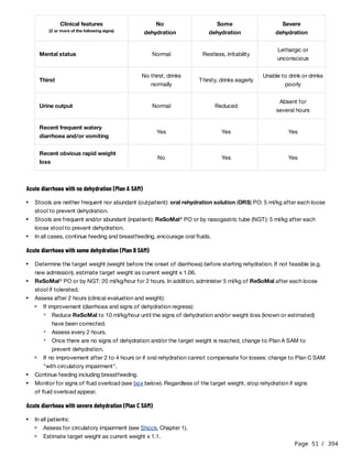
Classification of degree of dehydration (adapted from the WHO)
Treatment of dehydration
History of diarrhoea and/or vomiting and concomitant reduced urine output.
Clinical features depend on the degree of dehydration (see table below). Features such as dry mouth, absence of
tears may also be noted.
Patients with severe dehydration should be assessed for shock (tachycardia, low blood pressure and delayed
capillary refill time etc.).
Electrolyte disorders may cause tachypnoea, muscle cramps or weakness, cardiac arrhythmia (irregular heart rate,
palpitation), confusion and/or seizures.
[1][2]
Severe dehydration
At least 2 of the
following signs:
Some dehydration
At least 2
of the following signs:
No dehydration
No signs of "severe"
or "some" dehydration.
Mental status
Lethargic or
unconscious
Restless or irritable Normal
Radial pulse Weak or absent Palpable Easily palpable
Eyes(a) Sunken Sunken Normal
Skin pinch(b)
Goes back very slowly
(> 2 seconds)
Goes back slowly
(< 2 seconds)
Goes back quickly
(< 1 second)
Thirst
Drinks poorly
or not able to drink
Thirst,
drinks quickly
No thirst,
drinks normally
(a) Sunken eyes may be a normal feature in some children. Ask the mother if the child's eyes are the same as usual or if they are
more sunken than usual.
(b) Skin pinch is assessed by pinching the skin of the abdomen between the thumb and forefinger without twisting. In older
people this sign is not reliable as normal aging diminishes skin elasticity.](https://image.slidesharecdn.com/guideline-170-en-240313103309-343cfe20/85/guideline-170-en-pdf-44-320.jpg)
![Page 45 / 394
Severe dehydration
WHO Treatment Plan C
Some dehydration
WHO Treatment Plan B
Treat shock if present (see Shock, Chapter 1).
If able to drink, administer oral rehydration solution (ORS) PO whilst obtaining IV access.
according to WHO Treatment Plan C, monitoring infusion rate closely:
Insert peripheral IV line using large caliber catheter (22-24G in children or 18G in adults) or intraosseous needle.
Administer Ringer lactate (RL)a
[1][2]
Age First, give 30 ml/kg over :
(c) Then, give 70 ml/kg over:
Children < 1 year 1 hour 5 hours
Children ≥ 1 year and adults 30 minutes 2 ½ hours
(c) Repeat once if radial pulse remains weak or absent after first bolus.
In case of suspected severe anaemia, measure haemoglobin and treat accordingly (see Anaemia, Chapter 1).b
As soon as the patient is able to drink safely (often within 2 hours), provide ORS as the patient tolerates. ORS
contains glucose and electrolytes which prevent development of complications.
Monitor ongoing losses closely. Assess clinical condition and degree of dehydration at regular intervals to ensure
continuation of appropriate treatment.
If over the course of treatment the patient:
remains or becomes lethargic: measure blood glucose level and/or treat hypoglycaemia (see Hypoglycaemia,
Chapter 1).
develops muscle cramps/weakness and abdominal distention: treat for moderate hypokalaemia with 7.5%
potassium chloride syrup (1 mmol of K /ml) PO for 2 days:
Children under 45 kg: 2 mmol/kg (2 ml/kg) daily (according to weight, the daily dose is divided into 2 or 3 doses)
Children 45 kg and over and adults: 30 mmol (30 ml) 3 times daily
This treatment should only be given as an inpatient .
+
c
develops peri-orbital or peripheral oedema: reduce the infusion rate to a minimum, auscultate the lungs, re-
evaluate the stage of dehydration and the necessity of continuing IV rehydration. If IV rehydration is still required,
continue the infusion at a slower rate and observe the patient closely. If IV rehydration is no longer required,
change to oral treatment with ORS.
develops dyspnoea, cough and bibasal crepitations are heard on auscultation of the lungs: sit the patient up,
reduce the infusion rate to a minimum and administer one dose of furosemide IV (1 mg/kg in children; 40 mg in
adults). Monitor the patient closely over 30 minutes and assess for underlying cardiorespiratory or renal disease.
Once the patient is stabilised, reassess the degree of dehydration and the necessity of continuing IV rehydration.
If IV rehydration is still required, re-start at half the previous infusion rate and monitor closely. If IV rehydration is
no longer required, change to oral treatment with ORS.
Administer ORS according to WHO Treatment Plan B which equates to 75 ml/kg ORS given over 4 hours.
[1]d](https://image.slidesharecdn.com/guideline-170-en-240313103309-343cfe20/85/guideline-170-en-pdf-45-320.jpg)
![Page 46 / 394
No dehydration
Prevent dehydration:
WHO Treatment Plan A
Treatment of diarrhoea
In addition to the WHO treatment plan corresponding to patient's degree of dehydration:
Age
< 4
months
4 to
11 months
12 to
23 months
2 to 4 years
5 to
14 years
≥ 15 years
Weight < 5 kg
5 to
7.9 kg
8 to
10.9 kg
11 to 15.9
kg
16 to 29.9
kg
≥ 30 kg
Quantity of ORS over
4 hours
200 to 400
ml
400 to
600 ml
600 to
800 ml
800 to 1200
ml
1200 to
2200 ml
2200 to
4000 ml
Encourage additional age-appropriate fluid intake, including breastfeeding in young children. Give additional ORS
after each loose stool (see below).
Monitor ongoing losses closely. Assess clinical condition and degree of dehydration at regular intervals to ensure
continuation of appropriate treatment.
Encourage age-appropriate fluid intake, including breastfeeding in young children.
Administer ORS according to WHO Treatment Plan A after any loose stool.
[1][2]
Age Quantity of ORS
Children < 2 years 50 to 100 ml (10 to 20 teaspoons)
Children 2 to 10 years 100 to 200 ml (½ to 1 glass)
Children > 10 years and adults at least 250 ml (at least 1 glass)
Administer aetiologic treatment if required.
Administer zinc sulfate to children under 5 years (see Acute diarrhoea, Chapter 3).
Footnotes
(a) If RL not available, 0.9% sodium chloride can be used.
(b) If transfusion is required, it should be provided in parallel to IV fluids, using a separate IV line. The blood volume administered
should be deducted from the total volume of Plan C.
(c) If available, take blood tests to monitor urea and electrolyte levels.
(d) For more detailed information on ORS recommendations by age and weight, refer to the guide Management of a cholera
epidemic, MSF.](https://image.slidesharecdn.com/guideline-170-en-240313103309-343cfe20/85/guideline-170-en-pdf-46-320.jpg)

![Page 50 / 394
Management of complications
Infections
Severe anaemia
Diarrhoea and dehydration
Respiratory, cutaneous and urinary infections are common. However, classic signs of infection, such as fever, may
be absent .
[1]
Severe infection or sepsis should be suspected in children that are lethargic or apathetic or suffering from an acute
complication such as hypothermia, hypoglycaemia, seizures, difficulty breathing, or shock. Immediately
administer ampicillin IV 50 mg/kg every 8 hours + gentamicin IV 7.5 mg/kg once daily. Continue this treatment
unless the source of infection is identified and different antibiotic treatment is required.
If circulatory impairment or shock, immediately administer ceftriaxone IV, one dose of 80 mg/kg, then assess the
source of infection to determine further antibiotic treatment. See also Shock, Chapter 1. Transfuse urgently as for
severe anaemia (see below) if haemoglobin (Hb) is < 6 g/dl.
In less severe infections, assess the source of infection (see Fever, Chapter 1) and treat accordingly.
If fever is present and causes discomfort, undress the child. If insufficient, administer paracetamol PO in low dose:
10 mg/kg, up to 3 times maximum per 24 hours. Encourage oral fluids (including breast milk).
If hypothermia is present, place the child skin-to-skin against the mother's body and cover with a warm blanket.
Treat for infection as above. Check blood glucose level and treat for hypoglycaemia if necessary (see
Hypoglycaemia, Chapter 1).
In children with kwashiorkor, infection of cutaneous lesions is common and may progress to soft tissue or systemic
infection. If cutaneous infection is present, stop amoxicillin and start amoxicillin/clavulanic acid PO. Use
formulations in a ratio of 8:1 or 7:1. The dose is expressed in amoxicillin: 50 mg/kg 2 times daily for 7 days.
Children with Hb < 4 or < 6 with signs of decompensation (such as respiratory distress) or ongoing blood loss require
transfusion within the first 24 hours. See Anaemia (Chapter 1) for volume to be transfused and patient monitoring
during and after transfusion.
Preferably use packed red blood cells (PRBC), if available. Monitor closely for signs of fluid overload (see box
below).
Diarrhoea is common. Therapeutic foods facilitate the recovery of physiological function of the gastrointestinal
tract. Amoxicillin administered as part of routine treatment reduces intestinal bacterial overgrowth. Diarrhoea
generally resolves without additional treatment. If an aetiological treatment is necessary, see Acute diarrhoea,
Chapter 3.
Zinc supplementation is not needed if children consume recommended amounts of therapeutic foods.
The diagnosis of dehydration is based on history and clinical features.
Clinical assessment is difficult in children with SAM as delayed skin pinch test and sunken eyes are often present
even in the absence of dehydration.
For classification of degree of dehydration adapted for children with SAM, see table below:](https://image.slidesharecdn.com/guideline-170-en-240313103309-343cfe20/85/guideline-170-en-pdf-50-320.jpg)

![Page 53 / 394
Discharge criteria
In general:
Discharge criteria vary with context. Refer to national recommendations.
References
Children can be discharged from hospital and be treated as outpatients if the following criteria are met:
clinically well;
medical complications controlled;
able to eat RUTF (observed during appetite test);
reduction or absence of oedema;
caregiver feels able to provide care as outpatient;
vaccinations up to date or referral to vaccination service organised.
Children can be discharged from nutritional treatment if the following criteria are met:
co-existing medical conditions stable and outpatient treatment organised if necessary (e.g. dressing changes,
follow-up for chronic diseases);
vaccinations up to date or referral to vaccination service organised;
absence of oedema and WHZ > –2 or MUAC > 125 mm for at least 2 weeks.
Footnotes
(a) MUAC is measured at the mid-point of the left upper arm. The arm should be relaxed. The measuring tape should be in
contact with the skin all around the arm, without exerting pressure.
(b) For WHZ, see WHO simplified field tables in z-scores for girls and for boys:
https://www.who.int/tools/child-growth-standards/standards/weight-for-length-height
(c) ReSoMal is a specific oral rehydration solution for malnourished children, containing less sodium and more potassium than
standard ORS. It should be administered under medical supervision to avoid overdosing and hyponatremia.
(d) Remove 50 ml of Ringer lactate (RL) from a 500 ml RL bottle or bag, then add 50 ml of 50% glucose to the remaining 450 ml
of RL to obtain 500 ml of 5% glucose-RL solution.
1. Jones KDJ, Berkley JA. Severe acute malnutrition and infection. Paediatrics and International Child Health 2014; 34(sup1):
S1-S29.
https://www.tandfonline.com/doi/full/10.1179/2046904714Z.000000000218 [Accessed 24 August 2022]](https://image.slidesharecdn.com/guideline-170-en-240313103309-343cfe20/85/guideline-170-en-pdf-53-320.jpg)

![Page 61 / 394
Acute pharyngitis
Last updated: November 2020
Acute inflammation of the tonsils and pharynx. The majority of cases are of viral origin and do not require antibiotic
treatment. Group A streptococcus (GAS) is the main bacterial cause, and mainly affects children aged 3 to 14 years.
Acute rheumatic fever (ARF), a serious late complication of GAS pharyngitis, can be prevented with antibiotic
treatment.
One of the main objectives of assessing acute pharyngitis is to identify patients requiring antibiotic treatment.
Clinical features
Common forms:
Centor criteria
In patients over 14 years, the probability of GAS pharyngitis is low. Infectious mononucleosis (IM) due to the
Epstein-Barr virus should be suspected in adolescents and young adults with extreme fatigue, generalized
adenopathy and often splenomegaly.
Erythematous or exudative pharyngitis may also be associated with gonococcal or primary HIV infection. In
these cases, the diagnosis is mainly prompted by the patient's history.
Other forms of pharyngitis:
Features common to all types of pharyngitis: throat pain, dysphagia (difficulty swallowing), inflammation of the
tonsils and pharynx, tender anterior cervical lymph nodes, with or without fever.
Specific features, depending on the cause:
Erythematous (red throat) or exudative (red throat and whitish exudate) pharyngitis: this appearance is
common to both viral and GAS pharyngitis. Centor criteria help assessment and decrease the empirical use of
antibiotics in settings where rapid testing for GAS is not available. A Centor score of less than 2 rules out GAS
infection . Nevertheless, in patients with risk factors (immunosuppression, personal or family history of ARF)
for poststreptococcal complications, or for local or general complications, do not use Centor score and
prescribe empirical antibiotic treatment.
[1][2]
Criteria Score
Temperature > 38 °C 1
Absence of cough 1
Tender anterior cervical lymph node(s) 1
Tonsillar swelling or exudate 1
Pseudomembranous pharyngitis (red tonsils/pharynx covered with an adherent greyish white false membrane):
see Diphtheria, Chapter 2.
Vesicular pharyngitis (clusters of tiny blisters or ulcers on the tonsils): always viral (coxsackie virus or primary
herpetic infection).
Ulcero-necrotic pharyngitis: hard and painless syphilitic chancre of the tonsil; tonsillar ulcer soft on palpation in
a patient with poor oral hygiene and malodorous breath (Vincent tonsillitis).](https://image.slidesharecdn.com/guideline-170-en-240313103309-343cfe20/85/guideline-170-en-pdf-61-320.jpg)
![Page 62 / 394
Peritonsillar, retropharyngeal or lateral pharyngeal abscess: fever, intense pain, dysphagia, hoarse voice, trismus
(limitation of mouth opening), unilateral deviation of the uvula.
Treatment
Spots on oral mucosa (Koplik’s spots) accompanied by conjunctivitis and skin rash (see Measles, Chapter 8).
“Strawberry” (red and bumpy) tongue accompanied by a skin rash: scarlet fever caused by GAS.
Local complications:
General complications:
Complications due to the toxin: diphtheria (see Diphtheria, Chapter 2).
Poststreptococcal complications: ARF, acute glomerulonephritis.
Signs of serious illness in children: severe dehydration, severe difficulty swallowing, upper airway compromise,
deterioration of general condition.
Differential diagnosis: epiglottitis (see Epiglottitis, Chapter 2).
Symptomatic treatment (fever and pain): paracetamol or ibuprofen PO (Fever, Chapter 1).
Centor score ≤ 1: viral pharyngitis, which typically resolves within a few days (or weeks, for IM): no antibiotic
treatment.
Centor score ≥ 2 or scarlet fever: antibiotic treatment for GAS infections :
[3]
If single-use injection equipment is available, benzathine benzylpenicillin is the drug of choice as streptococcus
A resistance to penicillin remains rare; it is the only antibiotic proven effective in reducing the incidence of
rheumatic fever; and the treatment is administered as a single dose.
benzathine benzylpenicillin IM
Children under 30 kg (or under 10 years): 600 000 IU single dose
Children 30 kg and over (or 10 years and over) and adults: 1.2 MIU single dose
Penicillin V is the oral reference treatment, but poor adherence is predictable due to the length of treatment.
phenoxymethylpenicillin (penicillin V) PO for 10 days
Children 1 to < 6 years: 250 mg 2 times daily
Children 6 to < 12 years: 500 mg 2 times daily
Children 12 years and over and adults: 1 g 2 times daily
Children under 1 year: 125 mg 2 times daily
Amoxicillin is an alternative and the treatment has the advantage of being relatively short. However, it can
cause adverse skin reactions in patients with undiagnosed IM and thus should be avoided when IM has not been
excluded
amoxicillin PO for 6 days
Children: 25 mg/kg 2 times daily
Adults: 1 g 2 times daily
Macrolides should be reserved for penicillin allergic patients as resistance to macrolides is frequent and their
efficacy in the prevention of rheumatic fever has not been studied.
azithromycin PO for 3 days
Children: 20 mg/kg once daily (max. 500 mg daily)
Adults: 500 mg once daily
Gonococcal or syphilitic pharyngitis: as for genital gonorrhoea (Chapter 9) and syphilis (Chapter 9).
Diphtherial pharyngitis: see Diphtheria (Chapter 2).
Vincent tonsillitis: metronidazole or amoxicillin.
Peritonsillar retropharyngeal or lateral pharyngeal abscess: refer for surgical drainage.](https://image.slidesharecdn.com/guideline-170-en-240313103309-343cfe20/85/guideline-170-en-pdf-62-320.jpg)
![Page 63 / 394
References
If signs of serious illness or epiglottitis are present in children: hospitalise.
1. Fine AM, Nizet V, Mandl KD. Large-scale validation of the Centor and McIsaac scores to predict group A streptococcal
pharyngitis. Arch Intern Med. 2012;172(11):847-852.
https://www.ncbi.nlm.nih.gov/pmc/articles/PMC3627733/ [Accessed 20 October 2020]
2. National Institute for Health and Care Excellence. Sore throat (acute): antimicrobial prescribing. 2018.
http://www.nice.org.uk/ng84 [Accessed 20 October 2020]
3. Group A Streptococcal Disease, Centers for Disease Control and Prevention. Atlanta (GA): CDC; 2020.
https://www.cdc.gov/groupastrep/diseases-hcp/strep-throat.html [Accessed 20 October 2020]](https://image.slidesharecdn.com/guideline-170-en-240313103309-343cfe20/85/guideline-170-en-pdf-63-320.jpg)
![Page 64 / 394
Diphtheria
Last updated: October 2022
Diphtheria is a bacterial infection due to Corynebacterium diphtheriae, spread from person to person through
inhalation of infected respiratory droplets of symptomatic or asymptomatic individuals, or direct contact with
contaminated objects or diphtheria skin lesions .
After infection, C. diphtheriae has an incubation period of 1 to 5 days (max. 10 days) during which time it multiplies in
the upper respiratory tract. The bacteria secretes a toxin which causes severe local as well as systemic effects. Death
can occur from airway obstruction or as a result of systemic complications, including damage to the myocardium and
nervous system, caused by the toxin.
Cases can remain infectious up to 8 weeks after initial infection . Antibiotic treatment can reduce infectiousness to 6
days .
Vaccination is the key to prevention and control of diphtheria. It protects individuals from severe disease (fewer and
less severe symptoms) but does not prevent the spread of C. diphtheriae. Clinical disease does not confer protective
immunity and vaccination is an integral part of case management.
Clinical features
Laboratory
Treatment
[1][2]a
[1]
[2]
[3]
During clinical examination respect standard, contact, and droplet precautions (handwashing, gloves, gown, mask,
etc.). Conduct a careful examination of the throat.
Signs of respiratory diphtheria :
a
pharyngitis, rhinopharyngitis, tonsillitis or laryngitis with tough, greyish, firmly adherent pseudo-membranes of the
pharynx, nasopharynx, tonsils, or larynx;
dysphagia and cervical adenitis, at times progressing to massive swelling of the neck;
airway obstruction and possible suffocation when the infection extends to the nasal passages, larynx, trachea
and bronchi;
fever is generally low-grade .
[2]
Generalised signs due to effects of the toxin:
cardiac dysfunction (tachycardia, arrhythmias), severe myocarditis with heart failure and possibly cardiogenic
shock (see Shock, Chapter 1) 3 to 7 days or 2 to 3 weeks after onset of the disease;
neuropathies in 2 to 8 weeks after the onset of disease leading to nasal voice and difficulty with swallowing
(paralysis of the soft palate), vision (ocular motor paralysis), breathing (paralysis of respiratory muscles) and
ambulation (limb paralysis);
oliguria, anuria and acute renal failure.
Differential diagnoses: Epiglottitis and Acute pharyngitis, Chapter 2, Stomatitis, Chapter 3.
Diagnosis is confirmed by isolation of toxigenic C. diphtheriae by culture (and antibiotic susceptibility test) of swab
specimens collected from the affected areas: throat (tonsils, pharyngeal mucosa, soft palate, exudate, ulcer, etc.),
nasopharynx.
The presence of the toxin is confirmed by PCR testing (detection of diphtheria toxin gene).
Isolation of patients; standard, droplet, and contact precautions for medical staff.](https://image.slidesharecdn.com/guideline-170-en-240313103309-343cfe20/85/guideline-170-en-pdf-64-320.jpg)
![Page 65 / 394
There is a risk of anaphylactic reaction, especially in patients with asthma. Close monitoring of the patient is
essential, with immediate availability of equipment for manual ventilation (Ambu bag, face mask) and
intubation, Ringer lactate and epinephrine (see Shock, Chapter 1).
Besredka method: inject 0.1 ml SC and wait 15 minutes. If there is no allergic reaction (no erythema at the injection
site or a flat erythema of less than 0.5 cm in diameter), inject a further 0.25 ml SC. If there is no reaction after 15
minutes, inject the rest of the product IM or IV depending on the volume to be administered.
Doses are given as a function of the severity of illness, and the delay in treatment:
Never administer procaine benzylpenicillin by IV injection or infusion.
In penicillin-allergic patients, use erythromycin IV .
Diphtheria antitoxin (DAT) derived from horse serum:
Administer DAT as soon as possible after disease onset. Do not wait for bacteriological confirmation ; administer
DAT under close monitoring in a hospital setting, according to the Besredka method to assess possibility of allergy.
Any delay can diminish efficacy.
b
[1]
Clinical signs Dose in units Administration route
Laryngitis or pharyngitis
or duration < 48 hours
20 to 40 000
IM or IV infusion in 250 ml of 0.9% sodium
chloride in 2 to 4 hours for doses of more
than 20 000 units.
Rhinopharyngitis 40 to 60 000
Severe disease (respiratory distress,
shock), cervical oedema or duration ≥ 48
hours
80 to 100 000
Antibiotic treatment (as soon as possible without waiting for bacteriological confirmation ) for 14 days or according
to length of treatment recommended by the national protocol:
if the patient can swallow:
azithromycin PO (first-line)
Children: 10 to 12 mg/kg once daily (max. 500 mg daily)
Adults: 500 mg once daily
or
erythromycin PO
Children under 40 kg: 10 to 15 mg/kg (max. 500 mg) 4 times daily
Children 40 kg and over and adults: 500 mg 4 times daily
or
phenoxymethylpenicillin (penicillin V) PO
Children under 40 kg: 10 to 15 mg/kg (max. 500 mg) 4 times daily
Children 40 kg and over and adults: 500 mg 4 times daily
If the patient cannot swallow, start with one of the treatments below and change as soon as possible to oral
route with one of the oral treatments above to complete 14 days of treatment:
procaine benzylpenicillin IM
Children under 25 kg: 50 000 IU/kg (= 50 mg/kg) once daily (max. 1.2 MIU = 1.2 g daily)
Children 25 kg and over and adults: 1.2 MIU (= 1.2 g) once daily
c
Intubation/tracheotomy if necessary (airway obstruction, respiratory failure, etc.).
If the event of shock, see Shock, Chapter 1, for complementary treatment.
Update every patient's vaccination status before hospital discharge (or during first visit, if receiving home-based
care). If the patient has been administered DAT and can receive adequate home-based follow up after hospital
discharge, wait 3 weeks after administration of DAT before vaccination.](https://image.slidesharecdn.com/guideline-170-en-240313103309-343cfe20/85/guideline-170-en-pdf-65-320.jpg)
![Page 66 / 394
Management of close contacts
Close contacts include household members living under the same roof and people who were directly exposed (less than
one metre) to nasopharyngeal secretions of the patient on a regular basis (e.g. family or close friends, children in the
same class, medical personnel) during the 5 days or nights prior to onset of symptoms of the case .
Benzathine benzylpenicillin should never be administered by IV route.
or azithromycin PO or erythromycin PO as above for 7 days.
Outbreak surveillance measures
AND
Prevention
Administer 2 subsequent booster doses containing d at least 4 weeks apart .
[4]
Collect nasal and pharyngeal swabs for culture before starting antibiotic prophylaxis; temperature and throat
examination daily (10 days); exclusion from school or work until 48 hours after starting antibiotic prophylaxis. If
symptoms of respiratory infection appear: treat immediately as a case of diphtheria.
Antibiotic prophylaxis:
benzathine benzylpenicillin IM
Children under 30 kg: 600 000 IU single dose
Children 30 kg and over and adults: 1.2 MIU single dose
Check vaccination status:
if less than 3 injections received: complete vaccination schedule (see Prevention below);
if 3 injections received, with the last injection over one year ago: administer a booster dose immediately;
if 3 injections received, with the last injection less than one year ago: a booster dose is not immediately
necessary.
A suspected case of diphtheria is defined as a person with:
pharyngitis, rhinopharyngitis, tonsillitis and/or laryngitis
an adherent pseudo-membrane of the pharynx, nose, tonsils and/or larynx .
[1]
Isolate and treat suspect cases without delay. Collect swab samples before starting antibiotic treatment. Submit
case notification to the public health authorities within 24 hours .
[1]
Routine vaccination (EPI), for information: 3 doses of conjugate vaccine containing the higher potency (D)
formulation of diphtheria toxoid as soon as possible as of 6 weeks of age and at 4 week intervals; D booster
between 12 and 23 months, then between 4 and 7 years; booster with a vaccine containing a reduced dose (d) of
diphtheria toxoid between 9 and 15 years .
[5]
Catch-up vaccination (individuals who have not received routine vaccination), for information:
children 1 to 6 years: 3 doses of conjugate vaccine containing the higher potency (D) formulation of diphtheria
toxoid at least 4 weeks apart;
children 7 years and over and adults (including medical staff): 3 doses of conjugate vaccine containing a reduced
dose (d) of diphtheria toxoid. Administer with a minimum interval of 4 weeks between first and second dose and
an interval of at least 6 months between second and third dose (in the event of an outbreak this interval may be
reduced to 4 weeks to achieve protection quicker).
[5]
Footnotes
(a) This guide focuses on respiratory diphtheria and signs due to the toxin. It should be noted that cutaneous diphtheria is still
a significant reservoir of C. diphtheriae.](https://image.slidesharecdn.com/guideline-170-en-240313103309-343cfe20/85/guideline-170-en-pdf-66-320.jpg)
![Page 67 / 394
References
(b) DAT reduces mortality and should be given to all diphtheria patients. However, as supply is very limited, it may be necessary
to define criteria and reserve DAT for the treatment of patients who will benefit the most from it. DAT can be administered
to pregnant women.
(c) erythromycin IV infusion (60 minutes)
Children: 12.5 mg/kg every 6 hours (max. 2 g daily); adults: 500 mg every 6 hours
Erythromycin powder (1 g) should be reconstituted in 20 ml of water for injection only. Then, dilute each dose of
erythromycin in 10 ml/kg of 0.9% sodium chloride in children less than 20 kg and in a bag of 250 ml of 0.9% sodium chloride
in children 20 kg and over and in adults. Do not dilute in glucose.
1. World Health Organization. Diphtheria. Vaccine-Preventable Diseases Surveillance Standards. 2018.
https://www.who.int/immunization/monitoring_surveillance/burden/vpd/WHO_SurveillanceVaccinePreventable_04_Diphtheria
_R2.pdf?ua=1 [Accessed 11 August 2020]
2. Tiwari TSP
, Wharton M. Chapter 19: Diphtheria Toxoid. In: Plotkin SA, Orenstein WA, Offit PA, editors. Vaccines. 7th ed.
Philadelphia, PA: Elsevier; 2018. p. 261–275.
3. Truelove SA, Keegan LT, Moss WJ, Chaisson LH, Macher E, Azman AS, Lessler J. Clinical and Epidemiological Aspects of
Diphtheria: A Systematic Review and Pooled Analysis. Clin Infect Dis. 2020 Jun 24;71(1):89-97.
https://www.ncbi.nlm.nih.gov/pmc/articles/PMC7312233/ [Accessed 24 November 2020]
4. Pan American Health Organization, World Health Organization. Diphtheria in the Americas - Summary of the situation 2018.
Epidemiological Update Diphtheria. 16 April 2018.
https://www.paho.org/hq/index.php?option=com_docman&view=download&category_slug=diphtheria-
%098968&alias=44497-16-april-2018-diphtheria-epidemiological-update-497&Itemid=270&lang=en [Accessed 11 August
2020]
5. World Health Organization. Diphtheria vaccine: WHO position paper - August 2017. Weekly epidemiological record 2017;
92/(31):417–436.
https://www.who.int/immunization/policy/position_papers/wer_31_diphtheria_updated_position_paper.pdf?ua=1 [Accessed
11 August 2020]](https://image.slidesharecdn.com/guideline-170-en-240313103309-343cfe20/85/guideline-170-en-pdf-67-320.jpg)
![Page 69 / 394
Croup (laryngotracheitis and
laryngotracheobronchitis)
Last updated: December 2023
Common viral respiratory infection with peak incidence amongst children between 6 months and 3 years.
Clinical features
Treatment
Typical barking cough, hoarse voice or cry.
Inspiratory stridor (abnormal high pitched sound on inspiration):
Croup is considered mild if the stridor only occurs with agitation;
Croup is considered severe if there is stridor at rest, especially when it is accompanied by respiratory distress.
Wheezing may also be present if the bronchi are involved.
In the absence of inspiratory stridor or intercostal, subcostal or sternal retractions, treat symptomatically: ensure
adequate hydration, seek medical attention if symptoms worsen (e.g. respiratory difficulty, noisy breathing, inability
to tolerate oral fluids).
If stridor is only present with agitation (mild croup) :
[1]
Assure adequate hydration.
Corticosteroids:
dexamethasone PO: 0.15 to 0.6 mg/kg (max. 16 mg) single dose
a
or, if not available, prednisolone PO: 1 mg/kg single dose
Keep the child under observation at least 30 minutes after oral corticosteroid. Consider hospitalisation or longer
observation (> 4 hours) if the child is less than 6 months old, or is dehydrated, or lives far from health facility.
If danger signs are present (stridor at rest, respiratory distress, hypoxia) or the child is unable to drink, admit
to hospital :
[1]
Administer oxygen continuously if respiratory distress or SpO < 92%: maintain SpO between 94 and 98% (or if
SpO cannot be determined, at least 5 litres/minute).
2 2
2
Insert a peripheral IV line and provide IV hydration.
Epinephrine (adrenaline) via nebulizer: 0.5 mg/kg (max. 5 mg) to be repeated every 20 minutes if danger signs
persist (see table below).
Monitor heart rate during nebulization (if heart rate greater than 200, stop the nebulization).
Weight 6 kg 7 kg 8 kg 9 kg 10-17 kg
Dose in mg 3 mg 3.5 mg 4 mg 4.5 mg 5 mg
Dose in ml (1 mg/ml, 1 ml ampoule) 3 ml 3.5 ml 4 ml 4.5 ml 5 ml
NaCl 0.9%(a) 1 ml 1 ml – – –
(a) Add sufficient NaCl 0.9% to obtain a total volume of 4 to 4.5 ml in the nebulizing chamber.](https://image.slidesharecdn.com/guideline-170-en-240313103309-343cfe20/85/guideline-170-en-pdf-69-320.jpg)


![Page 95 / 394
Acute asthma (asthma attack)
Last updated: June 2023
Asthma attack is a substantial worsening of asthma symptoms. The severity and duration of attacks are variable and
unpredictable.
Assessment of the severity of asthma attack
The severity of the asthma attack must be rapidly evaluated by the following clinical criteria. Not all signs are
necessarily present.
Assessment of severity in children over 2 years and adults
Treatment
Reassure the patient. Treatment and follow-up depend on the severity of the attack and the patient’s response:
Mild to moderate attack
[1][2][3]
Mild or moderate attack Severe or life-threatening attack
Able to talk in sentences Cannot complete sentences in one breath
or
Too breathless to talk or feed
Mild or moderate increase of
respiratory rate (RR)
Very high RR
Children 2-5 years: > 40/minute
Children > 5 years and adults: > 30/minute
Normal or mild increase of heart rate (HR)
Children 2-3 years: ≤ 180/minute
Children 4-5 years: ≤ 150/minute
Children > 5 years and adults: ≤ 120/minute
Very high HR
Children 2-3 years: > 180/minute
Children 4-5 years: > 150/minute
Children > 5 years and adults: > 120/minute
SpO ≥ 90% (≥ 92% for children 2-5 years)
2 SpO < 90% (< 92% for children 2-5 years)
2
and
No criteria of severe or life-threatening attack Signs of life-threatening attack:
Altered level of consciousness (drowsiness, confusion, coma)
Exhaustion
Silent chest
Cyanosis
Arrhythmia or hypotension in adults
Place the patient in a 1/2 sitting position.
Administer:](https://image.slidesharecdn.com/guideline-170-en-240313103309-343cfe20/85/guideline-170-en-pdf-95-320.jpg)

![Page 97 / 394
Notes:
References
Children: 40 mg/kg (max. 2 g)
Adults: 2 g
If symptoms improve: continue salbutamol (solution for nebuliser) every 1 to 4 hours (depending on symptoms) and
oxygen as above. Assess symptoms at the end of each nebulisation. When possible, switch to salbutamol MDI
and continue as for mild to moderate attack.
If the attack is completely resolved, observe the patient for at least 4 hours. Continue the treatment with
salbutamol (MDI) and prednisolone PO and reassess as for a mild to moderate attack.
In pregnant women, treatment is the same as for adults. In mild or moderate asthma attacks, administering oxygen
reduces the risk of foetal hypoxia.
For all patients, irrespective of the severity of the asthma attack, look for underlying lung infection and treat
accordingly.
Footnotes
(a) If a conventional spacer is not available, use a 500 ml plastic bottle: insert the mouthpiece of the inhaler into a hole made in
the bottom of the bottle (the seal should be as tight as possible). The patient breathes from the mouth of the bottle in the
same way as they would with a spacer. The use of a plastic cup instead of a spacer is not recommended (ineffective).
(b) If pulse oxymetry is not available, administer oxygen continuously in case of moderate, severe or life-threatening attack.
(c) If signs of life-threatening attack, transfer to intensive care unit as soon as possible.
1. British guideline on the management of asthma. A national clinical guideline First published 2003. Revised edition published
July 2019.
https://www.sign.ac.uk/our-guidelines/british-guideline-on-the-management-of-asthma/ [Accessed 12 January 2023]
2. Global INitiative for Asthma. Global Strategy for Asthma Management and Prevention. 2022 update.
https://ginasthma.org/gina-reports/ [Accessed 12 January 2023]
3. WHO Pocket book of primary health care for children and adolescents: guidelines for health promotion, disease prevention
and management from the newborn period to adolescence. WHO Regional Office for Europe; 2022.
https://www.who.int/europe/publications/i/item/9789289057622 [Accessed 12 January 2023]](https://image.slidesharecdn.com/guideline-170-en-240313103309-343cfe20/85/guideline-170-en-pdf-97-320.jpg)
![Page 98 / 394
Chronic asthma
Last updated: June 2023
Clinical features
Treatment
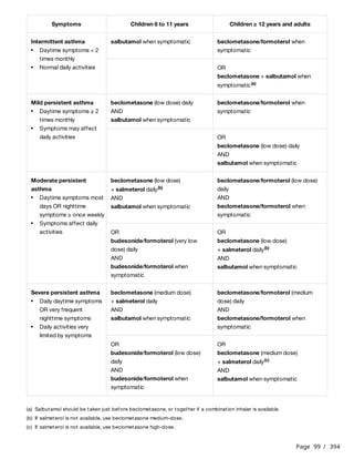
The mainstay of long-term treatment are inhaled corticosteroids (ICS) and long-acting beta-2 agonists (LABA). LABAs
should never be used alone but always in combination with an ICS. Combination inhalers are preferred, when available.
In addition to long-term treatment, salbutamol (short-acting beta-2 agonist, SABA) and combination inhalers can be
used to reduce bronchoconstriction if the patient is symptomatic.
Treatment is started at the step most appropriate to initial severity then, re-evaluated and adjusted according to
clinical response. An intervening severe asthma attack or loss of control necessitates treatment reassessment.
The inhaler is chosen according to age. In children, a spacer should be used. Instructions on inhaler technique and
information on asthma attack symptoms should be provided.
Long-term treatment of asthma according to severity in children 6 years and over and adults
Asthma should be suspected in patients with recurrent respiratory symptoms (wheezing, chest tightness, shortness
of breath and/or cough) of variable frequency, severity and duration, disturbing sleep, and causing the patient to sit
up to breathe. These symptoms may appear during or after exercise.
Chest auscultation may be normal or demonstrate diffuse sibilant wheezes.
A personal or family history of atopy (eczema, allergic rhinitis/conjunctivitis) or a family history of asthma increases
probability of asthma but their absence does not exclude asthma.
Patients with typical symptoms of asthma and a history of disease that is characteristic of asthma should be
considered as having asthma after exclusion of other diagnoses.
Any identified asthma risk factor (e.g. allergen, pollution, tobacco smoke exposure) should be eliminated where
possible. The assessment of the frequency of symptoms and limitations of daily activities determines the
treatment.
[1][2]](https://image.slidesharecdn.com/guideline-170-en-240313103309-343cfe20/85/guideline-170-en-pdf-98-320.jpg)
![Page 101
/ 394
If symptoms have not been well controlled for a period of 2 to 3 months, check inhalation technique and adherence
before changing to a stronger treatment.
If symptoms have been well controlled for a period of at least 3 months (the patient is asymptomatic or the asthma
attacks are well controlled): try a step-wise reduction in medication.
References
1. Global INitiative for Asthma. Global Strategy for Asthma Management and Prevention. 2022 update.
https://ginasthma.org/gina-reports/ [Accessed 23 January 2023]
2. WHO Pocket book of primary health care for children and adolescents: guidelines for health promotion, disease prevention
and management from the newborn period to adolescence. WHO Regional Office for Europe; 2022.
https://www.who.int/europe/publications/i/item/9789289057622 [Accessed 23 January 2023]](https://image.slidesharecdn.com/guideline-170-en-240313103309-343cfe20/85/guideline-170-en-pdf-101-320.jpg)
![Page 102
/ 394
Pulmonary tuberculosis
Pulmonary tuberculosis is a bacterial infection due to Mycobacterium tuberculosis, spread from person to person
through inhalation of infected respiratory droplets.
After infection, M. tuberculosis multiplies slowly in the lungs and is usually eliminated spontaneously or lies dormant.
Only 10% of cases develop active tuberculosis. The risk of progressing to active tuberculosis is higher in
immunocompromised patients. In certain countries, half of newly diagnosed tuberculosis patients are co-infected with
HIV .
For more information on tuberculosis, refer to the guide Tuberculosis, MSF.
Clinical features
In an endemic area, the diagnosis of tuberculosis is to be considered, in any patient consulting for respiratory
symptoms for over 2 weeks who does not respond to non-specific antibacterial treatment.
Laboratory
Treatment
For pulmonary tuberculosis, the standard treatment is a combination of four antituberculosis drugs (isoniazid,
rifampicin, pyrazinamide, ethambutol). The regimen is organised into 2 phases (initial phase and continuation phase) and
lasts 6 months.
If the strain is drug-resistant, the treatment is longer and different drug combinations are used.
It takes significant investment to cure tuberculosis, both from the patient and the medical team. Only uninterrupted
treatment will lead to cure and prevent the development of resistance. It is essential that the patient understands the
importance of treatment adherence and has access to correct case management until treatment is completed.
Prevention
References
[1]
Prolonged cough (> 2 weeks) with or without sputum production and/or haemoptysis, prolonged fever, night sweats,
anorexia, weight loss, chest pain and fatigue.
Differential diagnosis includes pneumonia, chronic obstructive pulmonary disease (COPD), lung cancer, pulmonary
distomatosis (Flukes, Chapter 6) and melioidosis (Southeast Asia).
In the general population: Xpert® MTB/RIF test which simultaneously detects M. tuberculosis (MTB) in sputum and
resistance to rifampicin (RIF). If not available perform sputum smear microscopy .
[2]
If HIV co-infection suspected or diagnosed: Xpert® MTB/RIF test and point-of-care, urine LF-LAM (lateral flow
urine lipoarabinomannan assay) .
[2]
BCG vaccination in neonates: provides 59% protection against pulmonary tuberculosis .
[3]
Infection control in healthcare settings: standard precautions and airborne precautions for confirmed or suspected
cases.
Close contacts: isoniazid preventive therapy for 6 months.](https://image.slidesharecdn.com/guideline-170-en-240313103309-343cfe20/85/guideline-170-en-pdf-102-320.jpg)
![Page 103
/ 394
1. World Health Organization. Global tuberculosis report 2018.
https://apps.who.int/iris/handle/10665/274453 [Accessed 21 October 2019]
2. Global Laboratory Initiative. GLI model TB diagnostic algorithms. 2018.
http://www.stoptb.org/wg/gli/assets/documents/GLI_algorithms.pdf [Accessed 21 October 2019]
3. World Health Organization. Weekly epidemiological record/Relevé épidémiologique hebdomadaire 23rd February 2018,
93rd year/23 Février 2018, 93e année. No 8, 2018, 93, 73–96.
https://www.who.int/immunization/policy/position_papers/bcg/en/ [Accessed 21 October 2019]](https://image.slidesharecdn.com/guideline-170-en-240313103309-343cfe20/85/guideline-170-en-pdf-103-320.jpg)

![Page 106
/ 394
Zinc sulfate is given in combination with oral rehydration solution in order to reduce the duration and severity of
diarrhoea, as well as to prevent further occurrences in the 2 to 3 months after treatment:
zinc sulfate PO
Children under 6 months: 10 mg (½ tablet) once daily for 10 days
Children from 6 months to 5 years: 20 mg (1 tablet) once daily for 10 days
Place the half-tablet or full tablet in a teaspoon, add a bit of water to dissolve it, and give the entire spoonful to the
child.
Antimicrobial treatment
Diarrhoea without blood
Most acute diarrhoeas are caused by viruses unresponsive to antimicrobials. Antimicrobials can be beneficial in the
event of cholera or giardiasis.
Diarrhoea with blood
Prevention
References
Cholera: the most important part of treatment is rehydration. In the absence of resistance (perform antibiotic-
sensitivity testing at the beginning of an outbreak), antibiotic treatment shortens the duration of diarrhoea. See the
guide Management of a cholera epidemic, MSF.
Giardiasis: see Intestinal protozoan infections, Chapter 6.
Shigellosis is the most frequent cause of bloody diarrhoea (amoebiasis is much less common). If there is no
laboratory diagnosis to confirm the presence of amoebae, first line treatment is for shigellosis (Chapter 3).
Amoebiasis: antiparasitic treatment only if motile Entamoeba histolytica amoebae are found in stools or if a
correct shigellosis treatment has been ineffective (see Amoebiasis, Chapter 3).
Breastfeeding reduces infant morbidity and mortality from diarrhoea and the severity of diarrhoea episodes.
When the child is weaned preparation and storage of food are associated with the risk of contamination by faecal
micro-organisms: discourage bottle-feeding; food must be cooked well; milk or porridge must never be stored at
room temperature.
Access to sufficient amounts of clean water and personal hygiene (washing hands with soap and water before food
preparation and before eating, after defecation etc.) are effective methods of reducing the spread of diarrhoea.
In countries with a high rotavirus diarrhoea fatality rate, the WHO recommends routine rotavirus vaccination in
children between 6 weeks and 24 months of age .
[1]
1. Weekly epidemiological record/Relevé épidémiologique hebdomadaire 1st February 2013, 88th year/1er Février 2013, 88e
année No. 5, 2013, 88, 49–64.
https://www.who.int/wer/2013/wer8805.pdf [Accessed 02 January 2019]](https://image.slidesharecdn.com/guideline-170-en-240313103309-343cfe20/85/guideline-170-en-pdf-106-320.jpg)
![Page 108
/ 394
azithromycin PO for 5 days
Children: one dose of 12 mg/kg on D1 then 6 mg/kg once daily from D2 to D5
Adults: one dose of 500 mg on D1 then 250 mg once daily from D2 to D5
or
cefixime PO for 5 days
Children: 8 mg/kg once daily (max. 400 mg daily)
Adults: 400 mg once daily
If there is no improvement 48 hours after starting second-line treatment, treat for amoebiasis .
Shigellosis in an epidemic context
References
[1][2]
For pain and/or fever:
paracetamol PO (see Pain, Chapter 1). All opioid analgesics are contra-indicated as they slow peristalsis.
Supportive therapy:
nutrition: nutritional supplement with frequent meals
+ 2500 kcal daily during hospitalisation
+ 1000 kcal daily as outpatients
rehydration: administration of ORS according to WHO protocols (see Dehydration, Chapter 1).
zinc supplement in children under 5 years (see Acute diarrhoea, Chapitre 3).
Never give loperamide or any other antidiarrhoeal.
Management of complications: rectal prolapse reduction, septicaemia (see Septic shock, Chapter 1), etc.
Isolation of hospitalised patients; school exclusion of children treated as outpatients.
Hygiene (handwashing, hygienic preparation and storage of food, home hygiene, etc.).
Management if signs worsen or bloody diarrhoea in entourage (seek medical attention).
Footnotes
(a) This definition excludes: blood detected on microscope examination; stool containing digested blood (melaena); streaks of
blood on the surface of normal stool (haemorrhoids, anal or rectal lesion, etc.).
(b) Ciprofloxacin should be avoided in pregnant women. Nevertheless, if ceftriaxone is not available, the other antibiotics can
be used, including ciprofloxacin if necessary.
1. Karen L. Kotloff et al. Seminar: Shigellosis. The Lancet, Volume 391, ISSUE 10122, P801-812, February 24, 2018.
2. Word Health Organization. Pocket book for hospital care in children: guidelines for the management of common childhood
illnesses, 2013.
http://apps.who.int/iris/bitstream/handle/10665/81170/9789241548373_eng.pdf;jsessionid=CE5C46916607EF413AA9FCA89B
84163F?sequence=1 [Accessed 20 September 2018]](https://image.slidesharecdn.com/guideline-170-en-240313103309-343cfe20/85/guideline-170-en-pdf-108-320.jpg)
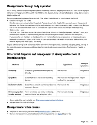
![Page 115
/ 394
Dyspepsia
Last updated: December 2020
Clinical features
Epigastric pain or discomfort following meals, often accompanied by bloating, sensation of fullness and nausea.
Dyspepsia is most commonly functional. The diagnosis of functional dyspepsia is based on clinical assessment after
ruling out organic causes (Gastro-oesophageal reflux, Gastric and duodenal ulcers, drug-induced symptoms, gastric
cancer). If possible, test for Helicobacter pylori.
Treatment
In adults:
Note: consider and treat possible intestinal parasites (see Intestinal protozoan infections, Cestodes, Nematode
infections, Chapter 6; Amoebiasis, Chapter 3).
References
In case of patients who test positive for H. pylori, see Eradication of Helicobacter pylori .
[1]
Omeprazole PO (10 mg once daily) for 4 weeks may help even in H. pylori-negative patients .
[2][3]
1. Ford AC, Mahadeva S, Carbone MF, Lacy BE, Talley NJ. Functional dyspepsia. Lancet. 2020 Nov 21;396(10263):1689-1702.
2. Moayyedi PM, Lacy BE, Andrews CN, et al. ACG and CAG clinical guideline: management of dyspepsia. Am J Gastroenterol.
2017 Jul;112(7):988-1013.
http://www.cag-acg.org/images/publications/CAG_CPG_Dyspepsia_AJG_Aug2017.pdf [Accessed 24 November 2020]
3. National Institute for Health and Care Excellence. Gastro-oesophageal reflux disease and dyspepsia in adults: investigation
and management. Sept 2014.
https://www.nice.org.uk/guidance/CG184/chapter/1-Recommendations#interventions-for-functional-
dyspepsia [Accessed 24 November 2020]](https://image.slidesharecdn.com/guideline-170-en-240313103309-343cfe20/85/guideline-170-en-pdf-115-320.jpg)


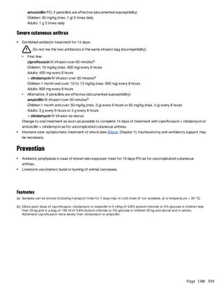
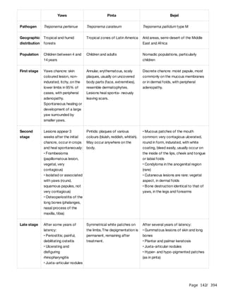
![Page 143
/ 394
Treatment
Yaws
azithromycin PO
Children and adults: 30 mg/kg single dose (max. 2 g)
or, if not available,
benzathine benzylpenicillin IM
Children under 10 years: 1.2 MIU single dose
Children 10 years and over and adults: 2.4 MIU single dose
Pinta and bejel
benzathine benzylpenicillin IM.
As for yaws.
For patients allergic to penicillin:
doxycycline PO (except in children under 8 years and pregnant or lactating women)
Children 8 years and over: 50 mg 2 times daily for 14 days
Adults: 100 mg 2 times daily for 14 days
Notes:
Treatment of contacts and latent cases
The same treatment should be administered to all symptomatic and asymptomatic contacts and to all latent cases
(asymptomatic individuals with positive serologic test for syphilis) in endemic zones.
References
[1]
[2][3]
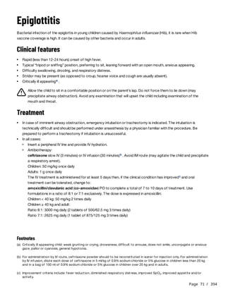
Antibiotic treatment will cure early stage cases and may relieve the pain of osteitis. It may be ineffective for late
stage infections.
Syphilis serology will remain positive despite clinical cure.
1. World Health Organization (
2012)
. Yaws: recognition booklet for communities. Reprinted with changes, 2014.
http://www.who.int/iris/handle/10665/75360 [Accessed 15 May 2018]
2. Oriol Mitjà, David Mabey. Yaws, bejel, and pinta (last updated. May 07, 2018). UpToDate [Accessed 15 May 2018].
3. Michael Marks, Anthony W Solomon, David C Mabey. Endemic treponemal diseases. Transactions of The Royal Society of
Tropical Medicine and Hygiene, Volume 108, Issue 10, 1 October 2014, Pages 601–607.
https://doi.org/10.1093/trstmh/tru128 [Accessed 15 May 2018]](https://image.slidesharecdn.com/guideline-170-en-240313103309-343cfe20/85/guideline-170-en-pdf-143-320.jpg)
![Page 144
/ 394
Leprosy
Leprosy is a chronic bacterial infection due to Mycobacterium leprae.
It is transmitted by frequent close contact, mainly between household members.
It mainly affects young adults. 94% of reported cases globally were in Bangladesh, Brazil, Democratic Republic of
Congo, Ethiopia, India, Indonesia, Madagascar, Myanmar, Nepal, Nigeria, the Philippines, Sri Lanka and the United
Republic of Tanzania.
Clinical features
Leprosy should be considered in any patient presenting with:
There are different clinical forms and classification systems of leprosy.
Ridley-Jopling classification
This classification differentiates 5 forms based on the bacteriological index. These forms correlate with the
immunological response to M. leprae. Patients with tuberculoid leprosy (TT) are resistant to the bacillus and infection
is localised. Patients with lepromatous leprosy (LL) are extremely sensitive to the bacillus and the infection is
disseminated. Borderline forms (BT, BB, BL) are between the two ends of the spectrum (TT and LL).
WHO classification
In order to simplify diagnosis and to promote rapid implementation of treatment, the WHO has simplified clinical
classification of leprosy and differentiates only 2 forms:
Multibacillary leprosy includes LL, BL and BB forms and paucibacillary leprosy includes the TT and BT forms of the
Ridley-Jopling classification system.
Laboratory
[1]
Hypopigmented or erythematous skin lesion(s) with partial or complete loss of sensation to touch, pain, heat;
Infiltrated pigmented nodules, initially with no sensory loss, on the face, ear lobes and the upper and lower limbs;
Tender, infiltrated and hypertrophied peripheral nerve (ulnar, radial, median, popliteal, tibial etc.) with possible
paraesthesia of the extremities, trophic changes (perforating ulcer of the foot) or paralysis (steppage gait,
deformaties of hands and feet, facial nerve paralysis).
Paucibacillary forms
(least contagious forms)
Multibacillary forms
(most contagious forms)
Tuberculoid Borderline
Tuberculoid
Borderline Borderline
Lepromatous
Lepromatous
T.T. B.T. B.B. B.L. L.L.
Multibacillary leprosy: more than 5 skin lesions
Paucibacillary leprosy: 1 to 5 skin lesions
Laboratory diagnosis is based on the detection of acid-fast bacilli in a Ziehl-Neelsen stained nasal smear and skin-
split smear taken from the ear lobe or from a skin lesion. In TT leprosy bacilli are not found.
In practice, in most endemic countries diagnosis is based on the WHO clinical classification (number of lesions).](https://image.slidesharecdn.com/guideline-170-en-240313103309-343cfe20/85/guideline-170-en-pdf-144-320.jpg)
![Page 145
/ 394
Treatment
Countries where leprosy is endemic have a control programme. Check national recommendations.
First-line treatment regimens recommended by the WHO
Note: the monthly doses of rifampicin and clofazimine are administered under direct observation by medical staff
whereas the daily doses of clofazimine and dapsone are taken by the patient at home. Rifampicin should be taken on
an empty stomach to improve absorption.
Teach the patient to recognise and quickly report a lepra reaction or relapse in order to modify or restart treatment.
Leprosy reactions
These reactions usually occur during the course of treatment in patients with multibacillary leprosy (BL and LL). They
are associated with the immunological response to M. leprae antigens. Urgent treatment is required to avoid
irreversible disability. Do not interrupt ongoing leprosy treatment.
Clinical features
Treatment
Age
Multibacillary leprosy
(more than 5 skin lesions)
Paucibacillary leprosy
(1 to 5 skin lesions)
Children 10 to 14 years rifampicin PO: 450 mg once monthly
+ clofazimine PO: 150 mg once monthly
and 50 mg on alternate days
+ dapsone PO: 50 mg once daily
rifampicin PO: 450 mg once monthly
+ clofazimine PO: 150 mg once monthly
and 50 mg on alternate days
+ dapsone PO: 50 mg once daily
Children 15 years and
over and adults
rifampicin PO: 600 mg once monthly
+ clofazimine PO: 300 mg once monthly
and 50 mg once daily
+ dapsone PO: 100 mg once daily
rifampicin PO: 600 mg once monthly
+ clofazimine PO: 300 mg once monthly
and 50 mg once daily
+ dapsone PO: 100 mg once daily
Duration 12 months 6 months
Reversal reactions:
Exacerbation of the skin lesions that become erythematous and oedematous and risk or ulceration. Onset or
worsening of numbness of skin lesions;
Onset of acute painful hypertrophic neuritis.
Erythema nodosum leprosum:
Fever, asthenia, alteration of the general state;
Crops of purplish-red, tender subcutaneous nodules, warmer than the surrounding skin.
Reversal reactions:
prednisolone (or prednisone) PO: 0.5 to 1 mg/kg once daily for 2 weeks. Re-examine the patient every 2 weeks
and decrease the dosage if the neurological signs recede. According to clinical response, treatment may last 3 to 6
months.
For example, for an adult:
Week 1 and 2: 40 mg once daily
[2]
[3]](https://image.slidesharecdn.com/guideline-170-en-240313103309-343cfe20/85/guideline-170-en-pdf-145-320.jpg)
![Page 146
/ 394
References
Week 3 and 4: 30 mg once daily
Week 5 and 6: 20 mg once daily
Week 7 and 8: 15 mg once daily
Week 9 and 10: 10 mg once daily
Week 11 and 12: 5 mg once daily
Erythema nodosum leprosum:
prednisolone (or prednisone) PO as for reversal reactions, for 3 months.[2]
Fever: paracetamol PO (see Fever, Chapter 1)
1. World Health Organization. Global Leprosy Programme. Global leprosy strategy 2016-2020. Accelerating towards a leprosy-
free world, 2016.
http://apps.who.int/iris/bitstream/handle/10665/208824/9789290225096_en.pdf?sequence=14&isAllowed=y [Accessed 17
October 2018]
2. World Health Organization. WHO Expert Committee on Leprosy. Eighth report. WHO technical report series, n° 968. Geneva,
2012.
http://www.searo.who.int/entity/global_leprosy_programme/publications/8th_expert_comm_2012.pdf [Accessed 17 October
2018]
3. World Health Organization. A guide to eliminating leprosy as a public health problem. Leprosy Elimination Group, 2000.
http://apps.who.int/iris/bitstream/handle/10665/66612/WHO_CDS_CPE_CEE_2000.14.pdf?sequence=1 [Accessed 17
October 2018]](https://image.slidesharecdn.com/guideline-170-en-240313103309-343cfe20/85/guideline-170-en-pdf-146-320.jpg)

![Page 154
/ 394
Pellagra
Pellagra is a dermatitis resulting from niacin and/or tryptophane deficiency (in persons whose staple food is sorghum;
patients with malabsorption, or during famine).
Clinical features
Classically, disease of the ‘three Ds’: dermatitis, diarrhoea and dementia.
Treatment
Children and adults: 100 mg 3 times daily, give with a diet rich in protein until the patient is fully cured.
References
Dark red plaques, well demarcated, symmetric, located on exposed areas of the body (forehead, neck, forearms,
legs). The skin becomes very scaly, pigmented, sometimes with haemorrhagic bullae.
Gastrointestinal (glossitis, stomatitis and diarrhoea) and neuropsychiatric symptoms are seen in more serious forms.
nicotinamide (vitamin PP) PO[1]
In the event of an epidemic of pellagra, for example in a refugee camp, it is vital that the food ration be modified
(add groundnuts or dry vegetables) in order to meet the daily requirements (approximately 15 mg daily for adults).
1. World Health Organization, United Nations High Commissions for Refugees. Pellagra and its prevention and control in major
emergencies. World Health Organization, 2000.
http://www.who.int/nutrition/publications/en/pellagra_prevention_control.pdf [Accessed 23 May 2018]](https://image.slidesharecdn.com/guideline-170-en-240313103309-343cfe20/85/guideline-170-en-pdf-154-320.jpg)
![Page 159
/ 394
Neonatal conjunctivitis
Conjunctivitis due to Neisseria gonorrhoeae and/or Chlamydia trachomatis in neonates born to mothers with genital
gonococcal and/or chlamydial infections at the time of delivery.
Neonatal conjunctivitis is a medical emergency. Without prompt treatment, risk of corneal lesions and visual
impairment.
Clinical features
Treatment
If symptoms persist 48 hours after parenteral treatment alone, administer azithromycin PO (or erythromycin PO
as above).
Notes:
Prevention
Apply as soon as possible and preferably within one hour after birth:
1% tetracycline eye ointment: application of 1 cm in each eye.
References
Unilateral or bilateral purulent conjunctivitis in the first 28 days of life.
Clean eyes with isotonic sterile solution (0.9% sodium chloride or Ringer lactate) 4 times daily to remove secretions.
Antibiotic treatment:
for all neonates with conjunctivitis in the first 28 days of life
for all neonates born to mothers with a genital infection (purulent cervical discharge) at the time of delivery
0 to 7 days 8 to 28 days
First line ceftriaxone IM: 50 mg/kg single dose
(max. 125 mg)
ceftriaxone IM: 50 mg/kg single dose
(max. 125 mg)
+
azithromycin PO: 20 mg/kg once daily for
3 days
Alternatives If ceftriaxone contra-indicated:
cefotaxime IM: 100 mg/kg single dose
If azithromycin unavailable:
erythromycin PO: 12.5 mg/kg 4 times daily for
14 days
When systemic treatment is not immediately available, clean both eyes and apply 1% tetracycline eye
ointment every hour, until systemic treatment is available.
In all cases, treat the genital infection of the mother and partner (see Genital infections, Chapter 9).
Azithromycin and erythromycin are associated with an increased risk of pyloric stenosis in neonates. The risk is
higher with erythromycin . Adverse effects should be monitored.
[1][2][3]](https://image.slidesharecdn.com/guideline-170-en-240313103309-343cfe20/85/guideline-170-en-pdf-159-320.jpg)
![Page 160
/ 394
1. Lund M et al. Use of macrolides in mother and child and risk of infantile hypertrophic pyloric stenosis: nationwide cohort
study. BMJ. 2014; 348: g1908.
https://www.bmj.com/content/348/bmj.g1908 [Accessed 16 April 2021]
2. Murchison L et al. Post-natal erythromycin exposure and risk of infantile hypertrophic pyloric stenosis: a systematic review
and meta-analysis. Pediatr Surg Int. 2016 Dec; 32(12): 1147-1152.
https://www.ncbi.nlm.nih.gov/pmc/articles/PMC5106491/ [Accessed 16 April 2021]
3. Almaramhy HH et al. The association of prenatal and postnatal macrolide exposure with subsequent development of
infantile hypertrophic pyloric stenosis: a systematic review and meta-analysis. Ital J Pediatr. 2019 Feb 4; 45(1)20.
https://ijponline.biomedcentral.com/articles/10.1186/s13052-019-0613-2 [Accessed 16 April 2021]](https://image.slidesharecdn.com/guideline-170-en-240313103309-343cfe20/85/guideline-170-en-pdf-160-320.jpg)
![Page 162
/ 394
Trachoma
Trachoma is a highly contagious keratoconjunctivitis due to Chlamydia trachomatis. The disease is endemic in the
poorest rural areas of Africa, Asia, Central and South America and the Middle East.
Infection is usually first contracted early in childhood by direct or indirect contact (dirty hands, contaminated towels,
flies). In the absence of hygiene and effective treatment, the inflammation intensifies with successive infections,
causing scars and deformities on the upper tarsal conjunctiva. The resulting ingrowing eyelashes (trichiasis) cause
corneal lesions followed by permanent blindness, usually in adulthood.
The WHO classifies trachoma into 5 stages. Early diagnosis and treatment of first stages is essential to avoid the
development of trichiasis and associated complications.
Clinical features
Several stages can occur simultaneously :
Treatment
[1][2]
Stage 1: trachomatous inflammation - follicular (TF)
Presence of five or more follicles in the upper tarsal conjunctiva. Follicles are whitish, grey or yellow elevations,
paler than the surrounding conjunctiva.
Stage 2: trachomatous inflammation - intense (TI)
The upper tarsal conjunctiva is red, rough and thickened. The blood vessels, normally visible, are masked by a
diffuse inflammatory infiltration or follicles.
Stage 3: trachomatous scarring (TS)
Follicles disappear, leaving scars: scars are white lines, bands or patches in the tarsal conjunctiva.
Stage 4: trachomatous trichiasis (TT)
Due to multiple scars the margin of the eyelid, usually the upper lid, turns inwards (entropion); the eyelashes rub
against the cornea and cause ulcerations and chronic inflammation.
Stage 5: corneal opacity (CO)
Cornea gradually loses its transparency, leading to visual impairment and blindness.
Stages 1 and 2:
Clean eyes and face several times per day.
Antibiotic treatment :
The treatment of choice is azithromycin PO:
Children: 20 mg/kg single dose
Adults: 1 g single dose
Failing the above, 1% tetracycline eye ointment: one application 2 times daily for 6 weeks, or, as a last resort,
erythromycin PO: 20 mg/kg (max. 1 g) 2 times daily for 14 days.
[3]
Stage 3: no treatment
Stage 4: surgical treatment
While waiting for surgery, if regular patient follow-up is possible, taping eyelashes to the eyelid is a palliative
measure that can help protect the cornea. In certain cases, this may lead to permanent correction of the trichiasis
within a few months.
The method consists in sticking the ingrowing eyelashes to the external eyelid with a thin strip of sticking-plaster,
making sure that the eyelid can open and close perfectly. Replace the plaster when it starts to peel off (usually once
a week); continue treatment for 3 months.](https://image.slidesharecdn.com/guideline-170-en-240313103309-343cfe20/85/guideline-170-en-pdf-162-320.jpg)
![Page 163
/ 394
Prevention
Cleaning of the eyes, face and hands with clean water reduces direct transmission and the development of secondary
bacterial infections.
References
Note: epilation of ingrowing eyelashes is not recommended since it offers only temporary relief and regrowing
eyelashes are more abrasive to the cornea.
Stage 5: no treatment
1. Solomon AW et al. The simplified trachoma grading system, amended. Bull World Health Organ. 2020;98(10):698-705.
https://www.ncbi.nlm.nih.gov/pmc/articles/PMC7652564/ [Accessed 20 April 2021]
2. Thylefors B et al. A simple system for the assessment of trachoma and its complications. Bull World Health Organ.
1987;65(4):477–83.
https://www.ncbi.nlm.nih.gov/pmc/articles/PMC2491032/ [Accessed 20 April 2021]
3. Evans JR et al. Antibiotics for trachoma. Cochrane Database Syst Rev. 2019 Sep 26;9:CD001860.
https://www.cochranelibrary.com/cdsr/doi/10.1002/14651858.CD001860.pub4/full [Accessed 20 April 2021]](https://image.slidesharecdn.com/guideline-170-en-240313103309-343cfe20/85/guideline-170-en-pdf-163-320.jpg)
![Page 172
/ 394
Malaria
Malaria is a parasitic infection due to protozoa of the genus Plasmodium, transmitted to humans by the bite of
Anopheles mosquitoes. Transmission by transfusion of parasite infected blood and transplacental transmission are
also possible.
5 species of Plasmodium cause malaria in humans: P
. falciparum, P
. vivax, P
. ovale, P
. malariae and P
. knowlesi. All
species may cause uncomplicated malaria. Severe malaria (defined by the presence of complications) is almost always
due to P
. falciparum. and, less frequently, P
. vivax and P
. knowlesi.
Uncomplicated malaria can rapidly progress to severe malaria, and severe malaria may cause death within a few hours
if left untreated.
Clinical features
Malaria should always be considered in patients living in or coming from, an endemic area, who presents with fever (or
history of fever in the previous 48 hours).
Uncomplicated malaria
Fever is frequently associated with chills, sweating, headache, muscular ache, malaise, anorexia or nausea. In children,
fever may be associated with abdominal pain, diarrhoea and vomiting. Mild to moderate anaemia is frequent in children
and pregnant women.
Severe malaria
In addition to the above, patients presenting with one or more of the following complications should be hospitalised
immediately:
Laboratory
Parasitological diagnosis
Diagnosis of malaria should be confirmed, whenever possible. If testing is not available, treatment of suspected malaria
should not be delayed.
Rapid diagnostic tests (RDTs)
Rapid tests detect parasite antigens. They give only a qualitative result (positive or negative) and may remain positive
several days or weeks following elimination of parasites.
[1]
Impaired consciousness, including coma.
Seizures: more than 2 episodes of generalised or focal (e.g. abnormal eye movements) seizures within 24 hours.
Prostration: extreme weakness; in children: inability to sit or drink/suck.
Respiratory distress: rapid, laboured breathing or slow, deep breathing.
Shock: cold extremities, weak or absent pulse, capillary refill time ≥ 3 seconds, cyanosis.
Jaundice: yellow discolouration of mucosal surfaces of the mouth, conjunctivae and palms.
Haemoglobinuria: dark red urine.
Abnormal bleeding: skin (petechiae), conjunctivae, nose, gums; blood in stools.
Acute renal failure: oliguria (urine output < 12 ml/kg/day in children and < 400 ml/day in adults) despite adequate
hydration.
[2]
a](https://image.slidesharecdn.com/guideline-170-en-240313103309-343cfe20/85/guideline-170-en-pdf-172-320.jpg)
![Page 173
/ 394
Microscopy
Thin and thick blood films enable parasite detection, species identification, quantification and monitoring of
parasitaemia.
Blood films may be negative due to sequestration of the parasitized erythrocytes in peripheral capillaries in severe
malaria, as well as in placental vessels in pregnant women.
Note: even with positive diagnostic results, rule out other causes of fever.
Additional examinations
Haemoglobin (Hb) level
To be measured routinely in all patients with clinical anaemia, and in all patients with severe malaria.
Blood glucose level
To be measured routinely to detect hypoglycaemia in patients with severe malaria and those with malnutrition (see
Hypoglycaemia, Chapter 1).
Treatment of malaria due to P. vivax, P. ovale, P. malariae, P. knowlesi
chloroquine (CQ) PO
Children and adults:
Day 1: 10 mg base/kg
Day 2: 10 mg base/kg
Day 3: 5 mg base/kg
In general P
. vivax remains sensitive to CQ but resistance is found in several countries. Where such resistance is high
(>10%), or in countries which have de-registered CQ due to P
. falciparum resistance, an artemisinin-based combination
therapy (ACT) should be used instead . For dosing information, see Treatment of uncomplicated falciparum malaria.
Relapses can occur with P
. vivax and P
. ovale due to activation of dormant parasites in the liver. Primaquine PO for 14
days (0.25 to 0.5 mg/kg once daily in children ≥ 15 kg; 15 mg once daily in adults) can be given to eliminate these
parasites, after the initial treatment with CQ or an ACT. However, this treatment is only recommended for patients living
in areas where reinfection is unlikely, i.e. non-endemic, low transmission areas or in countries aiming for elimination of
malaria. This treatment is contra-indicated in individuals with G6PD deficiency. If G6PD deficiency cannot be tested
individually, the decision to prescribe primaquine must take into account the prevalence of deficiency in the population.
Treatment of uncomplicated falciparum malaria
Antimalarial treatment
During pregnancy, see Antimalarial treatment in pregnant women.
Treatment is an artemisinin-based combination therapy (ACT) given by the oral route for 3 days . The first-line ACT
is chosen according to therapeutic efficacy in the area where the patient is living. If the first line ACT is unavailable,
contra-indicated or has failed despite being correctly administered, use another ACT. For dosing information, see table
below.
b
c [1]
c [1]](https://image.slidesharecdn.com/guideline-170-en-240313103309-343cfe20/85/guideline-170-en-pdf-173-320.jpg)
![Page 174
/ 394
Treatment of uncomplicated falciparum malaria
In low malaria endemic areas, in addition to ACT, all individuals (except in children < 30 kg, pregnant women or
breastfeeding women of infants aged < 6 months) diagnosed with P
. falciparum malaria, should be given a single dose
of 0.25 mg/kg primaquine to reduce the risk of transmission .
Notes:
Quinine PO is not recommended as standard treatment, however still remains in some national protocols:
quinine PO for 7 days
Children and adults under 50 kg: 10 mg/kg 3 times daily
Adults 50 kg and over: 600 mg 3 times daily
Symptomatic treatment
Paracetamol PO in the event of high fever only (Fever, Chapter 1).
Treatment of severe malaria
The patient must be hospitalised.
Antimalarial treatment
b
[3]
In infants below the age/weight mentioned in the table above, there is little data on efficacy and safety of ACTs.
The combinations AL, AS/AQ and DHA/PPQ can be used. The dose should be calculated so as to correspond to
10-16 mg/kg/dose of lumefantrine; 10 mg/kg daily of amodiaquine; 20 mg/kg daily of piperaquine.
Clinical condition of young children can deteriorate rapidly; it may be preferable to start parenteral treatment
straight away (see below).
b](https://image.slidesharecdn.com/guideline-170-en-240313103309-343cfe20/85/guideline-170-en-pdf-174-320.jpg)
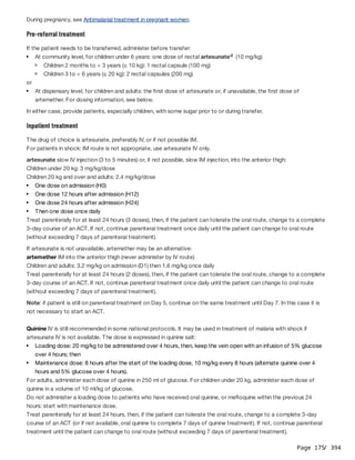
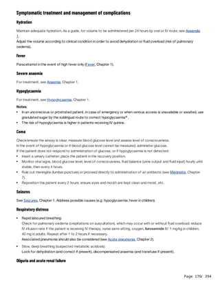
![Page 177
/ 394
Look first for dehydration (Dehydration, Chapter 1), especially due to inadequate fluid intake or excessive fluid losses
(high fever, vomiting, diarrhoea). Treat dehydration if present. Be aware of the risk of fluid overload and acute
pulmonary oedema. Monitor for the return of urine output.
Acute renal failure (ARF) is found almost exclusively in adults and is more common in Asia than Africa. Insert a urinary
catheter, measure output. Restrict fluids to 1 litre/day (30 ml/kg/day in children), plus additional volume equal to urine
output. Renal dialysis is often necessary.
Antimalarial treatment in pregnant women
Uncomplicated P. vivax, P. ovale, P. malariae, P. knowlesi malaria
As other patients.
Primaquine should not be given in pregnancy.
Uncomplicated falciparum malaria
All ACT included in the table Treatment of uncomplicated falciparum malaria can be used in all trimesters.
If ACTs are not available, quinine PO (for dosing, see Treatment of uncomplicated falciparum malaria)
combined with clindamycin PO if possible (10 mg/kg 2 times daily for 7 days) may be an alternative to ACT.
Primaquine should not be given in pregnancy.
Severe malaria
Artesunate, or if unavailable artemether, is recommended in all trimesters.
Quinine IV is not recommended as standard treatment, however it still remains in some national protocols.
Prevention
For pregnant women in areas with high risk of infection with P
. falciparum, refer to the guide Essential obstetric and
newborn care, MSF.
In areas with seasonal malaria transmission (in particular across the Sahel sub-region), seasonal malaria
chemoprevention in children < 5 years reduces mortality: administer amodiaquine + SP at monthly intervals for 4
months during the transmission period .
[4]
In malaria endemic areas and in epidemic-prone contexts, all in-patient facilities (including HIV treatment centres
and feeding centres), should be furnished with long-lasting insecticidal nets (LLINs). For more information, refer to
the guide Public health engineering, MSF.
See specialised literature for information regarding anti-vector measures and prevention in travellers.
Footnotes
(a) Most rapid tests detect the following antigens alone or in combination: HRP2 protein specific to P
. falciparum; an enzyme
(Pf pLDH) specific to P
. falciparum; an enzyme (pan pLDH) common to all 4 plasmodium species. HRP2 may continue to be
detectable for 6 weeks or more after parasite clearance; pLDH remains detectable for several days (up to 2 weeks) after
parasite clearance.
Use pan pLDH tests as first choice in hyper and holo-endemic areas, as well as in areas of intense seasonal transmission and
during outbreaks or complex emergencies. In other contexts, HRP2 tests (P
. falciparum > 95%) or HRP2 + pLDH combination
tests (P
. falciparum < 95%) are preferred.
(b) If the patient vomits within 30 minutes after administration: re-administer the full dose. If the patient vomits between 30
minutes and 1 hour after administration, re-administer half of the dose. If severe vomiting precludes oral therapy, manage as
severe malaria, see Treatment of severe malaria.](https://image.slidesharecdn.com/guideline-170-en-240313103309-343cfe20/85/guideline-170-en-pdf-177-320.jpg)
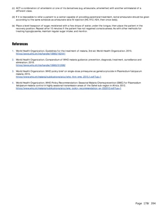
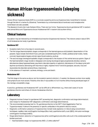
![Page 182
/ 394
American trypanosomiasis (Chagas disease)
Chagas disease is a zoonosis caused by the protozoa Trypanosoma cruzi. It is transmitted to humans by contact of
triatomine bug faeces with a break in the skin (often caused by a bite from the triatomine bug), or with mucous
membranes. Transmission by contaminated blood transfusion, accidental exposure to blood, mother-to-child (during
pregnancy or childbirth) or consumption of contaminated food and water is also possible.
Chagas disease has two phases: an acute phase, which lasts approximately 4 to 6 weeks, and a chronic phase, which
is lifelong if left untreated.
The disease is primarily found on the American continent . It is significantly underdiagnosed.
Clinical features
Acute phase
Chronic phase
Diagnosis
Laboratory
Other investigations
Treatment
a [1]
Most cases are asymptomatic.
If transmitted through a break in the skin: a red swelling on the skin (chagoma) or unilateral painless purplish
periorbital oedema (Romaña's sign) with local lymphadenopathy, headache and fever.
Rarely: multiple lymphadenopathies, hepatosplenomegaly, myocarditis (chest pain, dyspnoea), meningoencephalitis
(seizures, paralysis).
Many cases remain asymptomatic (indeterminate phase).
Up to 30% of cases develop organ damage:[2]
cardiac lesions (conduction disorders, dilated cardiomyopathy): arrhythmia, dyspnoea, chest pain, heart failure;
gastrointestinal lesions (dilation of the oesophagus or colon i.e. megaoesophagus, megacolon): difficulty
swallowing, severe constipation.
Individuals with immunosuppression have a higher risk of developing organ damage than the general population.
[1]
Acute phase:
Identification of Trypanosoma cruzi by direct microscopy of fresh blood or blood concentrated by
microhematocrit method.
In case of strong clinical suspicion despite no definitive diagnosis from direct microscopy, perform serologic
tests after a delay of approximately 1 month (see "Chronic phase").
Chronic phase:
Identification of anti-Trypanosoma cruzi antibodies by serologic tests, e.g. enzyme-linked immunosorbent assay
(ELISA), hemagglutination inhibition assay (HAI), indirect immunofluorescence (IIF) or rapid diagnostic test (RDT).
For a definitive diagnosis, two different serological tests should be performed simultaneously; in case of
conflicting results, a third test is recommended.b
ECG may demonstrate conduction disorders.
Chest or abdominal x-ray may demonstrate cardiomegaly, megaoesophagus or megacolon.](https://image.slidesharecdn.com/guideline-170-en-240313103309-343cfe20/85/guideline-170-en-pdf-182-320.jpg)
![Page 183
/ 394
Aetiologic treatment
For information:
Symptomatic treatment
See Seizures (Chapter 1), Pain (Chapter 1) and Heart failure (Chapter 12).
Prevention
References
Acute or chronic Chagas disease can be treated with either benznidazole or nifurtimox. However, treatment is not
recommended if patient has already developed cardiac or digestive complications.
Close clinical monitoring should be provided due to the frequent occurrence of adverse effects. Where available,
blood tests (complete blood count, liver and renal function tests) should be performed before, during and after
treatment.
Protocols vary according to the country, follow national recommendations.
Age Dose and duration
benznidazole
PO((a)
2 to 12 years[3] 5 to 8 mg/kg daily in 2 divided doses for 60 days
> 12 years and adults[4] 5 to 7 mg/kg daily in 2 divided doses for 60 days
nifurtimox
PO(b) [3]
≤ 10 years 15 to 20 mg/kg daily in 3 to 4 divided doses for 90 days
11 to 16 years 12.5 to 15 mg/kg daily in 3 to 4 divided doses for 90 days
≥ 17 years and adults 8 to 10 mg/kg daily in 3 to 4 divided doses for 90 days
(a) Benznidazole is contra-indicated in pregnancy, breastfeeding and in patient with severe hepatic/renal impairment.
(b) Nifurtimox is contra-indicated in pregnancy, breastfeeding, patients with severe hepatic/renal impairment or history of
severe mental disorders or seizures. Adverse effects (gastrointestinal disturbances, agitation, sleeping disorders, seizure)
are frequent and reversible and should not necessarily result in discontinuation of treatment. Avoid alcohol and fatty meals
during treatment.
Individual protection against bite from triatomine bugs: use of long-lasting insecticidal net.
In healthcare settings: standard precautions to avoid contamination with soiled materials or potentially infected
body fluids.
Blood transfusions: advise patients with Chagas disease not to donate blood. In endemic areas, screen donor
blood for Trypanosoma cruzi antibodies.
Footnotes
(a) For more information on geographical distribution of cases of T. cruzi infection:
http://gamapserver.who.int/mapLibrary/Files/Maps/Global_chagas_2009.png
(b) If resources are limited, ELISA alone can be performed. If the result is positive, a second serologic test should then be
performed to confirm the diagnosis before starting treatment.](https://image.slidesharecdn.com/guideline-170-en-240313103309-343cfe20/85/guideline-170-en-pdf-183-320.jpg)
![Page 184
/ 394
1. Pan American Health Organization. Guidelines for diagnosis and treatment of Chagas disease. Washington, D.C. 2019.
http://iris.paho.org/xmlui/bitstream/handle/123456789/49653/9789275120439_eng.pdf?sequence=6&isAllowed=y
2. Rassi A, Marin-Neto J. Seminar: Chagas disease. The Lancet, Volume 375, ISSUE 9723, P1388-1402, April 17, 2010.
3. Centers for Disease Control and Prevention. Parasites - American Trypanosomiasis.
https://www.cdc.gov/parasites/chagas/ [Accessed 17 February 2020]
4. World Health Organization. WHO Model Prescribing Information: Drugs Used in Parasitic Diseases - Second Edition. Geneva.
1995.
https://apps.who.int/iris/handle/10665/41765 [Accessed 6 May 2020]](https://image.slidesharecdn.com/guideline-170-en-240313103309-343cfe20/85/guideline-170-en-pdf-184-320.jpg)


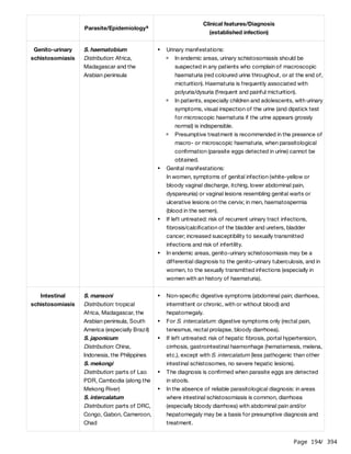
![Page 195
/ 394
Treatment
praziquantel PO
Children 4 years and over and adults :
References
[1][2]
b
S. haematobium, S. mansoni, S. intercalatum: 40 mg/kg single dose or 2 doses of 20 mg/kg administered 4 hours
apart
S. japonicum, S. mekongi: 2 doses of 30 mg/kg or 3 doses of 20 mg/kg administered 4 hours apart
Footnotes
(a) For more information on geographic distribution of schistosomiasis:
https://www.who.int/schistosomiasis/Schistosomiasis_2012-01.png?ua=1
(b) For the treatment of schistosomiasis, praziquantel may me administered to pregnant women.
1. Treatment Guidelines from The Medical Letter. Vol. 11 (Suppl). Drugs for Parasitic Infections. 2013.
https://www.uab.edu/medicine/gorgas/images/docs/syllabus/2015/03_Parasites/RxParasitesMedicalLetter2013.pdf [Accesse
d 25 May 2020]
2. Centers for Disease Control and Prevention (CDC). Schistosomiasis. Resources for Health Professionals. 2018.
https://www.cdc.gov/parasites/schistosomiasis/health_professionals/index.html#tx [Accessed 25 May 2020]](https://image.slidesharecdn.com/guideline-170-en-240313103309-343cfe20/85/guideline-170-en-pdf-195-320.jpg)
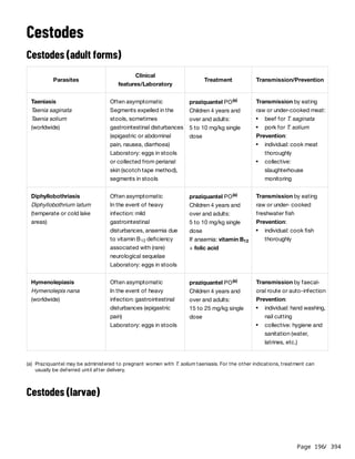
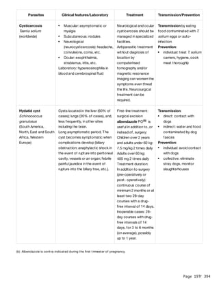
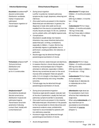
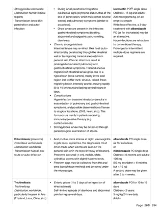
![Page 201
/ 394
Roundworms, whipworms and hookworms frequently co-infect the same host. This should be taken into account
when prescribing antihelminthic treatment.
The migrating larvae of Ancylostoma braziliense and caninum (hookworms of cats and dogs) also present as a
pruritic, inflammatory, creeping eruption in humans (cutaneous larva migrans) but with a slower rate of progression and a
longer duration (several weeks or months). Treatment is with albendazole (400 mg single dose or once daily for 3 days
in children > 6 months and adults; 200 mg in children > 6 months but < 10 kg) or ivermectin (200 micrograms/kg single
dose).
Transmission: consumption
of raw or undercooked
meat containing trichinella
larvae (pork, wart-hog, bear,
dog, etc.)
Muscular phase (about 1 week after ingestion)
High fever; muscular pain (ocular [pain on eye
movement], masseters [limitation of mouth
opening], throat and neck [pain with swallowing
and speech], trunk and limbs); facial or bilateral
peri-orbital oedema; conjunctival haemorrhage,
subungual haemorrhage; headache. Typical
features are not always present and the patient
may present with a non-specific flu-like
syndrome.
Other features, such as dietary habits
(consuming pork/raw meat), suggestive
symptoms (fever > 39 °C and myalgia and facial
oedema) in several individuals who have shared
the same meal (e.g. ceremony) or
hypereosinophilia > 1000/mm , reinforce the
clinical suspicion.
3
Definitive diagnosis: muscle biopsy; serology
(ELISA, Western Blot).
400 mg 2 times daily
or
mebendazole PO for 10 to 15
days
Children > 2 years:
2.5 mg/kg 2 times daily
Adults:
200 mg 2 times daily
plus, regardless of which anti-
helminthic is chosen:
prednisolone PO
0.5 to 1 mg/kg once daily for
the duration of treatment
(a)
(b)](https://image.slidesharecdn.com/guideline-170-en-240313103309-343cfe20/85/guideline-170-en-pdf-201-320.jpg)
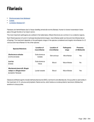
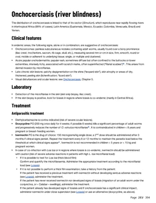
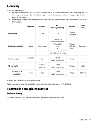
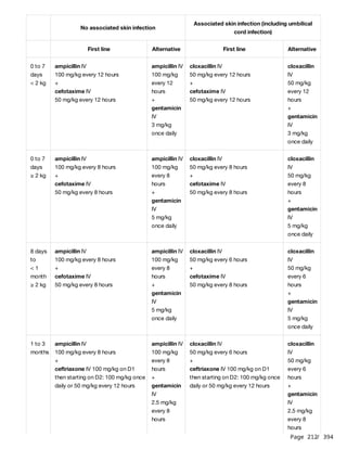
![Page 213
/ 394
Duration of antibiotherapy:
1) According to the pathogen:
2) If the pathogen is unknown:
Additional treatment
Treatment in an epidemic context
Antibiotic therapy
In this context, N. meningitidis is the most likely pathogen.
> 3
months
ceftriaxone IV
Children: 100 mg/kg on D1 then starting on D2: 100
mg/kg once daily or 50 mg/kg every 12 hours (max.
4 g daily)
cloxacillin IV
Children < 40 kg: 50 mg/kg every 6 hours
Children ≥ 40 kg: 2 g every 6 hours
+
ceftriaxone IV
Children: 100 mg/kg on D1 then starting on D2: 100
mg/kg once daily or 50 mg/kg every 12 hours (max.
4 g daily)
Adults ceftriaxone IV: 4 g once daily or 2 g every 12 hours cloxacillin IV: 2 g every 6 hours
+
ceftriaxone IV: 4 g once daily or 2 g every 12 hours
Haemophilus influenzae: 7 days
Streptococcus pneumonia: 10-14 days
Group B streptococcus and Listeria: 14-21 days
Gram-negative bacilli: 21 days
Neisseria meningitidis: see antibiotherapy in an epidemic context
Children < 3 months: 2 weeks beyond the first sterile CSF culture or 21 days
Children > 3 months and adults: 10 days. Consider extending treatment or alternative diagnoses if fever persists
beyond 10 days. On the other hand, a 7-day course of ceftriaxone is sufficient in patients who are making an
uncomplicated recovery.
Dexamethasone reduces the risk of hearing loss in patients with H. influenzae or S. pneumoniae.
Early administration is indicated in meningitis caused by these pathogens or when the pathogen is unknown, except
in neonates (and in presumed meningococcal meningitis in an epidemic context).
dexamethasone IV
Children > 1 month: 0.15 mg/kg (max. 10 mg) every 6 hours for 2 to 4 days
Adults: 10 mg every 6 hours for 2 to 4 days
The treatment should be started before or with the first dose of antibiotic, otherwise, the treatment offers no
benefit.
[1][2]
Ensure that the patient is well fed and well hydrated (infusions or nasogastric tube if necessary).
Seizures (Chapter 1).
Coma: prevention of bed sores, care of the mouth and eyes, etc.](https://image.slidesharecdn.com/guideline-170-en-240313103309-343cfe20/85/guideline-170-en-pdf-213-320.jpg)
![Page 214
/ 394
Note:
A short treatment with a single dose of ceftriaxone IM can be used in children 2 years and older and in adults during a
meningococcal meningitis epidemic if 1) confirmed by a reliable laboratory 2) the number of cases exceeds
management capacities with the 5-day treatment. Check national recommendations. Nevertheless, it is essential to
ensure a monitoring of cases after 24 hours.
ceftriaxone IM
Children 2 to < 12 years: 100 mg/kg single dose
Children ≥ 12 years and adults: 4 g single dose
If there is no clinical improvement (fever > 38.5 °C, repeated seizures, appearance or aggravation of a reduced level of
consciousness or of neurological signs) 24 hours after the injection, continue the treatment with ceftriaxone for 5 days.
Additional treatment
References
Age Treatment[3]
Children
< 2 months
ceftriaxone IV or IM for 7 days
100 mg/kg once daily
(a) (b)
Children ≥ 2 months
and adults
ceftriaxone IV or IM or 5 days
Children 2 months to < 5 years: 100 mg/kg once daily (max. 2 g daily)
Children ≥ 5 years and adults: 2 g once daily
(a) (b)
(a) The solvent of ceftriaxone for IM injection contains lidocaine. Ceftriaxone reconstituted using this solvent must never be
administered by IV route. For IV administration, water for injection must always be used.
(b) For IM administration, divide the dose into 2 injections if needed, half-dose in each buttock.
a
Ensure that the patient is well fed and well hydrated (infusions or nasogastric tube if necessary).
Seizures (Chapter 1).
Coma: prevention of bed sores, care of the mouth and eyes, etc.
Dexamethasone in not indicated.
Footnotes
(a) For IM administration, divide the dose into 2 injections if needed, half-dose in each buttock.
1. D. van de Beek, C. Cabellos, O. Dzupova, S. Esposito, M. Klein, A. T. Kloek, S. L. Leib, B. Mourvillier, C. Ostergaard, P
. Pagliano,
H.W. Pfister, R. C. Read, O. Resat Sipahi, M.C. Brouwer. ESCMID guideline: diagnosis and treatment of acute bacterial
meningitis, 2016.
https://www.clinicalmicrobiologyandinfection.com/article/S1198-743X(16)00020-3/pdf
2. Sheldon L Kaplan, MD. Bacterial meningitis in children: Dexamethasone and other measures to prevent neurologic
complications. UpToDate [Accessed 25 February 2019].
3. World Health Organization. Managing meningitis epidemics in Africa. A quick reference guide for health authorities and
health-care workers. 2015.
https://apps.who.int/iris/bitstream/handle/10665/154595/WHO_HSE_GAR_ERI_2010.4_Rev1_eng.pdf?sequence=1](https://image.slidesharecdn.com/guideline-170-en-240313103309-343cfe20/85/guideline-170-en-pdf-214-320.jpg)
![Page 215
/ 394
Tetanus
Last updated: August 2022
Tetanus is a severe infection due to the bacillus Clostridium tetani, found in soil, and human and animal waste. The
infection is noncontagious.
Clostridium tetani is introduced into the body through a wound and produces a toxin whose action on the central
nervous system is responsible for the symptoms of tetanus.
Tetanus is entirely preventable by vaccination. It occurs in people who have not been fully vaccinated before exposure
or have not received adequate post-exposure prophylaxis. In these individuals, most breaks in the skin or mucous
membranes carry a risk of tetanus, but the wounds with the greatest risk are: the stump of the umbilical cord in
neonates, puncture wounds, wounds with tissue loss or contamination with foreign material or soil, avulsion and crush
injuries, sites of non-sterile injections, chronic wounds (e.g. lower extremity ulcers), burns and bites. Surgical or
obstetrical procedures performed under non-sterile conditions also carry a risk of tetanus.
Clinical features
Generalised tetanus is the most frequent and severe form of the infection. It presents as muscular rigidity, which
progresses rapidly to involve the entire body, and muscle spasms, which are very painful. Level of consciousness is not
altered.
Children and adults
Neonates
Treatment
Hospitalisation is needed and usually lasts 3 to 4 weeks. Correct management can reduce mortality even in hospitals
with limited resources.
General measures
Average time from exposure to onset of symptoms is 7 days (3 to 21 days).
Muscular rigidity begins in the jaw muscles (difficulty with then inability to open mouth [trismus] preventing the patient
from speaking, eating), spreading to the face (fixed smile), neck (difficulty with swallowing), to the trunk (restriction
of respiratory muscles; hyperextension of spine [opisthotonus]), to the abdomen (guarding) and to the limbs (flexion
of the upper limbs and extension of the lower limbs).
Muscle spasms, which are very painful, appear at the onset or when muscular rigidity becomes generalised. They
are triggered by stimuli (noise, light, touch) or arise spontaneously. Spasms of the thoracic and laryngeal muscles
may cause respiratory distress or aspiration.
In 90% of cases, initial symptoms appear within 3 to 14 days of birth.
The first signs are significant irritability and difficulty sucking (rigidity of the lips, trismus) then rigidity becomes
generalised, as in adults. Any neonate, who initially sucked and cried normally, presenting with irritability and difficulty
sucking 3 to 28 days after birth and demonstrating rigidity and muscle spasms should be assumed to have neonatal
tetanus.
Ensure intensive nursing care.
The patient should be in a dark, quiet room. Blindfold neonates with a cloth bandage.](https://image.slidesharecdn.com/guideline-170-en-240313103309-343cfe20/85/guideline-170-en-pdf-215-320.jpg)
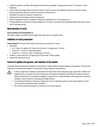
![Page 217
/ 394
Count the volume of the infusion of diazepam as part of the patient’s daily fluid intake.
Neonates diazepam emulsion for injection (10 mg ampoule, 5 mg/ml, 2 ml)
Example:
Neonate weighing 3 kg (administration by electric syringe)
0.1 mg/kg/hour x 3 kg = 0.3 mg/hour
Dilute one 10 mg ampoule of diazepam emulsion for injection in 50 ml of 10% glucose to obtain a
solution containing 0.2 mg of diazepam per ml.
Administer 1.5 ml/hour [dose (in mg/hour) ÷ dilution (in mg/ml) = dose in ml/hour i.e. 0.3 (mg/hour) ÷ 0.2
(mg/ml) = 1.5 ml/hour].
If an electric syringe is not available, diluting the diazepam emulsion in an infusion bag for continuous
infusion may be considered. Weigh the risks associated with this mode of administration (accidental
bolus or insufficient dose). The infusion should be monitored closely to avoid any change, however
small, of the prescribed rate.
0.1 to 0.3 mg/kg by slow IV injection (3 to 5 minutes) every 1 to 4 hours depending on the severity
and the persistence of the spasms as long as the RR is ≥ 30.
If despite hourly diazepam the spasms persist, start a continuous infusion of diazepam with an
electric syringe: 0.1 to 0.5 mg/kg/hour (2.4 to 12 mg/kg every 24 hours). Start with 0.1 mg/kg/hour
and if symptoms persist, increase by 0.1 mg/kg/hour as long as RR is ≥ 30.
If in spite of 0.5 mg/kg/hour symptoms persist, the dose can be increased up to 0.8 mg/kg/hour as
long as the RR ≥ 30.
Diluted diazepam emulsion does not keep for more than 6 hours.
Children
> 1
month
and
adults
Same doses and protocol as in neonates but:
Examples:
Use diazepam solution for injection 5 mg/ml: (10 mg ampoule, 5 mg/ml, 2 ml).(a)
These doses can be administered as long as the RR is:
≥ 30 in children under 1 year
≥ 25 in children 1 to 4 years
≥ 20 in children 5 to 12 years
≥ 14 in children over 12 years
≥ 12 in adults
Child weighing 6 kg (continuous IV infusion using a pediatric infusion set; 1 ml = 60 drops)
0.1 mg/kg/hour x 6 kg = 0.6 mg/hour
Dilute one 10 mg ampoule of diazepam solution for injection in 50 ml of 5% glucose (10%
glucose if child < 3 months) to obtain a solution containing 0.2 mg of diazepam per ml.
Administer 3 ml/hour [dose (in mg/hour) ÷ dilution (in mg/ml) = dose in ml/hour i.e. 0.6 (mg/hour) ÷ 0.2
(mg/ml) = 3 ml/hour] or 3 drops/minute (in a paediatric infusion set ml/hour = drops/minute).
Adult weighing 60 kg (standard adult infusion set, 1 ml = 20 drops)
0.1 mg/kg/hour x 60 kg = 6 mg/hour
Dilute 5 ampoules of 10 mg of diazepam solution (50 mg) in 250 ml of 0.9% sodium chloride or
5% glucose to obtain a solution containing 0.2 mg of diazepam per ml.
Administer 30 ml/hour [dose (in mg/hour) ÷ dilution (in mg/ml) = dose in ml/hour e.g. 6 (mg/hour) ÷ 0.5
(mg/ml) = 30 ml/hour] or 10 drops/minute.
(a) Administer the first dose rectally if an IV cannot be placed immediately.](https://image.slidesharecdn.com/guideline-170-en-240313103309-343cfe20/85/guideline-170-en-pdf-217-320.jpg)
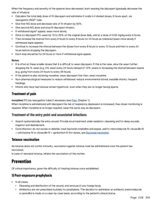
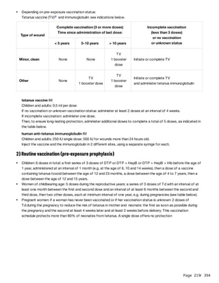
![Page 221
/ 394
Enteric (typhoid and paratyphoid) fevers
Last updated: September 2022
Enteric fevers include typhoid fever, due to Salmonella enterica serotype Typhi (S. Typhi) and paratyphoid fever, due
to Salmonella enterica serotype Paratyphi A, B or C (S. Paratyphi).
Enteric fevers are acquired by the ingestion of water or food contaminated with excreta of symptomatic or
asymptomatic carriers or by direct contact (dirty hands).
Enteric fevers are endemic in South, Central and Southeast Asia, sub-saharan Africa, Oceania and, to a lesser extent, in
Latin America.
Effective treatment significantly reduces the risk of complications and death.
Clinical features
Clinical manifestations of typhoid and paratyphoid fevers are the same. Enteric fevers have insidious onset and vary
from mild to severe.
Clinical diagnosis is difficult as enteric fevers resembles other infections present in regions where they are
endemic. The main differential diagnoses are: malaria, brucellosis, leptospirosis, typhus, rickettsiosis,
sepsis and dengue.
Laboratory
Treatment
In all cases
The characteristic sign is prolonged fever, which gradually increases during the first week, plateaus the second
week then decreases between the third and fourth week.
Non-specific signs and symptoms are frequently associated: gastrointestinal disturbances (abdominal pain,
constipation or diarrhoea, vomiting), headache, malaise, chills, fatigue, non productive cough and/or
hepatosplenomegaly.
Erythematous maculopapular rash on the trunk extreme fatigue and/or relative bradycardia (heart rate-temperature
dissociation) may be present.
Serious complications affect about 27% of hospitalised patients and usually occur during the second or third
week of illness. These may incude decreased level of consciousness, intestinal haemorrhage or perforation
or peritonitis, shock, or nephritis. In pregnant women, severe infection may lead to foetal complications
(miscarriage, preterm delivery, intrauterine death).
[1]
Relapse may occur 2 to 3 weeks after recovery. It is usually not due to antibiotic resistance, and re-treatment is
required.
Culture of S. Typhi or Paratyphi and drug susceptibility test (blood and stool specimens).
In all cases, rapid test for malaria in endemic regions (and antimalarial treatment if needed, see Malaria, Chapter 6).
Widal agglutination test, other serologic tests, and rapid diagnostic tests are not recommended (low sensitivity and
specificity).
Hydrate and treat fever (Chapter 1). Fever usually resolves 4 to 5 days after starting effective antibiotic treatment.
Choice of antibiotic treatment depends on the susceptibility of the strain, or when susceptibility is unknown, on
recent data on susceptibility of strains in the region. Check national recommendations. For information:](https://image.slidesharecdn.com/guideline-170-en-240313103309-343cfe20/85/guideline-170-en-pdf-221-320.jpg)
![Page 222
/ 394
Uncomplicated cases (outpatients)
Uncomplicated cases (the vast majority of cases) can be treated with an oral antibiotic treatment.
Severe cases (inpatients)
These cases should be treated under close monitoring. Antibiotic treatment is initially parenteral, then oral when
there is decreasing fever, clinical improvement and the patient can tolerate oral treatment.
Additional measures
Prevention
Strains resistant to chloramphenicol, ampicillin/amoxicillin and co-trimoxazole (multidrug-resistant, MDR
strains) are present in most parts of the world.
Ciprofloxacin is used as first-line treatment in some countries, however fluoroquinolone resistance is endemic in
Asia and is increasing in several parts of the world .
[2]
Ceftriaxone resistance has been identified in several regions .
[2]
MDR strains also resistant to fluoroquinolones and third-generation cephalosporins (extensively drug-resistant,
XDR strains) have developed .
[3]
First-line antibiotics:
azithromycin PO for 7 days (including for MDR and XDR cases, and pregnant women)
Children: 10 to 20 mg/kg (max. 1 g) once daily
Adults: 500 mg to 1 g once daily or 1 g on D1 then 500 mg once daily
or
cefixime PO for 10 to 14 days (except for third-generation cephalosporin resistance and XDR cases)
Children: 10 mg/kg (max. 200 mg) 2 times daily
Adults: 200 mg 2 times daily
Alternatives include, only if recent data show susceptibility of strains to these antibiotics in the region:
amoxicillin PO for 14 days
Children: 30 mg/kg (max. 1 g) 3 times daily
Adults: 1 g 3 times daily
or
co-trimoxazole PO for 14 days
Children: 20 mg SMX + 4 mg TMP/kg (max. 800 mg SMX + 160 mg TMP) 2 times daily
Adults: 800 mg SMX + 160 mg TMP 2 times daily
Severe cases include:
toxic appearance or decreased level of consciousness or medical or surgical complication;
oral administration not possible due to persistent vomiting.
Start with ceftriaxone IV (including for pregnant women)
Children: 50 to 100 mg/kg (max. 4 g) once daily
Adults: 2 g once daily or 2 times daily
Then change to azithromycin PO (as above) to complete at least 7 days of treatment.
a
For suspected or confirmed ceftriaxone resistance or XDR stains, use meropenem IV, including for pregnant
women, then change to azithromycin PO to complete at least 7 days of treatment.
In case of decreased level of consciousness or shock, dexamethasone IV: 3 mg/kg then 1 mg/kg every 6 hours for
2 days (total of 8 doses)
Treat in intensive care unit patients with shock, significant intestinal haemorrhage or suspected
perforation/peritonitis. If suspected perforation/peritonitis, get urgent surgical review and add metronidazole to
ceftriaxone for anaerobic bacterial coverage .
b](https://image.slidesharecdn.com/guideline-170-en-240313103309-343cfe20/85/guideline-170-en-pdf-222-320.jpg)
![Page 223
/ 394
References
Hygiene measures common to all diarrhoeas: handwashing; consumption of treated water (chlorinated, boiled,
bottled, etc.); washing/cooking of food, etc.
In hospitals: for patients with watery diarrhoea, consider disinfection of excreta with chlorinated solution, if stools
are collected in buckets.
Vaccination with the typhoid conjugate vaccine in endemic regions . This vaccine can be used to control typhoid
outbreaks. It does not protect against paratyphoid fever.
c
Footnotes
(a) The solvent of ceftriaxone for IM injection contains lidocaine. Ceftriaxone reconstituted using this solvent must NEVER be
administered by IV route. For IV administration, water for injection must always be used.
(b) Do not add metronidazole if the patient receives meropenem (meropenem already covers anaerobic bacteria).
(c) For more information, see Typhoid vaccines: WHO position paper:
http://apps.who.int/iris/bitstream/handle/10665/272272/WER9313.pdf?ua=1
1. Cruz Espinoza LM, McCreedy E, Holm M, et al. Occurrence of typhoid fever complications and their relation to duration of
illness preceding hospitalization: a systematic literature review and meta-analysis. Clin Infect Dis 2019;69(Suppl 6):S435-48.
https://www.ncbi.nlm.nih.gov/pmc/articles/PMC6821330/ [Accessed 28 June 2022]
2. Browne AJ, Hamadani BHK, Kumaran EAP
, Rao P
, et al. Drug-resistant enteric fever worldwide, 1990 to 2018: a systematic
review and meta-analysis. BMC Medicine 2020;18:1+22.
https://doi.org/10.1186/s12916-019-1443-1 [Accessed 23 February 2022]
3. Klemm EJ, Shakoor S, Page AJ, Qamar FN, et al. Emergence of an Extensively Drug-Resistant Salmonella enterica Serovar
Typhi Clone Harboring a Promiscuous Plasmid Encoding Resistance to Fluoroquinolones and Third-Generation
Cephalosporins. mBio. 2018 Jan-Feb; 9(1): e00105-18. https://www.ncbi.nlm.nih.gov/pmc/articles/PMC5821095/ [Accessed 26
June 2022]](https://image.slidesharecdn.com/guideline-170-en-240313103309-343cfe20/85/guideline-170-en-pdf-223-320.jpg)

![Page 228
/ 394
Forms of plague Clinical features Antibiotic treatment[1]
Bubonic
AND
Fever, chills, malaise,
headache
Lymph node (bubo), painful,
usually inguinal, (one or
more)
Children (including < 8 years) and adults:
doxycycline PO:
or
gentamicin IM or IV:
or
ciprofloxacin PO:
Under 45 kg: 4.4 mg/kg (max. 200 mg) on D1, then
2.2 mg/kg (max. 100 mg) 2 times daily
45 kg and over: 200 mg on D1, then 100 mg 2 times
daily
(a)
Children: 4.5 to 7.5 mg/kg once daily
Adults: 5 mg/kg once daily
Children: 15 mg/kg 2 to 3 times daily (max. 750 mg 2
times daily or 500 mg 3 times daily)
Adults: 750 mg 2 times daily
Pneumonic
AND
Fever, chills, malaise,
headache
Dyspnea, chest pain,
productive cough with
purulent or blood-stained
sputum
Respiratory distress or
failure and sepsis in severe
or advanced, untreated
disease
Children and adults:
If mild disease:
gentamicin IM or IV (as above)
or
ciprofloxacin PO (as above) or IV
If severe disease:
gentamicin + ciprofloxacin (as above)
or, if not available, gentamicin + doxycycline (as
above)
After clinical improvement, change to ciprofloxacin or
doxycycline PO (as above).
(a)
(b)
Septicaemic Frequently no localizing
symptoms or signs
Gastrointestinal
disturbances (abdominal
pain, vomiting, diarrhoea,
etc.) often present
As severe pneumonic plague
Meningitis(c) Signs of meningitis. Children and adults:
chloramphenicol IV:
+ ciprofloxacin PO or IV (as above)
or, if not available,
gentamicin + ciprofloxacin (as above)
Children 1 to 12 years: 25 mg/kg (max. 1 g) every 8
hours
Children 13 years and over and adults: 1 g every 8
hours](https://image.slidesharecdn.com/guideline-170-en-240313103309-343cfe20/85/guideline-170-en-pdf-228-320.jpg)
![Page 230
/ 394
References
1. Nelson CA, Meaney-Delman D, Fleck-Derderian S, Cooley KM, et al. Antimicrobial treatment and prophylaxis of plague:
recommendations for naturally acquired infection and bioterrorism response. MMWR Recomm Rep 2021;70(No. RR-3):1-27.
https://www.cdc.gov/mmwr/volumes/70/rr/rr7003a1.htm?s_cid=rr7003a1_w [Accessed 25 January 2022]](https://image.slidesharecdn.com/guideline-170-en-240313103309-343cfe20/85/guideline-170-en-pdf-230-320.jpg)
![Page 231
/ 394
Leptospirosis
Last update: October 2022
Leptospirosis is a zoonosis that affects many domestic and wild animals, mainly rodents (particularly rats) but also
dogs and cattle, etc.
It is transmitted to humans by contact through skin lesions or mucous membranes (e.g. eyes, mouth) with:
It is caused by bacteria (spirochetes) of the genus Leptospira.
Leptospirosis occurs worldwide, particularly in tropical and subtropical regions. There are often outbreaks after heavy
rainfall or flooding.
Clinical features
Approximately 90% of cases are asymptomatic or mild with a favourable outcome. 5 to 15% of cases present a
severe form with multiple organ dysfunction and a high mortality rate without prompt treatment.
Mild form
Severe or ictero-haemorrhagic form
The onset is the same but a few days later the symptoms worsen: renal disorders (oliguria or polyuria), hepatic disorder
(jaundice), widespread haemorrhages (purpura, ecchymoses, epistaxis, haemoptysis, etc.), pulmonary signs (chest pain)
or cardiac signs (myocarditis, pericarditis).
Other conditions to consider include a wide range of acute febrile illnesses, e.g.:
freshwater or moist soil contaminated with urine of an infected animal (indirect contact);
urine, blood and other body fluids or tissues of an infected animal (direct contact).
Acute phase (septicaemic):
Sudden onset of high fever with chills, headache, myalgia (especially calf and lumbar pain), photophobia, ocular
pain. Bilateral conjunctival suffusion affecting the bulbar conjunctiva (redness without discharge) is a
characteristic sign, but not always present.
May be associated with: gastrointestinal symptoms (anorexia, abdominal pain, nausea, vomiting), non-
productive cough, lymphadenopathy, hepatomegaly, and sometimes, skin rash.
Immune phase:
The signs of the acute phase regress after 5 to 7 days then reappear for a few days usually in a milder form
(milder fever, less severe myalgia) then disappear.
Signs of meningitis (thought to be of immune origin) are however very common during this phase.
Diagnosis is difficult because of the broad spectrum of clinical manifestations. Patients that present the following
should be considered as suspected cases of leptospirosis :
and
[1]
abrupt onset of fever, chills, conjunctival suffusion, headache, myalgia and jaundice
one or more risk factors for infection: exposure to contaminated freshwater (e.g. swimming, fishing, rice fields,
flooding) or infected animals (e.g. crop and livestock farmers, veterinarians, butchers and slaughterhouse
workers).
Viral haemorrhagic fevers, dengue, chikungunya, Zika, influenza, measles, viral hepatitis, other causes of meningitis](https://image.slidesharecdn.com/guideline-170-en-240313103309-343cfe20/85/guideline-170-en-pdf-231-320.jpg)

![Page 233
/ 394
Antibiotic treatment
Children: 80 to 100 mg/kg (max. 2 g) once daily
Adults: 2 g once daily
or
Children: 50 000 IU (30 mg)/kg (max. 2 MIU or 1200 mg) every 6 hours
Adults: 1 to 2 MIU (600 to 1200 mg) every 6 hours
Prevention
References
Specific management according to organs affected. Oliguria generally responds to correction of hypovolaemia.
Rest and treatment of pain and fever: paracetamol PO (Chapter 1). Avoid or use paracetamol with caution in
patients with hepatic involvement.
ceftriaxone IV for 7 daysa
benzylpenicillin IV for 7 days
Avoid bathing in freshwater in endemic areas.
Disinfect laundry and objects soiled by urine of infected animal or patient.
Vaccination and protective clothing (only for professionals at risk of exposure).
Footnotes
(a) For IV administration of ceftriaxone, dilute with water for injection only.
1. World Health Organization. Human leptospirosis: guidance for diagnosis, surveillance and control. World Health Organization,
2003.
https://apps.who.int/iris/bitstream/handle/10665/42667/WHO_CDS_CSR_EPH_2002.23.pdf?%20sequence=1&isAllowed=y
[Accessed 5 September 2022]](https://image.slidesharecdn.com/guideline-170-en-240313103309-343cfe20/85/guideline-170-en-pdf-233-320.jpg)
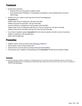

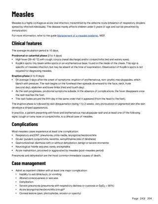
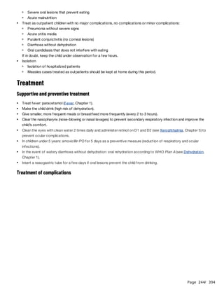
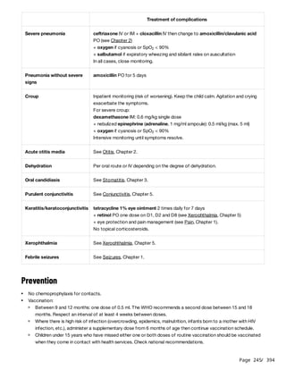
![Page 246
/ 394
Footnotes
(a) Symptoms (hoarse crying or voice, difficulty breathing, a high-pitched inspiratory wheeze [inspiratory stridor], characteristic
"barking" cough) are caused by inflammation and narrowing of the larynx. Croup is considered benign or “moderate” if the
stridor occurs when the child is agitated or crying, but disappears when the child is calm. The child should be monitored
during this period, however, because his general and respiratory status can deteriorate rapidly. Croup is severe when the
stridor persists at rest or is associated with signs of respiratory distress.](https://image.slidesharecdn.com/guideline-170-en-240313103309-343cfe20/85/guideline-170-en-pdf-246-320.jpg)
![Page 247
/ 394
Poliomyelitis
Poliomyelitis is an acute viral infection due to a poliovirus (serotypes 1, 2 or 3). Human-to-human transmission is direct
(faecal-oral) or indirect (ingestion of food and water contaminated by stool). Humans are the only reservoir of the virus.
In principle the disease can be eradicated by mass vaccination.
In endemic areas, poliomyelitis mainly affect children under 5 years not (or not fully) vaccinated, but the infection can
affect persons of any age, especially in areas where population immunity is low.
Clinical features
Laboratory
Look for the polio virus in stool samples. The virus is excreted for one month after infection, but only intermittently;
therefore, 2 samples must be collected with an interval of 24-48 hours, and within 14 days of onset of symptoms
. Send the stool samples to a reference laboratory, with a clinical description of the patient. The stool samples must be
stored and transported between 0 °C and 8 °C.
Treatment
Outbreak control in case of acute flaccid paralysis (AFP)
Up to 90% of cases are asymptomatic or present mild symptoms .
[1]
Non-paralytic form: a non-specific febrile illness with muscle pain, headache, vomiting, backache; no neurological
involvement. As spontaneous recovery usually occurs within 10 days, diagnosis is rarely made outside epidemic
contexts.
Paralytic form: in less than 1% of cases, after the non-specific signs, the patient develops rapid onset (from the
morning to the evening) asymmetrical acute flaccid paralysis, predominantly of the lower limbs, with ascending
progression. The muscles become soft with diminished reflexes. Sensation is maintained. The disease is life
threatening if paralysis involves the respiratory muscles or muscles of swallowing. Initial urinary retention is common.
Gastrointestinal disturbances (nausea, vomiting, diarrhoea), muscle pain and meningeal symptoms may also occur.
[2]
Hospitalise patients with the paralytic form: rest, prevent bed sores in bedridden patients, give analgesics (do not
give IM injections to patients in the febrile phase), ventilate patients with respiratory paralysis.
Physiotherapy once the lesions are stable to prevent muscle atrophy and contractures.
Care for sequelae: physiotherapy, surgery and prosthetics.
Consider any patient with AFP as a suspected case of poliomyelitis.
Send stool samples to a reference laboratory to confirm the diagnosis.
Organize vaccination of all children under 5 years living in the area (from the same village or neighbouring villages)
irrespective of their vaccination status, within 14 days of laboratory confirmation and with the available vaccine
(round 0) .
[3]
Organize two mass vaccination campaigns within 8 weeks of the laboratory confirmation. The type of vaccine, the
area and the age groups are determined by epidemiological data.
Organize a mop-up (door-to-door) vaccination campaign wherever monitoring suggests that children have been
missed, to ensure interruption of transmission.
Surveillance: for each case of AFP there are between 100 and 200 subclinical cases. Therefore, active surveillance
to detect new cases is essential for epidemic control.](https://image.slidesharecdn.com/guideline-170-en-240313103309-343cfe20/85/guideline-170-en-pdf-247-320.jpg)
![Page 248
/ 394
Prevention
In children who start routine vaccination late (after the age of 3 months), the dose of IPV is administered together with
the first dose of bOPV, followed by 2 doses of bOPV alone administered 4 weeks apart.
There is also an ‘IPV only’ schedule: 3 doses administered at least 4 weeks apart (e.g. at 6, 10 and 14 weeks) and a
booster dose at least 6 months later.
IPV should eventually completely replace bOPV.
References
3 types of vaccines exist:
a trivalent injectable inactivated poliovirus vaccine (IPV),
a bivalent oral live attenuated poliovirus vaccine (bOPV), containing serotypes 1 and 3,
a monovalent oral type 2 vaccine (mOPV or nOPV) exclusively used for responding to epidemics.
Vaccination schedule: depends on the epidemiology of the virus.
Protocols vary according to the country, follow national recommendations. For information, the WHO
recommends:
Schedule
Primary vaccination
Endemic or at risk zones(a) Other zones
Birth 1 dose bOPV(b) –
6 weeks 1 dose bOPV 1 dose bOPV
10 weeks 1 dose bOPV 1 dose bOPV
14 weeks 1 dose bOPV + 1 dose IPV 1 dose bOPV + 1 dose IPV
(a) Countries where poliomyelitis is endemic or zones at high risk of importation and subsequent spread of the virus.
(b) The first dose of bOPV is administered at birth, or as soon as possible, to optimise seroconversion rates after subsequent
doses and induce mucosal protection.
1. World Health Organization. Poliomyelitis (polio).
https://www.who.int/health-topics/poliomyelitis#tab=tab_1 [Accessed 08 June 2021]
2. Centers for Disease Control and Prevention. Poliomyelitis. 2020.
https://www.cdc.gov/vaccines/pubs/pinkbook/polio.html [Accessed 08 June 2021]
3. Global Polio Eradication Initiative. Standard operating procedures: responding to a poliovirus event or outbreak, version 3.1.
World Health Organization. 2020.
https://www.who.int/publications/i/item/9789240002999 [Accessed 08 June 2021]](https://image.slidesharecdn.com/guideline-170-en-240313103309-343cfe20/85/guideline-170-en-pdf-248-320.jpg)
![Page 249
/ 394
Rabies
Rabies is a viral infection of wild and domestic mammals, transmitted to humans by the saliva of infected animals
through bites, scratches or licks on broken skin or mucous membranes.
In endemic areas (Africa and Asia), 99% of cases are due to dog bites and 40% of cases are children under 15 years of
age .
Before symptoms develop, rabies can effectively be prevented by post-exposure prophylaxis.
Once symptoms develop, rabies is fatal. There is no curative treatment; care is palliative.
Clinical features
Diagnosis is often difficult: there may be no history of scratch or bite (exposure through licking) or wounds may have
healed; a reliable history may be difficult to obtain.
Post-exposure prophylaxis
Definitions of exposure categories (WHO)
Post-exposure prophylaxis is carried out for Category II and III exposures.
Treatment of the wound
In all cases
[1]
The incubation period averages 20 to 90 days from exposure (75% of patients), but can be shorter (in severe
exposure, e.g. bites to face, head and hands; multiple bites), or longer (20% of patients develop symptoms
between 90 days and 1 year, and 5% more than 1 year after exposure).
Prodromal phase: itching or paraesthesiae or neuropathic pain around the site of exposure, and non-specific
symptoms (fever, malaise, etc.).
Neurologic phase:
Encephalitic form (furious form): psychomotor agitation or hydrophobia (throat spasms and panic, triggered by
attempting to drink or sight/sound/touch of water) and aerophobia (similar response to a draft of air); sometimes
seizures. The patient is calm and lucid between episodes. Infection evolves to paralysis and coma.
Paralytic form (less common, 20% of cases): progressive ascending paralysis resembling Guillain-Barré
syndrome; evolves to coma.
Category I Contact with animal, or licks on intact skin No exposure
Category II
Nibbles on exposed skin
Minor bite(s) or scratch(es) without bleeding
Minor exposure
Category III
Transdermal bite(s) or scratch(es)
Licks on broken skin
Contamination of mucous membranes by animal’s saliva (licks)
Direct contact with batsa
Severe exposure](https://image.slidesharecdn.com/guideline-170-en-240313103309-343cfe20/85/guideline-170-en-pdf-249-320.jpg)
![Page 250
/ 394
Prolonged cleansing of the wound or contact site for 15 minutes to eliminate the virus, as soon as possible after
exposure, is of critical importance. For skin: use soap, rinse copiously with running water, remove all foreign material;
application of a disinfectant (povidone iodine 10% or other) is an additional precaution which does not take the place
of thorough wound washing. For mucous membranes (eye, mouth, etc.): rinse thoroughly with water or 0.9% sodium
chloride. Local cleansing is indicated even if the patient presents late.
According to condition/type of wound
In order to avoid inoculating virus deeper into the tissues, wounds are either not sutured at all (e.g. superficial, non-
mutilating or puncture wounds), or are left open and re-evaluated in 48-72 hours, with a view to possible closure. Highly
contaminated wounds, or wounds that may compromise function, require surgical management (exploration, removal
of foreign material, excision of necrotic tissue, copious irrigation with sterile 0.9% sodium chloride or Ringer lactate,
with local or general anaesthesia). When suturing is indicated (face), rabies immunoglobulin should be administered
several hours before wound closure (see below). Infected wounds are not sutured and reassessed daily.
Passive and active immunisation
Given the duration of incubation, administration of vaccine/immunoglobulin is always a priority, even for patients
exposed several months previously.
Anti-rabies serotherapy
Rabies immunoglobulin is indicated after:
It is intended to neutralize virus in the exposure site. It is given as a single dose on D0, with the first dose of rabies
vaccine.
human rabies immunoglobulin:
Children and adults: 20 IU/kg
or
highly purified rabies immunoglobulin F(ab')2 fragments:
Children and adults: 40 IU/kg
Infiltrate rabies immunoglobulin into and around the previously washed wound(s). Ensure it is not injected into a blood
vessel (risk of shock).
For finger wounds, infiltrate very cautiously to avoid increased pressure in the tissue compartment (compartment
syndrome).
In the event of multiple wounds, dilute the dose 2- to 3-fold with sterile 0.9% sodium chloride to obtain a sufficient
quantity to infiltrate all the sites exposed.
Infiltrate rabies immunoglobulin into the wound even if it has already healed.
For mucosal exposures with no wound, rinse with rabies immunoglobulin diluted in sterile 0.9% sodium chloride.
Monitor the patient during and after the injection (low risk of anaphylactic reaction).
If rabies immunoglobulin is not available on D0, the first dose of rabies vaccine is administered alone. Administer rabies
immunoglobulin as soon as possible between D0 and D7; from D8, it is not necessary to
administer rabies immunoglobulin as vaccine-induced antibodies begin to appear.
Post-exposure rabies prophylaxis
Category III exposures (except in patients who have received a full course of pre-exposure prophylaxis against
rabies, see Prevention);
Category II and III exposures in immunocompromised patients (even in patients who have received a full course of
pre-exposure prophylaxis against rabies).
b
[1]](https://image.slidesharecdn.com/guideline-170-en-240313103309-343cfe20/85/guideline-170-en-pdf-250-320.jpg)
![Page 251
/ 394
A complete prophylaxis series is indicated for Category II and III exposures. It should be started on D0 and continued to
completion if the risk of rabies has not been excluded . Several different types of rabies vaccines prepared from cell
cultures (CCEEV) exist. These vaccines must replace nerve tissue vaccines (NTV).
Prophylaxis schedules may vary from country to country, check national recommendations. The patient must be
administered the full course of doses indicated.
Main post-exposure prophylaxis regimens
Notes:
Other measures
Antibiotherapy/antibiotic prophylaxis
c
[1]
Date
No pre-exposure prophylaxis or unknown prophylaxis status
or incomplete pre-exposure prophylaxis or complete pre-exposure prophylaxis with an NTV
IM route
1 dose = 0.5 or 1 ml depending on the
manufacturer
(a)
ID route
1 dose = 0.1 ml
(b)
D0
2 doses
(1 dose in each arm or thigh)
(c)
1 dose(c) 2 doses
(1 dose in each arm)
(c)
D3 1 dose
2 doses
(1 dose in each arm)
D7 1 dose 1 dose
2 doses
(1 dose in each arm)
D14 1 dose(d)
D21 1 dose
(a) IM route: there are two possible schedules, the Zagreb regimen (2-0-1-0-1) over 21 days or the 4-dose Essen regimen (1-1-
1-1-0) over 14 to 28 days. The IM injection is administered into the anterolateral part of the thigh in children < 2 years; into
the deltoid muscle (arm) in children ≥ 2 years and adults; do not administer into the gluteal muscle.
(b) ID route: inject into the deltoid muscle (or the suprascapular region or the anterolateral part of the thigh). Incorrect ID
technique results in failure of post-exposure prophylaxis. If correct ID technique cannot be assured, use IM route.
(c) As well as a single dose of rabies immunoglobulin on D0 if indicated.
(d) The last injection can be administered between D14 and D28.
In immunocompromised patients: 1 dose on D0, 1 dose on D7 and 1 dose between D21 and D28.[1]
In patients that have received a full course of pre-exposure prophylaxis (see Prevention), the post-exposure
regimen is: 1 dose on D0 and 1 dose D3 by IM or ID route or 4 doses by ID route on D0.
[2]](https://image.slidesharecdn.com/guideline-170-en-240313103309-343cfe20/85/guideline-170-en-pdf-251-320.jpg)
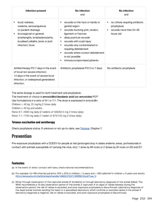
![Page 253
/ 394
References
(d) In penicillin-allergic patients:
• Children: co-trimoxazole (30 mg SMX + 6 mg TMP/kg 2 times daily) + clindamycin (10 mg/kg 3 times daily)
• Adults: co-trimoxazole (800 mg SMX + 160 mg TMP 2 times daily) or doxycycline (100 mg 2 times daily or 200 mg once
daily, except in pregnant and lactating women) + metronidazole (500 mg 3 times daily).
1. Weekly epidemiological record/Relevé épidémiologique hebdomadaire, 20 April 2018, 93th year/20 avril 2018, 93e année.
No 16, 2018, 93, 201–220.
http://apps.who.int/iris/bitstream/handle/10665/272371/WER9316.pdf?ua=1 [Accessed 25 october 2018]
2. Spencer O, Banerjee S. Animal bites. BMJ Best practice 2018 [Accessed 25 october 2018]](https://image.slidesharecdn.com/guideline-170-en-240313103309-343cfe20/85/guideline-170-en-pdf-253-320.jpg)
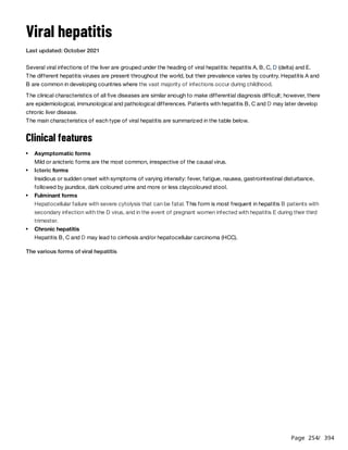
![Page 256
/ 394
Laboratory
Diagnosis
Other tests
Other investigations
Elastography (Fibroscan®): measures the elasticity of the liver to determine stage of liver fibrosis, scored from
F0 (absence of fibrosis) to F4 (cirrhosis).
Treatment
Treatment of chronic active hepatitis B
The goal of treatment is to reduce the risk of cirrhosis and HCC.
Individual
prevention
Polyvalent
immunoglobulin
Specific anti-HBs
immunoglobulin
Safe sex (condoms)
Specific anti-
HBs
immunoglobulin
may be
effective
As for hepatitis
B (the D virus
can only
develop with
B)
Cook meat
(pork)
Vaccination Anti-hepatitis
A
Anti-hepatitis B Does not exist Anti-hepatitis
B
Does not exist
Collective
prevention
Hygiene,
sanitation
Limit transfusion, screen blood prior to transfusion
Single use of disposable material
Hygiene,
sanitation
(a) Vertical transmission: transmission of the virus from the mother to the child during pregnancy, at the time of delivery, or
during the first 28 days after birth.
HAV, HDV and HEV infection: detection of IgM anti-HAV, anti-HDV and anti-HEV antibodies, respectively.
HBV infection: detection of HBsAg; chronic hepatitis B: presence of HBsAG for longer 6 months; chronic active
hepatitis B: detection of HBeAg and/or HBV DNA.
HCV infection: detection of anti-HCV antibodies and HCV RNA; chronic hepatitis C: viraemia persists for longer
than 6 months.
ALT (or AST) level, platelet count, creatinine, HCV diagnosis and HBV viral load to decide treatment of chronic
active hepatitis B.
APRI score (evaluation of liver fibrosis in chronic hepatitis): [(patient's AST level/normal AST level) x 100]/platelet
count (10 platelets/litre). An APRI score > 1 indicates probable severe fibrosis.
9
HIV test.
Rest, hydration, no special diet.
Do not administer drug therapy for symptomatic treatment (analgesics, antipyretics, antidiarrhoeals, antiemetics
etc.) during the acute phase as it may aggravate symptoms and the evolution of hepatitis. Corticosteroids are not
indicated.
Stop or reduce alcohol consumption.
Patients with HIV co-infection
Lifelong antiretroviral therapy of HIV that includes tenofovir. Do not administer tenofovir monotherapy or tenofovir](https://image.slidesharecdn.com/guideline-170-en-240313103309-343cfe20/85/guideline-170-en-pdf-256-320.jpg)
![Page 257
/ 394
Treatment of chronic hepatitis C
In case of decompensated cirrhosis (presence of ascites or jaundice or mental confusion or signs of gastrointestinal
haemorrhage): same treatment but for 24 weeks.
Treatment is contra-indicated during pregnancy and breastfeeding.
For women of childbearing age: provide a contraceptive; do not start treatment in women who do not want
contraception.
Vaccination
dual therapy with lamivudine or emtricitabine (risk of developing HIV drug resistance).
Patients without HIV co-infection
Treatment is indicated in the event of cirrhosis or advanced hepatic fibrosis (APRI score > 1.5 or Fibroscan F3-F4 >
10 kPa); HBsAg positive with persistently elevated ALT or AST > 2 times the normal values in 2 samples taken 3 or
6 months apart; or persistently elevated ALT or AST with a high viral load (> 20 000 IU/ml).
tenofovir PO (300 mg tab, equivalent to 245 mg of tenofovir disoproxil), lifelong therapy:
Children ≥ 12 years and adults, including pregnant women: one tablet once daily taken with a meal
[1]
Genotypes 1, 2, 3, 4, 5, 6 without cirrhosis
or with compensated cirrhosis
sofosbuvir/velpatasvir PO (400 mg SOF/100 mg VEL tablet)
1 tablet once daily for 12 weeks
Genotypes 1, 2, 4, 5, 6 without cirrhosis
or with compensated cirrhosis
Genotype 3 without cirrhosis
sofosbuvir/daclatasvir PO (400 mg SOF/60 mg DCV tablet)
1 tablet once daily for 12 weeks
Genotype 3 with compensated cirrhosis sofosbuvir/daclatasvir PO (400 mg SOF/60 mg DCV tablet)
1 tablet once daily for 24 weeks
Routine vaccination of neonates and infants (according to national vaccination schedule):
[2]
3 dose schedule: one dose as soon as possible after birth, preferably within the first 24 hours of life, then one
dose at 6 weeks and one dose at 14 weeksa
4 dose schedule: one dose as soon as possible after birth, preferably within the first 24 hours of life, then one
dose at 6 weeks, one dose at 10 weeks and one dose at 14 weeksa
Catch-up vaccination (unvaccinated individuals):
3 dose schedule (0-1-6): 2 doses 4 weeks apart, then a third dose 6 months after the first dose
Post-exposure prophylaxis:
One dose on D0, one dose on D7 and one dose between D21 and D30 then a booster dose 12 months after the
first dose
Footnotes
(a) At birth, only the monovalent hepatitis B vaccine can be used.
For the following doses, a monovalent or tetravalent (diphtheria, tetanus, pertussis, hepatitis B) or pentavalent (diphtheria,
tetanus, pertussis, hepatitis B and Haemophilus influenzae) vaccine can be used, depending on national recommendations.
If an infant was not administered the birth dose, this dose can be administered at anytime during the first contact with
health-care providers, up to the time of the next dose of the primary schedule.](https://image.slidesharecdn.com/guideline-170-en-240313103309-343cfe20/85/guideline-170-en-pdf-257-320.jpg)
![Page 258
/ 394
References
1. World Health Organization. Guidelines for the care and treatment of persons diagnosed with chronic hepatitis C virus
infection. July 2018.
http://apps.who.int/iris/bitstream/handle/10665/273174/9789241550345-eng.pdf?ua=1 [Accessed 21 December 2018]
2. Weekly epidemiological record/Relevé épidémiologique hebdomadaire 7 JULY 2017, 92th YEAR / 7 JUILLET 2017, 92e ANNÉE
No 27, 2017, 92, 369–392
http://apps.who.int/iris/bitstream/handle/10665/255841/WER9227.pdf?sequence=1 [Accessed 22 November 2018]](https://image.slidesharecdn.com/guideline-170-en-240313103309-343cfe20/85/guideline-170-en-pdf-258-320.jpg)
![Page 260
/ 394
Major differential diagnoses
Other conditions to consider include a wide range of acute febrile illnesses, e.g.:
Laboratory
Diagnosis
Symptoms according to severity (adapted from PAHO )
[1]
Dengue
without warning
signs
Fever + 2 of the following symptoms:
Nausea, vomiting
Rash resembling measles
Generalized aches (headache, retro-orbital pain, myalgias, arthralgias)
Benign mucocutaneous bleeding (petechiae, positive tourniquet test , epistaxis, gingival
bleeding)
(a)
Leucopenia
Dengue
with warning signs
Presence of at least one of these symptoms:
Intense abdominal pain
Persistent vomiting
Fluid accumulation (ascites, pleural effusion)
Mucosal bleeding
Hepatomegaly (> 2 cm)
Postural hypotension
Agitation or lethargy
Increasing haematocrit
Severe dengue Severe fluid accumulation (ascites, pleural effusion) with respiratory distress and/or shock
Severe mucocutaneous bleeding
Severe organ involvement (e.g.: transaminases > 1000 IU/litre, myocarditis, altered mental
status)
(a) Tourniquet test: inflate a blood pressure cuff on the upper arm to a point midway between the systolic and diastolic
pressure for 5 min. The test is positive when 20 or more petechiae per 2.5 cm square are observed.
Chikungunya, Zika, influenza, mononucleosis, measles, rubella, viral haemorrhagic fevers
Malaria
Meningococcemia, typhoid fever, leptospirosis, rickettsioses, other causes of sepsis
Leukaemia
NS-1 antigen detection during febrile phase with rapid diagnostic test or ELISA (serum, plasma or blood).
Antibody detection (complex interpretation):
IgM detection 5 to 6 days after onset of illness may support (but does not confirm) a diagnosis of recent
infection;
IgG detection may indicate prior infection by, or vaccination against, dengue virus or a closely related virus (e.g.
chikungunya, Zika, Japanese encephalitis, yellow fever).
PCR may also be available in reference laboratories.
In all cases, rapid test for malaria in endemic regions (and antimalarial treatment if needed, see Malaria, Chapter 6).](https://image.slidesharecdn.com/guideline-170-en-240313103309-343cfe20/85/guideline-170-en-pdf-260-320.jpg)
![Page 261
/ 394
Monitoring disease course
Treatment
Patients in Group A (outpatients)
Patients with no warning signs, able to drink sufficiently and with a normal urine output.
Patients in Group B (inpatients)
Patients with any of the following:
In all cases:
If poor oral intake:
If warning signs:
Haematocrit (Hct) or if available full blood count (FBC) at baseline, then daily if possible.
A progressive increase in Hct is a warning sign. It indicates haemoconcentration due to increased vascular
permeability (plasma leakage). Hct should be monitored frequently (before and after fluid administration) in
patients with warning signs up to the end of the fluid treatment.
Leukopenia and thrombocytopenia are common and improve as the recovery phase begins. Leukocytosis may
occur with severe bleeding.
Liver function tests if possible at baseline, then according to results.
Bed rest and good hydration.
Fever and pain: paracetamol PO at the usual doses (see Fever, Chapter 1), maintaining a 6 to 8 hour interval
between doses. Do not prescribe acetylsalicylic acid, ibuprofen or other NSAIDs.
Seek medical attention if: no clinical improvement, persistent vomiting, cold extremities, agitation or lethargy,
breathing difficulties or absence of urine output.
If follow-up is impossible or symptoms cannot be monitored at home (patients living far from the health care
facility/living alone), hospitalise for observation.
Warning sign(s)
Acute (e.g. severe dehydation or malaria) or chronic (e.g. diabetes, cardiovascular, renal or haemolytic disease,
obesity) co-morbidities
Risk factors for bleeding (e.g. anticoagulation, coagulopathy, peptic ulcer or gastritis, treatment with NSAIDs)
Pregnant women, patients under 1 year or 65 years and over or patients with difficulty drinking
Place the patient under a mosquito net; encourage oral fluid intake (including oral rehydration solution (ORS) if
needed).
Avoid invasive procedures (nasogastric tube, IM injections) to minimize the risk of bleeding.
Fever and pain: paracetamol PO with caution and without exceeding:
[2]
children: 10 mg/kg every 6 to 8 hours
adults: 500 mg every 6 to 8 hours
In case of elevated transaminases ≥ 10 times the upper limit of normal, do not administer paracetamol. Use tepid
sponging for reducing fever.
Monitor vital signs, fluid intake (infusion and oral) and urine output every 4 hours .
b
Place an intravenous line and administer:
children: 5% glucose + Ringer lactate solution as maintenance fluids, according to the Holliday-Segar
formula, i.e. 4 ml/kg/hour for first 10 kg of body weight + 2 ml/kg/hour for next 10 kg + 1 ml/kg/hour for each
additional kg above 20 kg.
c
adults: Ringer lactate, 2 to 3 ml/kg/hour
Encourage oral intake as soon as possible.](https://image.slidesharecdn.com/guideline-170-en-240313103309-343cfe20/85/guideline-170-en-pdf-261-320.jpg)
![Page 262
/ 394
Patients in Group C (intensive care unit)
Patients with severe dengue requiring emergency treatment for managing shock and other complications (e.g. severe
bleeding, acidosis, coagulopathy).
Prevention
References
Monitor clinical status (warning signs, general symptoms, vital signs, capillary refill time), IV and oral fluid intake, urine
output, hourly for at least 4 hours, then every 4 hours while the patient is on IV fluid treatment.
Place an intravenous line and administer a bolus of Ringer lactate:
children and adults: 10 ml/kg over one hour
patients 65 years and over or with co-morbidities : 5 ml/kg over one hour
Re-assess the patient:
If no improvement after first bolus: administer a second bolus as above. If necessary, a total of 3 boluses can
be administered. If still no improvement after 3 bolus, consider as severe dengue (patients in Group C) and
transfer to intensive care unit.
If improvement after the first, second, or third bolus, reduce Ringer lactate:
children and adults: 5 to 7 ml/kg/hour over 2 to 4 hours
patients 65 years and over or with co-morbidities: 5 ml/kg/hour over 2 to 4 hours
If continuing improvement, reduce Ringer lactate (then stop as soon as possible to reduce the risk of fluid
overload):
children and adults: 3 to 5 ml/kg/hour over 2 to 4 hours, then 2 to 4 ml/kg/hour over 24 to 48 hours
patients 65 years and over or with co-morbidities: 3 ml/kg/hour over 2 to 4 hours, then 2 ml/kg/hour over 24
to 48 hours
If the patient deteriorates after initial improvement, resume the bolus therapy with Ringer lactate (up to 3 bolus)
as above.
Individual protection: long sleeves and trousers, repellents, mosquito net (Aedes bites during the day).
Elimination of mosquito breeding sites (small collections of water in discarded tires, flower pots, and other
containers).
Footnotes
(a) For more information:
http://gamapserver.who.int/mapLibrary/Files/Maps/Global_DengueTransmission_ITHRiskMap.png?ua=1
(b) Adequate urine output: at least 1 ml/kg/hour in children and 0.5 ml/kg/hour in adults. If unavailable, ensure that the patient is
urinating at least every 4 hours.
(c) Remove 50 ml of Ringer lactate (RL) from a 500 ml RL bottle or bag, then add 50 ml of 50% glucose to the remaining 450 ml
of RL to obtain 500 ml of 5% glucose-RL solution.
1. Pan American Health Organization. Dengue: guidelines for patient care in the Region of the Americas, 2nd edition.
Washington, D.C.: PAHO, 2016.
https://iris.paho.org/bitstream/handle/10665.2/31207/9789275118900-eng.pdf?sequence=1&isAllowed=y [Accessed 23 Aug
2022]
2. Pan American Health Organization. Guidelines for the Clinical Diagnosis and Treatment of Dengue, Chikungunya, and Zika.](https://image.slidesharecdn.com/guideline-170-en-240313103309-343cfe20/85/guideline-170-en-pdf-262-320.jpg)
![Page 263
/ 394
Washington, D.C. : PAHO; 2022.
https://iris.paho.org/handle/10665.2/55867 [Accessed 16 Aug 2022]](https://image.slidesharecdn.com/guideline-170-en-240313103309-343cfe20/85/guideline-170-en-pdf-263-320.jpg)
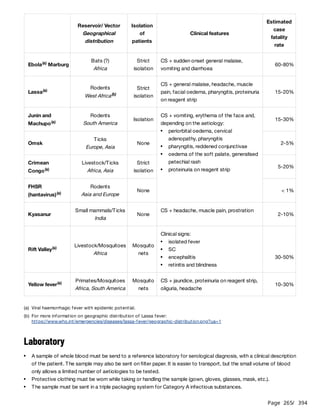

![Page 267
/ 394
Prevention
References
Aetiological treatment: ribavirine for Lassa fever and Crimean-Congo fever.
Symptomatic treatment:
Fever: paracetamol (Chapter 1). Acetylsalicylic acid (aspirin) is contra-indicated.
Pain: mild (paracetamol), moderate (tramadol), severe (sublingual morphine): see Pain, Chapter 1.
Dehydration: oral rehydration salts and/or IV rehydration with Ringer lactate, see Dehydration, Chapter 1.
Seizures (Chapter 1).
Vomiting: ondansetron PO
Children 6 months to < 2 years: 2 mg once daily
Children 2 to < 4 years: 2 mg 2 times daily
Children 4 to < 12 years: 4 mg 2 times daily
Children ≥ 12 years and adults: 4 to 8 mg 2 times daily
[1]
For Ebola and Marburg haemorrhagic fevers: invasive procedures must be strictly limited. Health care staff is at risk
of contamination when inserting and maintaining IV lines. An IV line must be well secured so that the patient, often
confused, cannot pull it out.
Vaccination against yellow fever :
Children and adults: 0.5 ml single dose
[2]
Routine vaccination : children from 9 months of age, along with the measles vaccine.
Mass vaccination campaign during an epidemic: children from 6 months and adults ; for pregnant women, only
administer during an epidemic.
Vaccination against Rift Valley fever: only during an epidemic.
Vector control programmes for known vectors.
Infection control measures are essential in all cases.
1. World Health Organization. Clinical management of patients with viral haemorrhagic fever. A pocket guide for front-line
health workers. Interim emergency guidance for country adaptation, February 2016.
http://apps.who.int/iris/bitstream/handle/10665/205570/9789241549608_eng.pdf;jsessionid=15E17DE39631519C2051413DD
CBBC8A7?sequence=1 [Accessed 11 January 2019]
2. Weekly epidemiological record-Relevé épidémiologique hebdomadaire 5 july 2013, 88th year / 5 juillet 2013, 88e année No. 27,
2013, 88, 269–284.
https://www.who.int/wer/2013/wer8827.pdf?ua=1 [Accessed 10 december 2018]](https://image.slidesharecdn.com/guideline-170-en-240313103309-343cfe20/85/guideline-170-en-pdf-267-320.jpg)
![Page 268
/ 394
HIV infection and AIDS
Last updated: January 2024
Acquired immune deficiency syndrome (AIDS) is the most advanced stage of infection with human immunodeficiency
virus (HIV).
Two subtypes of HIV have been identified. HIV-1 is more widespread than HIV-2, the latter mainly being found in West
Africa. HIV-2 is less virulent and less transmissible than HIV-1.
HIV weakens the immune system by causing a deficit in CD4 T lymphocytes.
Evolution of the disease
The World Health Organization (WHO) has proposed a clinical classification of HIV infection in 4 stages of severity for
adults and adolescents and for children.
Laboratory
Diagnosis of HIV infection
CD4 lymphocyte counts
CD4 cell depletion is a marker of the progression of immune depression. The level of the CD4 cell count is a predictor
of the development of opportunistic infections or neoplasms and can be used to orient their diagnosis, e.g. cerebral
toxoplasmosis or cryptococcal meningitis appear when the CD4 count is below 100 cells/mm in adults. If clinical
symptoms/signs are present suggesting one of these infections, but the CD4 count is greater than or equal to 200
cells/mm , it is unlikely that that particular infection is present.
Opportunistic infections
Primary infection or acute retroviral syndrome: 50 to 70% of newly infected individuals develop during
seroconversion (from 15 days to 3 months post exposure), a viral syndrome with fever, malaise, and
lymphadenopathy.
Asymptomatic HIV infection (after seroconversion): a period of clinical latency, but not viral latency. The time
period for progression from HIV infection to the development of severe immune deficiency in western countries is
approximately 10 years. This period appears to be shorter in developing countries.
Symptomatic HIV infection: with progressive destruction of the immune system, common and more severe
diseases occur more frequently, and with higher mortality, in seropositive individuals.
AIDS: this stage corresponds to the development of severe opportunistic infections and neoplasms. From a
biological point of view, AIDS is defined as a CD4 count < 200 cells/mm . Without treatment the disease
progresses rapidly towards death.
3
[1]
The diagnosis is made with serological (detection of antibodies against the virus) or virological (especially in infants)
testing.
Testing should always be done voluntarily with informed consent.
All HIV test results must be strictly confidential in order to avoid discrimination.
The individual should have access to services offering pre-test and post-test counselling, treatment and support.
A diagnosis of HIV infection can be made only after at least 2 different test results (2 different brands) are clearly
positive: the positive result of an initial (highly sensitive) test must be confirmed through use of a second (highly
specific) test. In areas where HIV prevalence is low, diagnosis is confirmed after 3 positive test results.
3
3](https://image.slidesharecdn.com/guideline-170-en-240313103309-343cfe20/85/guideline-170-en-pdf-268-320.jpg)
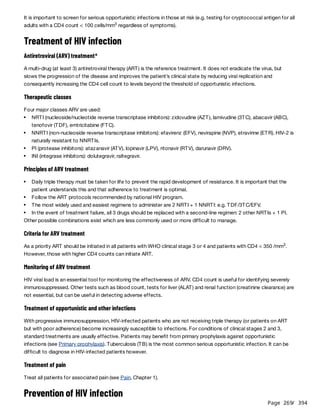
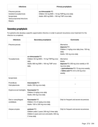

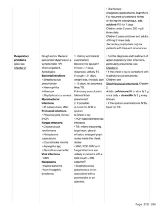
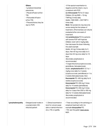
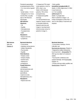
![Page 281
/ 394
If the LP is positive:
• Bacterial meningitis: see Chapter 7.
• TB meningitis: see the guide
Tuberculosis, MSF.
• Cryptococcal meningitis :
amphotericin B IV: 1 mg/kg once
daily + flucytosine PO: 25 mg/kg 4
times daily for 1 week
then fluconazole PO: 1200 mg once
daily for 1 week then 800 mg once
daily for 8 weeks
or, if not available
amphotericin B IV: 1 mg/kg once
daily + fluconazole PO: 1200 mg
once daily for 2 weeks
then fluconazole PO alone: 800 mg
once daily for 8 weeks
or
fluconazole PO: 1200 mg once daily
+ flucytosine PO: 25 mg/kg 4 times
daily for 2 weeks
then fluconazole PO alone: 800 mg
once daily for 8 weeks
During the induction phase: give
fluconazole IV (same doses) if the
patient cannot take oral treatment;
liposomal amphotericin B (3 mg/kg
daily 2 weeks) may be used instead
of conventional amphotericin B in
case of renal impairment.
A secondary prophylaxis is
recommended.
Note: intracranial pressure (ICP) is
often raised in cryptococcal
meningitis. To lower ICP, repeated
‘therapeutic’ punctures to drain CSF
may be necessary at the beginning
of treatment.
Neurosyphilis:
benzylpenicillin IV: 2 to 4 MIU (1.2
to 2.4 g) every 4 hours for 14 days
or ceftriaxone IV or IM: 2 g once
daily for 10 to 14 days
Headache of unknown origin:
symptomatic treatment starting with
a step 1 analgesic (see Pain,
Chapter 1).
[2]
Neurological Aetiologies: Good history taking as Positive malaria test: see Malaria,](https://image.slidesharecdn.com/guideline-170-en-240313103309-343cfe20/85/guideline-170-en-pdf-281-320.jpg)
![Page 283
/ 394
If the LP is positive:
• Bacterial meningitis: see Chapter 7.
• TB meningitis: see the guide
Tuberculosis, MSF.
• Cryptococcal meningitis (in order of
preference) :
amphotericin B IV: 1 mg/kg once
daily + flucytosine PO: 25 mg/kg 4
times daily for 1 week then
fluconazole PO: 12 mg/kg once
daily (max. 800 mg daily) for 1 week
then 6-12 mg/kg once daily (max.
800 mg daily) for 8 weeks
or, if not available
amphotericin B IV: 1 mg/kg once
daily + fluconazole PO: 12 mg/kg
once daily (max. 800 mg daily) for 2
weeks then fluconazole PO alone:
6-12 mg/kg once daily for 8 weeks
(max. 800 mg daily)
or
fluconazole PO: 12 mg/kg once
daily (max. 800 mg daily) +
flucytosine PO: 25 mg/kg 4 times
daily for 2 weeks then fluconazole
PO alone: 6-12 mg/kg once daily
(max. 800 mg daily) for 8 weeks
During the induction phase: give
fluconazole IV (same doses) if the
child cannot take oral treatment;
liposomal amphotericin B (3 mg/kg
daily, 2 weeks) may be used instead
of conventional amphotericin B in
case of renal impairment.
A secondary prophylaxis is
recommended.
[2]
Persistent or
recurrent fever
Temperature > 38 °C,
chronic (lasting more than
5 days) or recurrent
(multiple episodes in a
period of more than 5
days)
Aetiologies:
1. History and clinical
examination: look for a
ENT or urinary infection,
TB, skin infection,
enlarged lymph nodes
etc.
2. In endemic areas,
check for malaria.
3. Suspected TB: look
for AFB.
Positive malaria test: see Malaria,
Chapter 6.
If testing is not available: in endemic
areas, treat malaria.
Suspected meningitis: treat
according to the results of the LP.
If LP is not available, treat for
bacterial meningitis, see Chapter 7.](https://image.slidesharecdn.com/guideline-170-en-240313103309-343cfe20/85/guideline-170-en-pdf-283-320.jpg)
![Page 284
/ 394
References
Infections
• Common childhood
diseases
• Severe bacterial
infections (TB,
pneumonia, typhoid fever,
septicaemia, meningitis,
endocarditis, etc.)
• Occult bacterial
infections (sinusitis, otitis,
urinary tract infections)
• Opportunistic infections
(TB, mycosis,
toxoplasmosis)
• Malaria
Neoplasms
• Non-Hodgkin’s
lymphoma
HIV infection
Fever caused by
medication
4. Chest x-ray, CBC,
blood cultures, urinalysis,
stool culture, serology,
lumbar puncture (LP).
If the child is under
treatment, consider
adverse effects of
medication.
Identified or suspected focus of
infection:
• ENT: see Chapter 2; urinary: see
Chapter 9, etc.
• TB: see the guide Tuberculosis,
MSF.
Footnotes
(a) For more information: The use of antiretroviral drugs for treating and preventing HIV infection. Recommendations for a
public health approach. World Health Organization, second edition, 2016.
http://apps.who.int/iris/bitstream/10665/208825/1/9789241549684_eng.pdf?ua=1
1. World Health Organization. WHO case definitions of HIV for surveillance and revised clinical staging and immunological
classification de HIV-related disease in adults and children, 2007.
http://www.who.int/hiv/pub/guidelines/HIVstaging150307.pdf [Accessed 17 May 2018]
2. Word Health Organization. Guidelines for the diagnosis, prevention, and management of cryptococcal disease in HIV-
infected adults, adolescents and children, Geneva, 2018.
http://apps.who.int/iris/bitstream/handle/10665/260399/9789241550277-eng.pdf?sequence=1 [Accessed 17 May 2018]](https://image.slidesharecdn.com/guideline-170-en-240313103309-343cfe20/85/guideline-170-en-pdf-284-320.jpg)
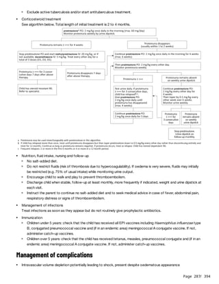
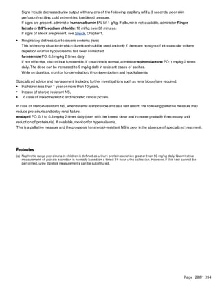
![Page 293
/ 394
Preferably use the combination ampicillin + gentamicin to cover enterococci.
Pyelonephritis with abscess formation or emphysematous pyelonephritis may require longer antibiotherapy.
Children 0 to 7 days (≥ 2 kg): 5 mg/kg once daily
Children 8 days to < 1 month: 5 mg/kg once daily
or
cefotaxime slow IV (3 minutes) for 7 to 10 days
Children 0 to 7 days (< 2 kg): 50 mg/kg every 12 hours
Children 0 to 7 days (≥ 2 kg): 50 mg/kg every 8 hours
Children 8 days to < 1 month: 50 mg/kg every 8 hours
Children one month and over
ceftriaxone IM or slow IV (3 minutes): 50 mg/kg once daily until the child's condition improves (at least 3 days)
then change to oral route to complete 10 days of treatment with:
amoxicillin/clavulanic acid PO (dose expressed in amoxicillin)
Children < 40 kg: 25 mg/kg 2 times daily
Children ≥ 40 kg:
Ratio 8:1: 2000 mg daily (2 tablets of 500/62.5 mg 2 times daily)
Ratio 7:1: 1750 mg daily (1 tablet of 875/125 mg 2 times daily)
b
Antibiotherapy in adults[1]
Uncomplicated pyelonephritis
ceftriaxone IM: 1 g single dose or gentamicin IM: 5 mg/kg single dose
+
ciprofloxacin PO: 500 mg 2 times daily for 7 days
or
amoxicillin/clavulanic acid PO (dose expressed in amoxicillin) for 10 to 14 days
Ratio 8:1: 2000 mg daily (2 tablets of 500/62.5 mg 2 times daily)
Ratio 7:1: 1750 mg daily (1 tablet of 875/125 mg 2 times daily)
or
cefixime PO: 200 mg 2 times daily or 400 mg once daily for 10 to 14 days
Pyelonephritis with criteria for hospital admission
ampicillin slow IV (3 minutes): 2 g every 6 hours for at least 3 days + gentamicin IM: 5 mg/kg once daily for 3
days
then change to amoxicillin/clavulanic acid PO (or another antibiotic depending on the antibiotic susceptibility
test) to complete 10 to 14 days of treatment
or
ceftriaxone IV : 1 g once daily for at least 3 days + gentamicin IM: 5 mg/kg once daily for 3 days in the event
of sepsis then change to amoxicillin/clavulanic acid PO (or another antibiotic depending on the antibiotic
susceptibility test) to complete 10 to 14 days of treatment
b
Treatment of fever and pain: do not administer NSAID (Fever, Chapter 1).
Maintain proper hydration (1.5 litres daily in adults), especially in children (risk of dehydration); treat dehydration if
present (see Dehydration, Chapter 1).
Management of septic shock if needed.
Footnotes
(a) Pyelonephritis is rare in men; bacterial prostatitis should be suspected in the event of febrile urinary tract infection.](https://image.slidesharecdn.com/guideline-170-en-240313103309-343cfe20/85/guideline-170-en-pdf-293-320.jpg)
![Page 294
/ 394
References
(b) The solvent of ceftriaxone for IM injection contains lidocaine. Ceftriaxone reconstituted using this solvent must never be
administered by IV route. For IV administration, water for injection must always be used.
1. Gupta K, Hooton TM, Naber KG, Wullt B, Colgan R, Miller LG, Moran GJ, Nicolle LE, Raz R, Schaeffer AJ, Soper DE, Infectious
Diseases Society of America, European Society for Microbiology and Infectious Diseases. International clinical practice
guidelines for the treatment of acute uncomplicated cystitis and pyelonephritis in women: A 2010 update by the Infectious
Diseases Society of America and the European Society for Microbiology and Infectious Diseases. Clin Infect Dis.
2011;52(5):e103.
https://academic.oup.com/cid/article/52/5/e103/388285 [Accessed 17 December 2018]](https://image.slidesharecdn.com/guideline-170-en-240313103309-343cfe20/85/guideline-170-en-pdf-294-320.jpg)
![Page 295
/ 394
Acute prostatitis
Prostatitis is an acute bacterial infection of the prostate.
The most common causative pathogen is Escherichia coli. Other pathogens include Proteus
mirabilis, Klebsiella sp, Pseudomonas aeruginosa and Enterococcus sp.
Progression to chronic prostatitis is possible.
Clinical features
On examination:
Treatment
References
Fever (often high) and chills.
Signs of cystitis (burning on urination and urinary frequency).
Perineal, urethral, penile or rectal pain.
Urinary retention.
Very painful digital rectal examination. Fluctuant mass in case of prostatic abscess.
Leukocyturia, pyuria, possible macroscopic haematuria.
Antibiotic therapy:
ciprofloxacin PO: 500 mg 2 times daily for 14 days then review the patient. Stop treatment if signs and symptoms
have completely resolved. If signs and symptoms are ongoing continue the same treatment for a further 14 days.[1]
Symptomatic treatment:
Ensure adequate hydration (1.5 litres daily).
Treat fever (Chapter 1) and pain (Chapter 1).
Refer to a surgeon in case of suspected prostatic abscess.
1. National Institute for Health and Care Excellence. NICE guideline [NG110] Prostatitis (acute): antimicrobial prescribing, 2018.
https://www.nice.org.uk/guidance/ng110/resources/visual-summary-pdf-6544018477 [Accessed 4 March 2020]](https://image.slidesharecdn.com/guideline-170-en-240313103309-343cfe20/85/guideline-170-en-pdf-295-320.jpg)
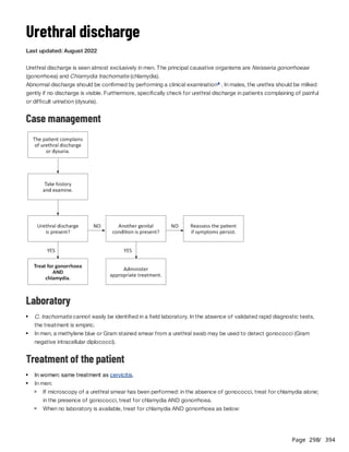
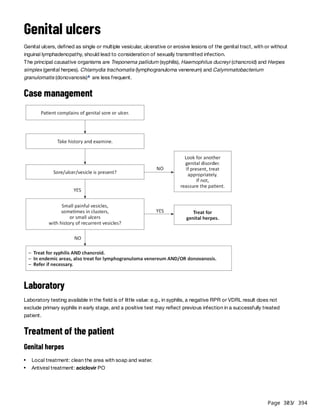
![Page 304
/ 394
In patients with a first episode, treatment may reduce the duration of symptoms when given within 5 days after the
onset of symptoms: 400 mg 3 times daily for 7 days.
In patients with recurrence, give the same dose for 5 days, but treatment is only effective if initiated during the
prodromal phase or within 24 hours after the onset of symptoms.
In patients with frequent recurrences (more than 6 episodes par year), see HIV infection and AIDS, Chapter 8.
Syphilis
benzathine benzylpenicillin IM: 2.4 MUI per injection (half the dose in each buttock) .
Early syphilis (primary, secondary, or early latent infection of less than 12 months duration): single dose
Late latent syphilis (infection of more than 12 months duration or of unknown duration): one injection weekly for 3
weeks
or, for penicillin-allergic patients or if penicillin is not available:
erythromycin PO: 1 g 2 times daily or 500 mg 4 times daily for 14 days (early syphilis) or 30 days (late latent syphilis)
or
doxycycline PO: 100 mg 2 times daily for 14 days (early syphilis) or 30 days (late latent syphilis)
or
azithromycin PO: 2 g single dose (only in cases of early syphilis and only if the strain is sensitive)
Chancroid
azithromycin PO: 1 g single dose
or
ceftriaxone IM: 250 mg single dose
or
erythromycin PO: 1 g 2 times daily or 500 mg 4 times daily for 7 days
Fluctuant lymph nodes may be aspirated through healthy skin as required. Do not incise and drain lymph nodes.
Note: treat simultaneously for syphilis AND chancroid as both are frequent, and cannot be correctly distinguished on
clinical grounds.
Lymphogranuloma venereum
erythromycin PO: 1 g 2 times daily or 500 mg 4 times daily for 14 days
or
doxycycline PO: 100 mg 2 times daily for 14 days
Fluctuant lymph nodes may be aspirated through healthy skin as required. Do not incise and drain lymph nodes.
Donovanosis
Treatment is given until the complete disappearance of the lesions (usually, several weeks; otherwise risk of
recurrence):
azithromycin PO: 1 g on D1 then 500 mg once daily
or
erythromycin PO: 1 g 2 times daily or 500 mg 4 times daily
or
doxycycline PO: 100 mg 2 times daily
In HIV infected patients, add gentamicin IM: 6 mg/kg once daily.
Treatment of the partner
Treatment of pain: paracetamol PO (Chapter 1).
[1]
b
[2]
b
b](https://image.slidesharecdn.com/guideline-170-en-240313103309-343cfe20/85/guideline-170-en-pdf-304-320.jpg)
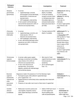
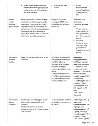
![Page 314
/ 394
Abnormal uterine bleeding (in the absence of
pregnancy)
Last updated: October 2021
For the management of pregnancy-related bleeding, refer to the guide Essential obstetric and newborn care, MSF.
In all events
According to clinical examination
Heavy menstrual bleeding or intermenstrual genital bleeding
In women of childbearing age:
assess if the bleeding is pregnancy-related;
perform a pregnancy test.
Rapidly assess the severity of bleeding.
Perform a pelvic examination:
speculum examination: determine the origin (vagina, cervix, uterine cavity) and cause of the bleeding; appearance
of the cervix; amount and intensity of bleeding;
bimanual examination: look for cervical motion tenderness, uterine enlargement or irregularity.
Assess for recent trauma or surgical history.
Measure haemoglobin, if possible, to prevent or treat anaemia.
In the event of signs of shock, see Shock, Chapter 1.
In the event of heavy bleeding:
start an IV infusion of Ringer lactate;
monitor vital signs (heart rate, blood pressure);
administer :
tranexamic acid IV: 10 mg/kg (max. 600 mg) every 8 hours. When bleeding has been reduced, switch to
tranexamic acid PO: 1 g 3 times daily, until bleeding stops (max. 5 days).
[1]
if bleeding persists and/or in case or contraindication to tranexamic acid, administer one of the following two
drugs (except if suspicion of cervical or endometrial cancer):
ethinylestradiol/levonorgestrel PO (0.03 mg/0.15 mg tab): one tablet 3 times daily for 7 days
or medroxyprogesterone acetate PO: 20 mg 3 times daily for 7 days
In case of massive haemorrhage and/or lack of response to medical management: surgical management (dilation
and curettage, intrauterine balloon, and as a last resort, hysterectomy).
In the event of referral to a surgical facility, difficult transport conditions may aggravate the bleeding: the patient
should have an IV line and/or be accompanied by family members who are potential blood donors.
If available, POCUS : perform FAST to evaluate for free fluid and/or urological abnormalities; perform pelvic views
to evaluate for uterine and/or adnexal pathologies.
a
Friable, hard, ulcerated, hypertrophic mass on the cervix: possible cervical cancer; surgical treatment,
chemotherapy, radiation therapy or palliative care is required depending on the stage of the cancer. While waiting
for appropriate treatment, tranexamic acid PO (1 g 3 times daily for 5 days max.) may be used to reduce bleeding.
Inflammation of the cervix, light or moderate bleeding, purulent cervical discharge, pelvic pain: consider cervicitis
(see Abnormal vaginal discharge) or salpingitis (see Upper genital tract infections).](https://image.slidesharecdn.com/guideline-170-en-240313103309-343cfe20/85/guideline-170-en-pdf-314-320.jpg)


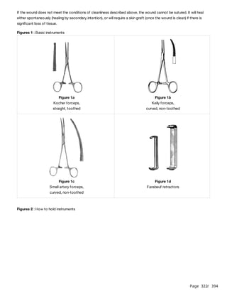
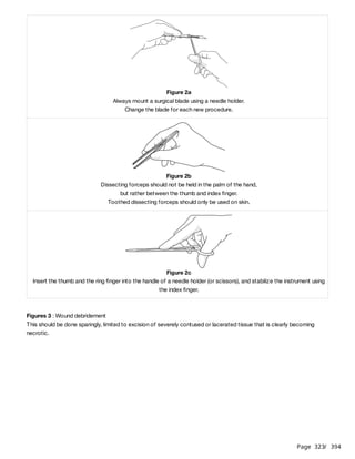
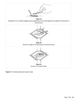
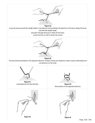
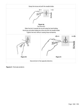
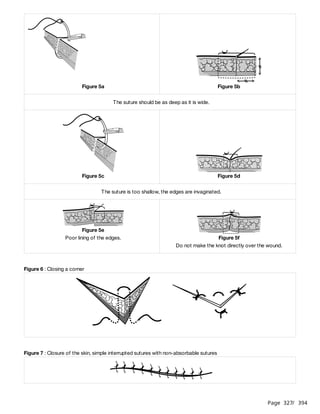
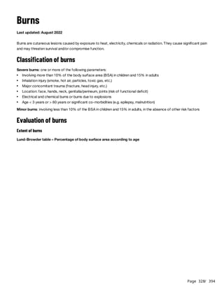
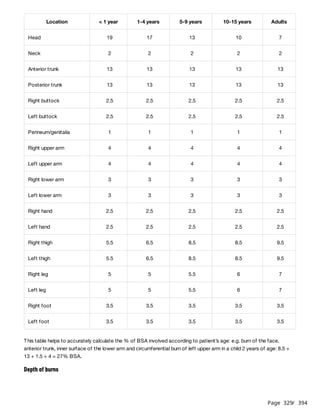
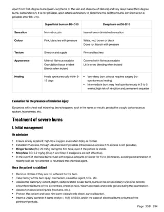

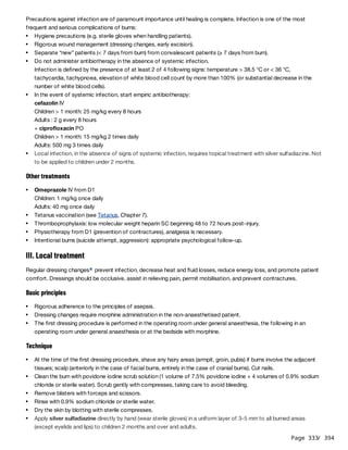
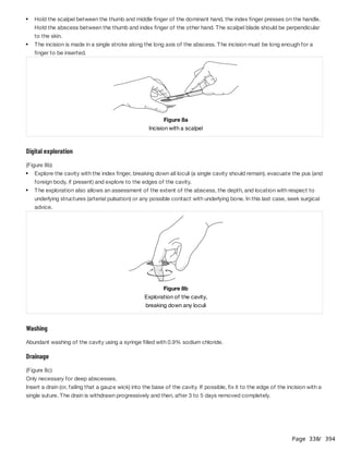
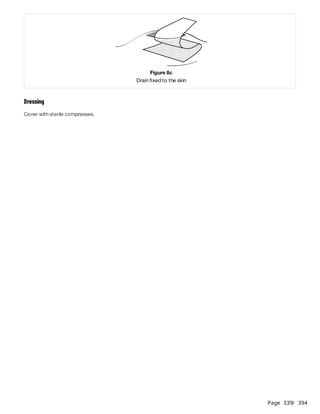
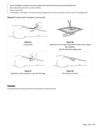
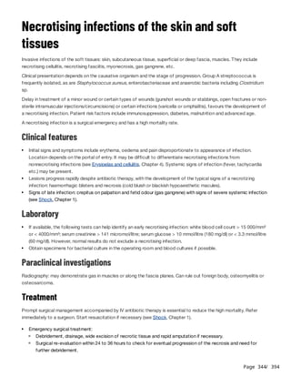
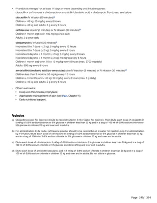
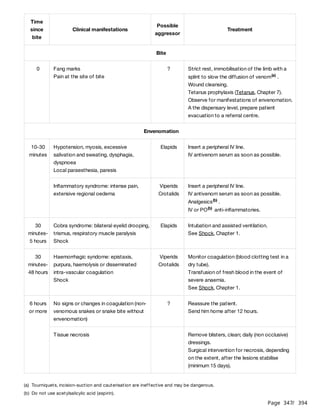


![Page 356
/ 394
Post-traumatic stress disorder
Last updated: November 2021
An event is “traumatic” when someone has been directly confronted with death, either by seeing another person being
killed or seriously injured as the result of violence, or by experiencing serious harm, such as a threat to his/her life or
physical integrity (e.g. rape, torture). Exposure to one or several of these events causes feelings of helplessness and
horror.
Immediate, transitory symptoms (disorientation, anxiety, sadness, fleeing, etc.) are to be distinguished from secondary,
long-lasting problems that appear and/or last several weeks or months after the event: post-traumatic stress, often
associated with depression (Depression), or sometimes acute psychosis (Psychotic disorders), even in people with no
history of psychotic symptoms.
Post-traumatic stress disorder is characterized by three types of psychological response, generally seen in
combination .
Hypervigilance (constant state of alert), exaggerated startle reaction, anxiety, insomnia, poor
concentration; sometimes somatic symptoms (sweating, shaking, tachycardia, headache, etc.).
Re-experiencing is highly distressing and causes disorders that may worsen over time; people isolate themselves,
behave differently, stop fulfilling their family/social obligations, and experience diffuse pain and mental exhaustion.
Management
Psychological intervention is essential to reduce the suffering, disabling symptoms and social handicaps resulting from
PTSD.
It is important to reassure the patient that their symptoms are a normal response to an abnormal event. Sessions
should be conducted with tact. The patient should be listened to. Avoid intensely questioning the patient about their
emotions: leave it to the patient to decide how far they want to go.
Associated symptoms (anxiety or insomnia), if persistent, can be relieved by symptomatic treatment (see Anxiety and
Insomnia) for no more than two weeks.
If the patient has severe symptoms (obsessive thoughts, pronounced hypervigilance, comorbid despression etc.), the
pharmacological treatment is fluoxetine PO (20 mg once daily) or paroxetine PO (10 to 20 mg once daily) or sertraline
PO (50 mg once daily), to be continued for 2 to 3 months after symptoms resolve then, stop gradually.
[1]
Persistent re-experiencing
The patient describes:
images, thoughts or perceptions related to the traumatic experience, which intrude despite efforts to block
them out, including at night in the form of distressing dreams;
flashbacks during which the patient “relives” parts of the traumatic scene.
Avoidance
The patient tries to avoid:
places, situations and people that might be associated with the trauma;
having thoughts or feelings related to the trauma; patients may use alcohol, drugs or any psychotropic agents
for this purpose.
Persistent perceptions of heightened current threat](https://image.slidesharecdn.com/guideline-170-en-240313103309-343cfe20/85/guideline-170-en-pdf-356-320.jpg)
![Page 357
/ 394
References
1. World Health Organization. Post traumatic stress disorder. International Classification of Diseases for Mortality and
Morbidity Statistics, Eleventh Revision (ICD-11).
https://icd.who.int/browse11/l-m/en#/http://id.who.int/icd/entity/2070699808 [Accessed 26 January 2021]](https://image.slidesharecdn.com/guideline-170-en-240313103309-343cfe20/85/guideline-170-en-pdf-357-320.jpg)

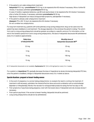
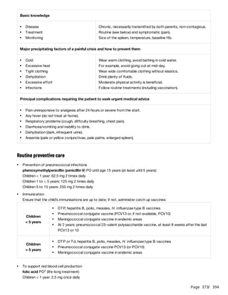
![Page 375
/ 394
Diabetes type 2 in adults
Last update: November 2023
Diabetes is a metabolic disorder that leads to hyperglycaemia.
Type 2 diabetes usually occurs in adults and accounts for 90 to 95% of diabetes cases worldwide.
Type 2 diabetes can lead to acute complications, as well as chronic complications that result in serious organ damage
(cardiovascular events; diabetic retinopathy, neuropathy, nephropathy).
Clinical features
Diagnosis
[1]
Few or no symptoms; symptoms of hyperglycaemia may be present: polyuria (frequent urination) and polydypsia
(excessive thirst and drinking).
In rare cases, patients may present with severe hyperglycaemia (impaired consciousness, coma or acute
dehydration).
Look for diabetes in the event of:
symptoms of hyperglycaemia;
cardiovascular disorders: stroke, myocardial infarction, hypertension;
peripheral neuropathies, foot ulcers, absence of tendon reflexes or peripheral pulse.
Diagnosis is made on one of the following results :
a[1][2]
Tests Symptomatic patient Asymptomatic patient
Fasting blood glucose(a) 1 fasting blood glucose
≥ 7 mmol/litre (≥ 126 mg/dl)
2 fasting blood glucose
≥ 7 mmol/litre (≥ 126 mg/dl)
Random blood glucose(b) 1 random blood glucose
≥ 11 mmol/litre (≥ 200 mg/dl)
See tablenote (b)
Post-load blood glucose(c) 1 post-load blood glucose:
on venous blood
≥ 11 mmol/litre (≥ 200 mg/dl)
2 post-load blood glucose:
on venous blood
≥ 11 mmol/litre (≥ 200 mg/dl)
or
on capillary blood
≥ 12.2 mmol/litre (≥ 220 mg/dl)
or
on capillary blood
≥ 12.2 mmol/litre (≥ 220 mg/dl)
Glycated Hb (HbA1c)(d) 1 HbA1c ≥ 6.5% 2 HbA1c ≥ 6.5%
(a) Fasting blood glucose test: performed on patient that has fasted at least 8 hours. Values are the same for venous and
capillary blood.
(b) Random blood glucose test: performed at any moment of the day. Values are for venous blood only.
For asymptomatic patients, it is not recommended to perform 2 random blood glucose tests. If the first test is a random
blood glucose, the second test should be a fasting blood glucose.
(c) Post-load blood glucose test: performed 2 hours after oral ingestion of 75 g glucose (one sachet of 75 g anhydrous
glucose powder dissolved in 200 to 300 ml of water, to be drunk within 10 minutes).](https://image.slidesharecdn.com/guideline-170-en-240313103309-343cfe20/85/guideline-170-en-pdf-375-320.jpg)
![Page 376
/ 394
or
Treatment
Glycaemic targets
Fasting blood glucose ≤ 7 mmol/litre (≤ 126 mg/dl) or HbA1c of 7%.
The closer blood glucose levels remain to these values, the more cardiovascular complications are prevented or
delayed.
Depending on the context (healthcare provision) or patient profile (older patient, history of severe hypoglycaemia or
long-standing poorly controlled diabetes), HbA1c ≤ 8% is acceptable.
Blood glucose should not fall < 4.5 mmol/litre (or < 80 mg/dl) or HbA1c < 6.5%.
Lifestyle and dietary advice
Pharmacological treatment
First-line treatment metformin PO .
The usual dose is 1 to 2 g daily. For information:
Week 1: 500 mg once daily in the morning at breakfast
Week 2: 500 mg 2 times daily (morning and evening) during meals
Increase in increments of 500 mg per week as long as the drug is well tolerated (max. 2 g daily, i.e. 1 g morning and
evening).
If glycaemic control is not acheived, administer metformin in combination with a sulfonylurea.
Sulfonylurea doses are adjusted in increments to avoid the risk of hypoglycaemia, based on blood glucose results.
The usual dose is 5 mg 2 times daily. For information:
Week 1: 2.5 mg once daily in the morning at breakfast
Week 2: 5 mg once daily in the morning at breakfast
Increase in increments of 2.5 mg weekly until fasting blood glucose reaches target levels (max.15 mg daily).
The usual dose is 40 to 80 mg 2 times daily. For information:
Weeks 1 and 2: 40 mg once daily in the morning at breakfast
Increase in increments of 40 mg every 2 weeks (weeks 3 and 4: 80 mg once daily in the morning at breakfast) until
fasting blood glucose reaches target levels (max. 240 mg daily, i.e. 120 mg morning and evening).
(d) Glycated haemoglobin (HbA1c): reflects average glycaemia over around 3 months.
Perform urine dipstick analysis for ketones if:
fasting blood glucose ≥ 15 mmol/litre (≥ 270 mg/dl) and symptoms of hyperglycemia,
fasting or random blood glucose ≥ 18 mmol/litre (≥ 325 mg/dl) even without symptoms.
Refer the patient if acute complications (such as hyperosmolar hyperglycaemia or ketoacidosis) are present.
[3]
[2]
b
Avoid sugared foods and drinks (but no excessive restriction of carbohydrates).
High fibre intake; limit animal fats and alcohol.
Physical activity.
Weight control. If BMI ≥ 25, try to reduce weight by 5 to 10%.
Stop smoking.
c
In patients under 60, glibenclamide PO:
In patients over 60, gliclazide PO (immediate release tablet):](https://image.slidesharecdn.com/guideline-170-en-240313103309-343cfe20/85/guideline-170-en-pdf-376-320.jpg)
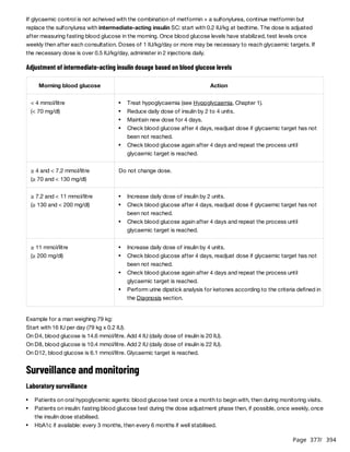
![Page 378
/ 394
Clinical monitoring
Patient education
References
Other necessary tests according to comorbidities and chronic complications.
Routine consultations: check blood pressure (should remain < 140/80 mmHg) and weight, examine feet.
Consultations once a month for the first 6 months, then individualised frequency of consultations depending on the
patient's characteristics (e.g. every 6 months if the diabetes is well controlled).
Annual check-up: check for cardiovascular and neurological complications, evaluate renal function (serum creatinine
and proteinuria dipstick test), examination of teeth and gums.
Management of diabetes complications.
b
Lifestyle and dietary measures (diet, physical activity, etc.).
Patients on sulfonylurea or insulin therapy: signs of hypoglycaemia/hyperglycaemia and management.
Patients on insulin therapy: auto-administration (schedule, injection sites and techniques); storage of insulin.
Patients on insulin therapy or presenting hypoglycaemic episodes : self-monitoring of blood glucose and
adjustment of doses at home using a glucometer.
[3]
Patients with sensory neuropathy or peripheral arterial disease: autoexamination of feet; prevention of foot lesions.
Footnotes
(a) Even in symptomatic patients, it is preferable to perform a second blood glucose test to confirm the result.
(b) These measures concern all patients regardless of medication prescribed. They can be sufficient alone to normalize blood
glucose levels in certain patients.
(c) If metformin is contraindicated or not tolerated, replace with a sulfonylurea.
1. World Health Organization. Classification of Diabetes Mellitus. World Health Organization; 2019 [Accessed October 19, 2023].
https://iris.who.int/handle/10665/325182
2. HEARTS D: diagnosis and management of type 2 diabetes [Accessed October 19, 2023].
https://www.who.int/publications-detail-redirect/who-ucn-ncd-20.1
3. Type 2 diabetes in adults: management | Guidance | NICE. Published December 2, 2015 [Accessed October 19, 2023].
https://www.nice.org.uk/guidance/ng28](https://image.slidesharecdn.com/guideline-170-en-240313103309-343cfe20/85/guideline-170-en-pdf-378-320.jpg)
![Page 379
/ 394
Essential hypertension in adults
Hypertension (or high blood pressure - HBP) is defined as elevated blood pressure (BP) at rest that persists over time
i.e. measured 3 times during 3 separate consultations over a period of three months.
Essential hypertension is defined as HBP of undetermined cause (the large majority of cases).
The global overall prevalence of HBP in adults aged 25 and over is around 40%.
Serious complications of HBP can be acute (hypertensive encephalopathy, left-sided heart failure, acute renal failure)
or delayed i.e. occur after a long period during which HBP has not been controlled (stroke, ischaemic heart disease,
peripheral arterial disease, chronic renal impairment).
For pregnancy-induced hypertension, see Essential obstetric and newborn care, MSF.
Clinical features
SBP ≥ 180 and/or DBP ≥ 110 and some symptoms (moderate headaches, epistaxis, dizziness, tinnitus, eye
floaters) but no signs of end-organ damage;
SBP ≥ 180 and/or DBP ≥ 110 and signs of end-organ damage:
Paraclinical investigations
[1]
HBP thresholds:
HBP classification
Blood pressure (BP) in mmHg
Systolic (SBP) Diastolic (DBP)
Mild 140 or over 90 or over
Moderate 160 or over 100 or over
Severe 180 or over 110 or over
Severe HBP is defined more by the presence of serious end-organ damage than the blood pressure reading:
Uncomplicated hypertensive crisis:
Hypertensive emergency:
intense headaches, nausea/vomitting, confusion, seizures, coma in the event of hypertensive
encephalopathy;
dyspnoea, chest pain in the event of heart failure or cardiac ischaemia;
rapid and/or irregular heart rate in the event of heart failure;
anuria, oliguria in the event of renal impairment.
History and clinical examination should look for:
medications being taken that can cause or aggravate HBP;a
focal neurological sign(s) suggestive of stroke;
comorbidities and risk factors: heart failure, diabetes, renal impairment; excessive smoking or consumption of
alcohol, excess weight (BMI ≥ 25), etc.
Blood test: ionogram (particularly serum potassium levels), serum creatinine.
Other necessary laboratory tests according to comorbidities (e.g. diabetes).](https://image.slidesharecdn.com/guideline-170-en-240313103309-343cfe20/85/guideline-170-en-pdf-379-320.jpg)
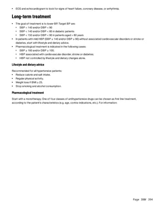
![Page 382
/ 394
Treatment of hypertensive crisis
Uncomplicated hypertensive crisis
Most frequent. Reassure the patient and prescribe rest. Check BP a few days later to start or adapt treatment.
Hypertensive emergency
Treat in an intensive care unit.
The aim is to reduce BP by 10 to 15% within the first hour and to not reduce it more than 25% during the first 24
hours.
labetalol IV (contra-indicated in patients with asthma :
20 mg over at least 1 minute. Administer another dose after 10 minutes if BP has not decreased. If necessary, 40
mg doses are administered every 10 minutes until hypertension is controlled (max. 300 mg total dose).
References
Lifestyle and dietary advice.
Treatment observance: do not stop treatment abruptly, particularly if taking beta blockers (risk of malaise, angina).
Consultation in the event of epistaxis, tinnitus, eye floaters; adverse effects of treatment (e.g. cough with AEC
inhibitors, erectile dysfunction with beta blockers, oedema with calcium channel blockers).
Hypertensive encephalopathy:
b
Stroke: do not try to decrease BP during the first 3 days unless SBP is ≥ 220 and/or DBP ≥ 120 (in this event
administer labetalol).
Acute pulmonary oedema: see Acute heart failure.
Footnotes
(a) Consider secondary hypertension caused by medications being taken, mainly NSAID, corticosteroids, opioids, oral
estroprogestogens, etc. Treatment, in this event, consists in stopping or replacing the causative drug.
(b) In patient with asthma, hydralazine IV: 5 to 10 mg diluted in 10 ml of 0,9% sodium chloride administered by slow IV, to be
repeated after 20 to 30 minutes if necessary.
1. World Health Organization. Media center. High blood pressure: a public health problem, 2018.
http://www.emro.who.int/media/world-health-day/public-health-problem-factsheet-2013.html [Accessed 12 September
2018]](https://image.slidesharecdn.com/guideline-170-en-240313103309-343cfe20/85/guideline-170-en-pdf-382-320.jpg)
![Page 384
/ 394
Chronic heart failure
Clinical features
Fatigue and/or progressive onset of dyspnoea, occurs on exertion and then at rest, accentuated by the decubitus
position, preventing the patient from lying down; peripheral oedema.
Oedema of the lower limbs, hepatomegaly, jugular vein distention, hepatojugular reflux; ascites in advanced stages.
Left and right-sided signs; right-sided signs are often the most prominent.
Evaluate severity of HF :
Identify causative or aggravating factors:
Paraclinical investigations
Treatment
Lifestyle and dietary advice
Left-sided HF (left ventricle failure; most frequent form)
Right-sided HF (right ventricle failure)
Global HF (failure of both ventricles)
[1]
Class I No limitation of physical activity. No symptoms during ordinary physical activity.
Class II
Slight limitation of physical activity. Comfortable at rest. Ordinary physical activity results in fatigue,
palpitation, dyspnoea.
Class III
Marked limitation of physical activity. Comfortable at rest. Less than ordinary activity causes fatigue,
palpitation, or dyspnoea.
Class IV Unable to carry on any physical activity without discomfort. Symptoms of heart failure at rest.
Coronary or valvular heart disease, hypertension, viral or toxic cardiopathy, pericarditis.
Anaemia, American trypanosomiasis, rheumatic fever, diabetes, thyroid disorder, drug/alcohol addiction.
Cardiac ultrasound: if available, method of choice to confirm cardiopathy.
Electrocardiogram (ECG): can diagnose left ventricular cardiomyopathy (left ventricular hypertrophy and/or left
bundle branch block) or arrhythmia and particularly atrial fibrillation (AF or Afib) or signs of myocardial ischemia or
infarction.
Chest x-ray: can exclude lung disease in patients with dyspnoea or can show cardiomegaly or pleural effusion (often
bilateral) and alveolar-interstitial syndrome.
Blood test: full blood count, ionogram, serum creatinine.
Other necessary laboratory tests according to comorbidities (e.g. diabetes, thyroid disorder).
Reduce salt intake to limit fluid retention.
Normal fluid intake except in cases of very severe oedema.
Stop smoking.
Physical activity adapted to the patient’s capacity.](https://image.slidesharecdn.com/guideline-170-en-240313103309-343cfe20/85/guideline-170-en-pdf-384-320.jpg)
![Page 387
/ 394
Acute heart failure (acute pulmonary oedema)
Last updated: April 2021
Clinical features
Signs of severity:
Paraclinical investigations
Diagnosis is mainly clinical.
If available:
Treatment
Systolic blood pressure is < 90 mmHg
See Shock, Chapter 1.
Systolic blood pressure is ≥ 90 mmHg
Sudden onset or exacerbation of dyspnoea
Fatigue, increased time to recover after exercise
Bilateral peripheral oedema
Cold extremities
Elevated jugular venous pressure
On auscultation: bilateral pulmonary crepitations and/or extra heart sound (gallop rhythm)
Severe respiratory distress (intercostal retractions, nasal flaring, see-saw breathing, SpO < 90%,
etc.), cyanosis, profuse sweating, confusion
2
Systolic blood pressure < 90 mmHg (cardiogenic shock)
Rapid and excessive increase in arterial blood pressure (hypertensive emergency)
Heart rate (HR) > 130/minute or < 40/minute
Respiratory rate (RR) > 30/minute or < 12/minute
Chest pain if underlying cardiac ischemia
ECG: look for signs of myocardial ischemia or arrhythmia.
Chest x-ray: signs vary depending on the severity of pulmonary oedema. In early stage, dilation of vessels in upper
lobes then perihilar haze and thickening of septa. In advanced stage, prominent opacities in hilar and perihilar regions
and pleural effusion. Can exclude other lung disease, such as pulmonary infection.
POCUS :
a
Perform 12-zone lung exam to evaluate for signs of bilateral pulmonary oedema and/or pleural effusions.
Perform 5-view cardiac exam to evaluate for signs of acute volume overload and/or decreased cardiac
function.
Monitoring: full blood count, electrolytes, serum creatinine; cardiac troponins if available.
[1][2]
The patient must be hospitalised.
Place patient in semi-seated position, legs down.
In patients with SpO < 90%, administer oxygen with a mask at the necessary flow rate to maintain SpO ≥ 95%. If
pulse oximetry is not available, administer oxygen at a flow rate of 6 to 10 litres/minute to patients with signs of
2 2](https://image.slidesharecdn.com/guideline-170-en-240313103309-343cfe20/85/guideline-170-en-pdf-387-320.jpg)
![Page 388
/ 394
isosorbide dinitrate sublingual (5 mg tablet)
5 mg per dose; if necessary up to 2 doses taken 10 minutes apart
or
isosorbide dinitrate IV (10 ml ampoule, 1 mg/ml)
2 mg (= 2 ml) by slow IV injection (over 2 minutes) then if necessary 2 to 10 mg/hour by continuous infusion with
an electric syringe pump
or
glyceryl trinitrate sublingual (0.5 mg tablet)
0.5 mg per dose; if necessary up to 3 doses taken 5 minutes apart
Subsequent treatment depends on the underlying pathology (chronic heart failure, hypertension, acute coronary
syndrome, etc.).
References
hypoxia.
Insert an IV line.
If there are signs of volume overload (and/or in case of hypertensive emergency): furosemide IV, 40 to 80 mg, may
be repeated if necessary according to urine output, signs of respiratory distress and SpO . If the patient was
already taking furosemide at doses of > 40 mg, administer pre-existing dose by IV route.
2
Add a short-acting nitrate (vasodilator) if systolic blood pressure is > 100 mmHg. The aim is to gradually lower
systolic blood pressure to near-baseline value. If the patient’s baseline value is unknown, for information, lower
systolic blood pressure to 120-150 mmHg and the diastolic pressure to under 110 mmHg.
Non-invasive ventilation using continuous positive airway pressure (CPAP) is recommended in patients with
persistent hypoxaemia, unless contraindicated (e.g. impaired consciousness) and on condition that appropriate
monitoring is available.
Monitoring: HR, RR, BP, SpO , mental status, urine output.
2
Footnotes
(a) POCUS should only be performed and interpreted by trained clinicians.
1. Ponikowski P et al. 2016 ESC Guidelines for the diagnosis and treatment of acute and chronic heart failure: The Task Force
for the diagnosis and treatment of acute and chronic heart failure of the European Society of Cardiology (ESC). Developed
with the special contribution of the Heart Failure Association (HFA) of the ESC. Eur J Heart Fail. 2016;18(8):891-975.
https://academic.oup.com/eurheartj/article/37/27/2129/1748921 [Accessed 23 March 2021]
2. Ezekowitz, Justin A. et al. 2017 Comprehensive Update of the Canadian Cardiovascular Society Guidelines for the
Management of Heart Failure. Can J Cardiol 2017;33:1342-1433.
https://www.onlinecjc.ca/action/showPdf?pii=S0828-282X%2817%2930973-X [Accessed 23 March 2021]](https://image.slidesharecdn.com/guideline-170-en-240313103309-343cfe20/85/guideline-170-en-pdf-388-320.jpg)
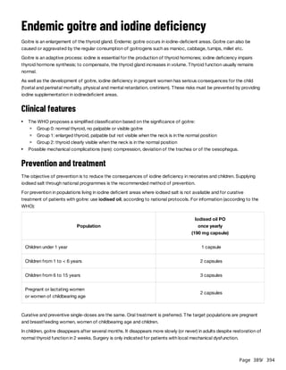
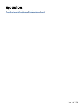
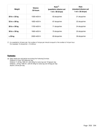


The document is a clinical guidelines manual developed by Médecins Sans Frontières (MSF) for medical professionals involved in curative care at dispensaries and primary hospitals. It covers various medical conditions, treatment protocols, and includes essential information from WHO and other specialists. The guide emphasizes practical responses to medical challenges faced in the field and contains details regarding symptoms, diseases, and management protocols.


![PEDIATRICS_SHOCK_MANAGMENT.pptx1114[1].pptx](https://cdn.slidesharecdn.com/ss_thumbnails/pediatricsshockmanagment-230702193650-dab388b8-thumbnail.jpg?width=640&height=640&fit=bounds)


