Osteomalacia: Causes, Symptoms, and Treatment
•Download as PPTX, PDF•
3 likes•1,301 views

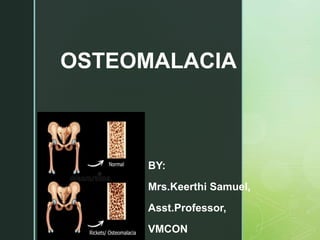
OSTEOMALACIA

Recommended
More Related Content
What's hot
What's hot (20)
Similar to Osteomalacia: Causes, Symptoms, and Treatment
Similar to Osteomalacia: Causes, Symptoms, and Treatment (20)
More from keerthi samuel
More from keerthi samuel (20)
Recently uploaded
Recently uploaded (20)
Osteomalacia: Causes, Symptoms, and Treatment
- 2. Osteomalacia in children is known as rickets, and because of this, use of the term "osteomalacia" is often restricted to the milder, adult form of the disease. Signs and symptoms can include diffuse body pains, muscle weakness, and fragility of the bones INTRODUCTION
- 4. z VITAMIN-D FUNCTIONS •Intestinal Calcium absorption •Interaction with PTH in regulation of blood Calcium Re-absorption of Calcium from Kidneys Mineralization of Bone
- 5. z Osteomalacia is the softening of the bones caused by defective bone mineralization secondary to inadequate levels of available phosphate and calcium, or because of overactive resorption of calcium from the bone which can be caused by hyperparathyroidism (which causes hypercalcemia). Metabolic disease characterized by inadequate and delayed mineralization of osteoid in mature compact and spongy bone. DEFINITION
- 6. z INCIDENCE The prevalence of Vitamin D deficiency ranged from 40% to 99%, with most of the studies reporting a prevalence of 80%–90%. It was prevalent in all the age groups and high-risk groups alike.
- 7. z ETIOLOGY Inadequate mineralization of the bone. There are two main causes of osteomalacia: Insufficient calcium absorption from the intestine because of lack of dietary calcium or a deficiency of, or resistance to, the action of vitamin D; and phosphate deficiency caused by increased
- 8. z ETIOLOGY –Contd.. Vitamin D deficiency. Sunlight produces vitamin D in your skin. Dietary vitamin D is usually from foods to which the vitamin has been added, such as cow's milk. People who live in areas where sunlight is limited, get little exposure to sunlight or eat a diet low in vitamin D can develop osteomalacia. Vitamin D deficiency is the most common cause of osteomalacia worldwide.
- 9. z ETIOLOGY – Contd.. Celiac disease. In this autoimmune disorder, foods containing gluten, a protein found in wheat, barley and rye, can damage the lining of your small intestine. A damaged intestinal lining doesn't absorb nutrients well, and can lead to vitamin D and calcium deficiency.
- 10. z ETIOLOGY – Contd.. chronic liver disease: hepatocellular: 25- hydroxylation vitamin D biliary: abnormal gut absorption malabsorption syndromes such as Crohn's partial gastrectomy (self- restriction of fatty foods)
- 11. z ETIOLOGY –Contd.. Administration of phenobarbital (alternate liver pathway) Decreased deposition of calcium in bone (high PTH) Diphosphonates-(Bisphosphonates are a group of medicines that slow down or prevent bone loss, strengthening bones) Other causes Tumour induced osteomalacia Cadmium poisoning like Itai-itai disease (Cadmium poisoning can also cause softening of the bones and kidney failure).
- 12. z ETIOLOGY-contd Drugs: Phenytoin Phenobarbital Carbamazepine Acetazolamide Aluminium Compound Flouride
- 13. z PATHOPHYSIOLOGY-I HYDROXAPETITE(mineral saltin the osteoid matrix, which is a crystalline complex of calcium and phosphate) is responsible for hardness of the bone Defeciency of hydroxypetite leads to softening of bone
- 14. z PATHOPHYSIOLOGY-II Deficiency of Vit-d hypocalcemia Osteomalcia/rickets Increase in bone resorption Increased plasma calcium and decreased phosporous Increased PTH
- 15. z CLINICAL MANIFESTATIONS Diffuse joint and bone pain (especially of spine, pelvis, and legs) Muscle weakness Difficulty walking, waddling gait Hypocalcemia (positive Chvostek sign) Compressed vertebrae and diminished stature Pelvic flattening, Bending of bones Weak, soft bones, Easy fracturing
- 17. z CLINICAL MANIFESTATIONS Osteomalacia in adults starts insidiously as aches and pains in the lumbar (lower back) region and thighs before spreading to the arms and ribs. The pain is symmetrical, non-radiating and accompanied by sensitivity in the involved bones. Proximal muscles are weak, and there is difficulty in climbing up stairs and getting up from a squatting position. Physical signs include deformities like triradiate pelvis and lordosis. The patient has a typical "waddling" gait.
- 18. z SIGNS AND SYMPTOMS However, these physical signs may derive from a previous osteomalacial state, since bones do not regain their original shape after they become deformed. Pathologic fractures due to weight bearing may develop. Most of the time, the only alleged symptom is chronic fatigue, while bone aches are not spontaneous but only revealed by pressure or shocks. It differs from renal osteodystrophy, where the latter shows hyperphosphatemia.
- 19. z CARDINAL SIGNS Pseudofracture s, also called Looser’s Zones. Codfish vertebrae. Protrusion Acetabuli, a hip joint disorder- Triradiant pelvis
- 20. z NORMAL PELVIS TRIRADIANT PELVIS Protrusion of hip and spine into soft pelvis and protrusion with protrusion of acetabuli
- 21. z NORMAL NECK OF THE FEMUR PSEUDO FRACTURE OF NECK OF THE FEMUR(Milkman's Syndrome) Lucent bands that occur due to a build-up of osteoid, the unmineralized organic component of one.
- 22. z z Codfish vertebra' refers to the biconcave deformity of the vertebral body and is more common than anterior vertebral wedging and flat vertebra signs. NORMAL VERTEBRAE COD FISH VERTEBRAE
- 23. z z RICKETS
- 24. z DIAGNOSTIC FINDINGS Biochemical features are similar to those of rickets. The major factor is an abnormally low vitamin D concentration in blood serum. Major typical biochemical findings include: Low serum and urinary calcium Low serum phosphate, except in cases of renal osteodystrophy
- 25. zDIAGNOSTIC FINDINGS Elevated serum alkaline phosphatase (due to an increase in compensatory osteoblast activity) Elevated parathyroid hormone (due to low calcium) Furthermore, a technetium bone scan will show increased activity (also due to increased osteoblasts).
- 26. z RADIOLOGY Diffuse demineralization: osteoporotic-like pattern may show a characteristic smudgy "erased" or "fuzzy" type of demineralization coarsened trabeculae insufficiency fractures Pseudofractures (looser’s zone) articular manifestations (uncommon) rheumatoid arthritis-like picture osteogenic synovitis ankylosing spondylitis-like picture
- 29. z TREATMENT Nutritional osteomalacia responds well to administration of 10,000 IU weekly of vitamin D for four to six weeks. Osteomalacia due to malabsorption may require treatment by injection or daily oral dosing of significant amounts of vitamin D. Calcitriol supplement for CKD.
- 30. z TREATMENT Exercise Exercise helps to strengthen the bones, especially weight- bearing exercise (anything that involves walking or running). However, you should avoid intensive exercise while any fractures or cracks in the bones are healing. Sunlight Where possible, going outside and exposing your arms and face to sunlight is the best way to get vitamin D. From June to August just 15 minutes a day is generally enough. Don’t allow your skin to go red and take care not to burn, particularly in strong sunshine and if you have fair or sensitive skin.
- 31. Diet and nutrition A diet that includes vitamin D and calcium can help, but this won’t prevent the condition by itself. Nevertheless, a diet that provides vitamin D is especially important if you don’t get enough exposure to sunlight.