SlideShare a Scribd company logo
1 of 39
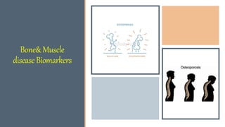
Bone&Muscle
diseaseBiomarkers
Introduction
Bone has three important functions:
Provides structural support to the body
Haemopoietic bone marrow
Metabolically active being essential for calcium and phosphate
homeostasis.
Bone is a metabolically active tissue.
In mature bone, there is a continuous cycle of bone resorption and replacement.
 Osteoblasts are responsible for the synthesis of bone matrix and osteoclasts for
bone resorption.
The important role of osteocytes is increasingly recognized.
Normal bone formation, metabolism and repair depend on the coordinated
activity of these cells and the integrity of the homoeostatic mechanisms for
calcium and phosphate, which primarily involve parathyroid hormone,
FGF23(fibroblast growth factor) and vitamin D
Bone remodeling (or bone metabolism) is a lifelong process where
mature bone tissue is removed from the skeleton (a process called
bone resorption) and new bone tissue is formed (a process called
ossification or new bone formation).
These processes also control the reshaping or replacement of bone
following injuries like fractures but also micro-damage, which occurs
during normal activity.
Remodeling responds also to functional demands of the mechanical
loading.
Two main types of cells are responsible for bone metabolism: osteoblasts (which
secrete new bone), and osteoclasts (which break bone down).
The structure of bones as well as adequate supply of calcium requires close
cooperation between these two cell types and other cell populations present at the
bone remodeling sites (eg. immune cells).
Bone metabolism relies on complex signaling pathways and control mechanisms
to achieve proper rates of growth and differentiation.
 These controls include the action of several hormones, including parathyroid
hormone (PTH), vitamin D, growth hormone, steroids, and calcitonin, as well as
several bone marrow-derived membrane and soluble cytokines and growth factors.
It is in this way that the body is able to
maintain proper levels of calcium
required for physiological processes.
Thus bone remodeling is not just
occasional "repair of bone damage" but
rather an active, continual process that is
always happening in a healthy body.
The finding that a patient has hypercalcaemia or hypocalcaemia does not
imply that there will be marked bone changes.
Conversely, severe bone disease can occur whilst serum calcium levels
appear quite normal.
The main bone diseases are:
■ osteoporosis
■ osteomalacia and rickets
■ Paget’s disease.
Bonemetabolism
Bone is constantly being broken down and reformed in the process of bone remodeling.
 The clinician looking after patients with bone disease will certainly need to know to what extent
bone is being broken down, and, indeed, if new bone is being made.
 Biochemical markers of bone resorption and bone formation can be useful in assessing the extent
of disease, as well as monitoring treatment.
Hydroxyproline, from the breakdown of collagen, can be used to monitor bone resorption.
 However, urinary hydroxyproline is markedly influenced by dietary gelatin.
Better markers of resorption are required.
 One candidate would seem to be another collagen degradation product: the fragments of the
molecule containing the pyridinium cross-links.
Deoxypyridinoline is one such cross-link that is specific for bone, and not metabolized or
influenced by diet.
The activity of the enzyme alkaline phosphatase has traditionally been used as an indicator of
bone turnover.
 The osteoblasts that lay down the collagen framework and the mineral matrix of bone have high
activity of this enzyme.
Increased osteoblastic activity is indicated by an elevated alkaline phosphatase activity in a serum
specimen.
Indeed, children who have active bone growth compared with adults have higher ‘normal’ alkaline
phosphatase activity in serum.
The bone isoenzyme of alkaline phosphatase may be measured, but there is need for a more
specific and more sensitive marker.
Osteocalcin meets some of these requirements.
 It is synthesized by osteoblasts and is an important non-collagenous constituent of
bone.
Not all of the osteocalcin that an osteoblast makes is incorporated into the bone
matrix.
Some is released into plasma, and provides a sensitive indicator of osteoblast
activity.
The test is available in specialized laboratories.
Commonbonedisorders
Osteoporosis
Osteoporosis is the commonest of bone disorders
Osteomalacia and rickets
Osteomalacia is the name given to defective bone mineralization in adults.
- Rickets is characterized by defects of bone and cartilage mineralization in children.
- Vitamin D deficiency was once the most common reason for rickets and osteomalacia, but the addition
of vitamin D to foodstuffs has reduced the condition except in the elderly or house-bound, the
institutionalized, and in certain ethnic groups.
- Although elderly Asian women with a predominantly vegetarian diet are particularly at risk, it should be
noted that there appears to be a increase of rickets and osteomalacia in the white population of lower
socio-economic status, due to poor diet and limited exposure to sunlight.
Rickets and Osteomalacia
Patients who have vitamin D deficiency or disturbed metabolism of vitamin D are all
liable to suffer from the bone disease osteomalacia or, in children, from rickets.
These patients have bone pain, with local tenderness, and may have a proximal
myopathy.
Skeletal deformity may be present, particularly in rickets.
Mineralisation of osteoid is defective, with absence of the calcification front.
Other causes of rickets or osteomalacia, unrelated to vitamin D deficiency or defects
in its metabolism, have also been described.
An inherited defect in the tubular reabsorption of phosphate,
hypophosphataemic vitamin D-resistant rickets, leads to similar bone deformities, but
without muscle weakness; there is a low serum [phosphate] and phosphaturia.
In Fanconi syndrome, tubular phosphate loss may also lead to low
serum [phosphate] associated with rickets or osteomalacia.
Hypophosphatasia is a hereditary disease in which vitamin D-
resistant rickets is the most prominent finding.
 Tissue and serum ALP activities are usually low, and excessive
amounts of phosphoryl ethanolamine are present in the urine.
Osteoporosis
Osteoporosis is a major public health problem and a major cause of morbidity and mortality in the
elderly.
Bone is in a constant state of turnover, which is kept in balance by opposing actions of osteoblasts
(bone formation) and osteoclasts (bone resorption).
 Osteoporosis results when, irrespective of the cause, this balance is disturbed and shifts in favour
of resorption.
It is defined as a progressive systemic skeletal disorder characterized by low bone mineral
density (BMD), deterioration of the microarchitecture of bone tissue, and susceptibility to
fracture.
The World Health Organization (WHO) proposed a clinical definition based on measurements of
BMD
Vitamin D status can be assessed by measurement of the main circulating
metabolite, 25-hydroxycholecalciferol, in a serum specimen.
In severe osteomalacia due to vitamin D deficiency, serum calcium will fall, and
there will be an appropriate increase in PTH secretion.
Serum alkaline phosphatase activity will also be elevated.
The bony features of osteomalacia and rickets are also shared by other bone
diseases
Paget’sdisease
Paget’s disease is common in the elderly and characterized by increased
osteoclastic activity, which leads to increased bone resorption.
 Increased osteoblastic activity repairs resorbed bone, but the new bone is laid
down in a disorganized way.
The clinical presentation is always bone pain, which can be particularly severe.
Serum alkaline phosphatase is high, and urinary hydroxyproline excretion is
elevated.
These provide a way of monitoring the progress of the disease.
Although a viral cause for Paget’s disease has been proposed, the aetiology
remains obscure.
Other Bone Diseases
Examples include:
■ Vitamin-D-dependent rickets, types 1 and 2.
These are rare bone diseases resulting from genetic disorders leading to the inability to make the
active vitamin D metabolite, or from receptor defects that do not allow the hormone to act.
■ Tumoral calcinosis. This is characterized by ectopic calcification around the joints.
■ Hypophosphatasia. This is a form of rickets or osteomalacia that results from a deficiency of
alkaline phosphatase.
- Hypophosphataemic rickets. This is believed to be a consequence of a renal tubular defect in
phosphate handling.
. ■ Osteogenesis imperfecta. Brittle bone syndrome is an inherited disorder which occurs around
once in every 20 000 births.
Diagnosis of these and other rare conditions may require help from specialized laboratories.
Biochemistry testing in calcium disorders or
bone disease
The role of the routine biochemistry laboratory in diagnosis and treatment of patients with
calcium disorders and bone disease is to provide measurements of calcium, albumin,
phosphate and alkaline phosphatase in a serum specimen as first line tests.
Follow-up tests that may be requested include:
■ PTH
■ magnesium
■ urine calcium excretion
■ 25-hydroxycholecalciferol
■ urine hydroxyproline excretion
■ osteocalcin.
Biochemical Profiles in Bone Diseases
Osteitis fibrosa cystica
(primary
hyperparathyroidism)
Calcium is elevated
Phosphate is low or normal
PTH is increased, or clearly detectable
Bone metastases Calcium may be high, low or normal
Phosphate may be high, low or normal
PTH is usually low
Alkaline phosphatase may be elevated or normal
Osteomalacia/rickets Calcium will tend to be low
PTH will be elevated
25-hydroxycholecalciferol will be decreased if the disease is due to vitamin D
deficiency
Paget’s disease Calcium is normal
Alkaline phosphatase is grossly elevated
Osteoporosis Biochemistry is unremarkable
Renal osteodystrophy Calcium is decreased, PTH is very high
Bone disease (summary)
Alkaline phosphatase is a marker for bone formation.
Urinary hydroxyproline is a marker for bone resorption.
Better markers for bone turnover are available in specialized laboratories, but have
limited utility.
 Osteomalacia due to vitamin D deficiency can be confirmed by finding a low 25-
hydroxycholecalciferol concentration. In severe disease, alkaline phosphatase will
be increased.
 Calcium may well fall and there will be an appropriate rise in PTH.
The characteristic biochemical marker of Paget’s disease is a grossly increased
alkaline phosphatase activity, as a consequence of increased bone turnover.
RiskFactors
Age and menopause are the two main non-modifiable risk factors.
Peak bone mass is reached around 25–30 years.
Contributing factors are genetic, dietary intake of calcium and vitamin D and
physical exercise.
 Other risk factors include:
 history of previous fracture,
family history of osteoporosis and hip fracture,
sex hormone deficiency,
smoking,
 alcoholism,
 immobility and sedentary lifestyle.
Osteoporosis may also be
 secondary to endocrine disorders including Cushing’s syndrome,
 primary hyperparathyroidism,
 hypo vitaminosis D (classically associated with osteomalacia),
hypogonadism and hyperthyroidism,
 systemic illnesses such as rheumatoid arthritis,
chronic kidney and liver diseases and malignancies.
The commonest cause of secondary osteoporosis is corticosteroid use.
Diagnosis
- A detailed clinical history helps to determine the presence of risk factors.
- Clinical examination is usually not very informative.
- Measurement of bone density by dual energy X-ray absorptiometry (DEXA) scan is
the mainstay of diagnosis.
- There is no role for routine biochemical tests in the diagnosis of osteoporosis.
- Biochemical markers of bone turnover have very limited use in the diagnosis of
osteoporosis, but an occasionally be helpful in determining the most appropriate type
of treatment.
Principles of treatment: Treatment is aimed at strengthening the
bone and preventing fractures.
The mainstay of drug treatment are oral bisphosphonates that inhibit
osteoclastic function, thereby slowing down bone loss.
However, in practice this does not always result in increased BMD on
DEXA scans.
Markers of bone turnover
Biochemical markers of bone turnover can be divided into those that reflect bone
formation and those that reflect bone resorption.
The formation markers include enzymes and peptides released by the osteoblast at the
time of bone formation, whereas the resorption markers are typically a measure of the
breakdown products of type 1 collagen.
While bone markers cannot reveal how much bone is present in the skeleton and cannot
substitute for the measurement of bone mineral density, they have the potential to be used in
the assessment of fracture risk and may be used to monitor the response of patients with
osteoporosis to anti-resorptive and anabolic therapies.
However, the clinical utility of routine bone marker measurements has not yet been fully
established and these assays remain available in specialist centers only.
Muscle Disease Biomarkers
Myopathies
Myopathies are conditions affecting the muscles that lead to weakness and/or atrophy.
 They may be caused by congenital factors (as in the muscular dystrophies), by viral infection or by acute
damage due to anoxia, infections, toxins or drugs.
Muscle denervation is a major cause of myopathy.
Muscle weakness can occur due to a lack of energy producing molecules or a failure in the balance of
electrolytes within and surrounding the muscle cell necessary for neuromuscular function.
Normal muscle that is overused will end up weak or in spasm until rested.
 In severe cases of overuse, especially where movements are strong and erratic as might occur during
convulsions, damage to muscle cells may result.
Severely damaged muscle cells release their contents, e.g. myoglobin, a condition known as
rhabdomyolysis.
Muscleweakness
Muscle weakness, which may or may not progress to rhabdomyolysis, has many
causes (Fig 73.1).
 Diagnosis of the condition will depend on the clinical picture and will include
investigation of genetic disorders by enzymic or chromosomal analysis, endocrine
investigations and the search for drug effects.
 Infective causes may be diagnosed by isolation of the relevant organism or its
related antibody, but often no organism is detected.
These cases, known as myalgic encephalitis (ME), post-viral syndrome or chronic
fatigue syndrome, are relatively common and are now regarded as true diseases,
whereas formally they were thought to be psychosomatic.
Metabolic causes of Myopathy
Fatty acid oxidation defects
Glycogen storage disorders Pompe’s and Fabry’s diseases
 Myoadenalate deaminase deficiency
Carnitine palmitoyl transferase deficiency
Respiratory chain (electrolyte transport) defects
Investigation
Investigation In all cases of muscle weakness, serum electrolytes should be checked along
with creatine kinase (CK).
A full drug history should be taken to exclude pharmacological and toxicological causes,
and a history of alcohol abuse should be excluded.
Neuromuscular electrophysiological studies should be performed to detect neuropathies.
Where a genetic or metabolic cause is suspected.
 Investigations include measurement of plasma (and CSF) lactate and specialist metabolic
tests in blood, CSF and urine; muscle biopsy for histopatological studies and measurement
of muscle enzymes may also be indicated.
In contrast to rhabdomyolysis, serum CK may sometimes be normal in myopathic disorders,
especially in the chronic setting, and if muscle mass decreases.
Rhabdomyolysis
Muscle cells that are damaged will leak creatine kinase into the plasma.
This enzyme exists in different isoforms.
CK-MM or total CK is used as an index of skeletal muscle damage.
Very high serum levels may be expected in patients who have been convulsing or have
muscular damage due to electrical shock or crush injury.
Creatine kinase concentrations may also be high in acute spells in muscular dystrophy.
 The damaged muscle cells will also leak myoglobin.
 This compound stores oxygen in the muscle cells for release under conditions of hypoxia,
as occurs during severe exercise.
When muscle cells become anoxic or are damaged by trauma,
myoglobin is released into the plasma.
It is filtered at the glomerulus and excreted in the urine, which appears
orange or brown coloured; on urine dipstick testing myoglobin gives a
false positive reaction for the presence of blood, which can lead to the
mistaken diagnosis of haematuria.
The damaged muscle cells also release large amounts of potassium and
phosphate ions giving rise to hyperkalaemia and hyperphosphataemia.
Severe muscle damage is frequently accompanied by a reduction in the blood volume.
This may occur directly as a result of haemorrhage in severe trauma, or indirectly because
of fluid sequestration in the damaged tissue.
The resultant shock frequently causes acute renal failure.
Myoglobin per se is not nephrotoxic, but the accompanying acidosis, and volume depletion
lead to acute tubular necrosis.
 Additionally, in acidic pH myoglobin is converted to ferrihaemate, which produces free
radicals and causes direct nephrotoxicity.
Children with muscular dystrophy do not develop renal failure despite having increased
levels of myoglobin in urine for many years.
Investigation and treatment
The biochemistry laboratory has a major role to play in the diagnosis and
investigation of rhabdomyolysis
This includes:
1. serum total creatine kinase, which allows the diagnosis to be made, and levels monitored
to assess recovery and prognosis
2. urea and electrolytes, to look for evidence of resulting renal impairment
3. alcohol and drugs of abuse screen, to look for specific causes.
Urine or plasma myoglobin would be a sensitive marker of muscle damage. It is, in fact,
too sensitive.
Even minor degrees of muscle damage that do not warrant investigation or treatment will
give rise to myoglobin release.
Treatment is directed towards maintaining tissue perfusion and the
control of electrolyte imbalances. It includes:
Cardiac monitoring
 Control of hyperkalaemia, hyperphosphataemia and hypocalcaemia.
Haemodialysis may be necessary where renal function is severely
compromised.
Skeletal muscle disorders (summary)
 Muscle weakness is a common complaint with a wide variety of causes.
Biochemical investigation of muscle weakness can provide rapid diagnosis and effective
treatment where ionic changes are the cause.
 Intracellular enzyme analysis from muscle biopsies can provide a diagnosis in some
inherited disorders.
Severely damaged muscle cells release potassium, creatine kinase, myoglobin and
phosphate.
Severe rhabdomyolysis, e.g. following injury, is an important cause of acute renal failure.

More Related Content

Similar to Bone & Muscle disease biomarkers.ppt

Similar to Bone & Muscle disease biomarkers.ppt (20)

1 mk-bone012
1 mk-bone0121 mk-bone012
1 mk-bone012
 
Osteoporosis
OsteoporosisOsteoporosis
Osteoporosis
 
osteoporosis 3.pptx
osteoporosis 3.pptxosteoporosis 3.pptx
osteoporosis 3.pptx
 
Osteoporosis
OsteoporosisOsteoporosis
Osteoporosis
 
OSTEOPOROSIS
OSTEOPOROSISOSTEOPOROSIS
OSTEOPOROSIS
 
Metabolic Bone Diseases Current Concept
Metabolic Bone Diseases Current ConceptMetabolic Bone Diseases Current Concept
Metabolic Bone Diseases Current Concept
 
Calsi soya ppt
Calsi soya pptCalsi soya ppt
Calsi soya ppt
 
Bone disorders
Bone disordersBone disorders
Bone disorders
 
Osteoporosis PPT for nursing students...
Osteoporosis PPT for nursing students...Osteoporosis PPT for nursing students...
Osteoporosis PPT for nursing students...
 
Osteoporosis clinical features and management
Osteoporosis clinical features and management Osteoporosis clinical features and management
Osteoporosis clinical features and management
 
Osteoporosis
OsteoporosisOsteoporosis
Osteoporosis
 
Osteomalacia 2nd-150704155942-lva1-app6892 [autosaved]
Osteomalacia 2nd-150704155942-lva1-app6892 [autosaved]Osteomalacia 2nd-150704155942-lva1-app6892 [autosaved]
Osteomalacia 2nd-150704155942-lva1-app6892 [autosaved]
 
Osteomalcia1 1
Osteomalcia1 1Osteomalcia1 1
Osteomalcia1 1
 
Orthopedic disorders of metabolic bone disease - البروفيسور فريح عوده ابوحسان
Orthopedic disorders of metabolic bone disease - البروفيسور فريح عوده ابوحسان Orthopedic disorders of metabolic bone disease - البروفيسور فريح عوده ابوحسان
Orthopedic disorders of metabolic bone disease - البروفيسور فريح عوده ابوحسان
 
Residual ridge resorption
Residual ridge resorption Residual ridge resorption
Residual ridge resorption
 
Rickets and osteomalacia
Rickets and osteomalacia Rickets and osteomalacia
Rickets and osteomalacia
 
The control of bone growth and repair
The control of bone growth and repairThe control of bone growth and repair
The control of bone growth and repair
 
1933864184
19338641841933864184
1933864184
 
Osteoporesis and Gout _RDP
Osteoporesis and Gout _RDPOsteoporesis and Gout _RDP
Osteoporesis and Gout _RDP
 
Bone physiology
Bone physiologyBone physiology
Bone physiology
 

More from Mohamed Afifi

1- Biochemical and molecular basis of lung diseases .ppt
1- Biochemical and molecular basis  of lung diseases .ppt1- Biochemical and molecular basis  of lung diseases .ppt
1- Biochemical and molecular basis of lung diseases .pptMohamed Afifi
 
CHO presentation.ppt
CHO presentation.pptCHO presentation.ppt
CHO presentation.pptMohamed Afifi
 
1- metabolism of the brain (I) 2012-13.pdf
1- metabolism of the brain (I) 2012-13.pdf1- metabolism of the brain (I) 2012-13.pdf
1- metabolism of the brain (I) 2012-13.pdfMohamed Afifi
 
Renal Disease biochemical investigation.pdf
Renal Disease biochemical investigation.pdfRenal Disease biochemical investigation.pdf
Renal Disease biochemical investigation.pdfMohamed Afifi
 
Cardiovascular Disorders.ppt
Cardiovascular Disorders.pptCardiovascular Disorders.ppt
Cardiovascular Disorders.pptMohamed Afifi
 
1610881717923_Tumour Markers.pdf
1610881717923_Tumour Markers.pdf1610881717923_Tumour Markers.pdf
1610881717923_Tumour Markers.pdfMohamed Afifi
 
CLINICAL BIOCHEM_2022.pdf
CLINICAL BIOCHEM_2022.pdfCLINICAL BIOCHEM_2022.pdf
CLINICAL BIOCHEM_2022.pdfMohamed Afifi
 
CLINICAL BIOCHEM_2023.pdf
CLINICAL BIOCHEM_2023.pdfCLINICAL BIOCHEM_2023.pdf
CLINICAL BIOCHEM_2023.pdfMohamed Afifi
 
automation-151203160733-lva1-app6892.pdf
automation-151203160733-lva1-app6892.pdfautomation-151203160733-lva1-app6892.pdf
automation-151203160733-lva1-app6892.pdfMohamed Afifi
 
KATO_THICK__SMEAR__TECHNIQUE_0.pptx
KATO_THICK__SMEAR__TECHNIQUE_0.pptxKATO_THICK__SMEAR__TECHNIQUE_0.pptx
KATO_THICK__SMEAR__TECHNIQUE_0.pptxMohamed Afifi
 

More from Mohamed Afifi (16)

1- Biochemical and molecular basis of lung diseases .ppt
1- Biochemical and molecular basis  of lung diseases .ppt1- Biochemical and molecular basis  of lung diseases .ppt
1- Biochemical and molecular basis of lung diseases .ppt
 
CHO.ppt
CHO.pptCHO.ppt
CHO.ppt
 
CHO presentation.ppt
CHO presentation.pptCHO presentation.ppt
CHO presentation.ppt
 
1-Hormone.pdf
1-Hormone.pdf1-Hormone.pdf
1-Hormone.pdf
 
1- metabolism of the brain (I) 2012-13.pdf
1- metabolism of the brain (I) 2012-13.pdf1- metabolism of the brain (I) 2012-13.pdf
1- metabolism of the brain (I) 2012-13.pdf
 
2.ppt
2.ppt2.ppt
2.ppt
 
Renal Disease biochemical investigation.pdf
Renal Disease biochemical investigation.pdfRenal Disease biochemical investigation.pdf
Renal Disease biochemical investigation.pdf
 
lecture 4+5.pptx
lecture 4+5.pptxlecture 4+5.pptx
lecture 4+5.pptx
 
Cardiovascular Disorders.ppt
Cardiovascular Disorders.pptCardiovascular Disorders.ppt
Cardiovascular Disorders.ppt
 
1610881717923_Tumour Markers.pdf
1610881717923_Tumour Markers.pdf1610881717923_Tumour Markers.pdf
1610881717923_Tumour Markers.pdf
 
2.ppt
2.ppt2.ppt
2.ppt
 
lecture 1.pptx
lecture 1.pptxlecture 1.pptx
lecture 1.pptx
 
CLINICAL BIOCHEM_2022.pdf
CLINICAL BIOCHEM_2022.pdfCLINICAL BIOCHEM_2022.pdf
CLINICAL BIOCHEM_2022.pdf
 
CLINICAL BIOCHEM_2023.pdf
CLINICAL BIOCHEM_2023.pdfCLINICAL BIOCHEM_2023.pdf
CLINICAL BIOCHEM_2023.pdf
 
automation-151203160733-lva1-app6892.pdf
automation-151203160733-lva1-app6892.pdfautomation-151203160733-lva1-app6892.pdf
automation-151203160733-lva1-app6892.pdf
 
KATO_THICK__SMEAR__TECHNIQUE_0.pptx
KATO_THICK__SMEAR__TECHNIQUE_0.pptxKATO_THICK__SMEAR__TECHNIQUE_0.pptx
KATO_THICK__SMEAR__TECHNIQUE_0.pptx
 

Recently uploaded

VIP Mumbai Call Girls Thakur village Just Call 9920874524 with A/C Room Cash ...
VIP Mumbai Call Girls Thakur village Just Call 9920874524 with A/C Room Cash ...VIP Mumbai Call Girls Thakur village Just Call 9920874524 with A/C Room Cash ...
VIP Mumbai Call Girls Thakur village Just Call 9920874524 with A/C Room Cash ...Garima Khatri
 
(8264348440) 🔝 Call Girls In Shaheen Bagh 🔝 Delhi NCR
(8264348440) 🔝 Call Girls In Shaheen Bagh 🔝 Delhi NCR(8264348440) 🔝 Call Girls In Shaheen Bagh 🔝 Delhi NCR
(8264348440) 🔝 Call Girls In Shaheen Bagh 🔝 Delhi NCRsoniya singh
 
Call Girls in Karachi | +923081633338 | Karachi Call Girls
Call Girls in Karachi  | +923081633338 | Karachi Call GirlsCall Girls in Karachi  | +923081633338 | Karachi Call Girls
Call Girls in Karachi | +923081633338 | Karachi Call GirlsAyesha Khan
 
call girls in G.T.B. Nagar (DELHI) 🔝 >༒9953330565🔝 genuine Escort Service 🔝✔️✔️
call girls in  G.T.B. Nagar (DELHI) 🔝 >༒9953330565🔝 genuine Escort Service 🔝✔️✔️call girls in  G.T.B. Nagar (DELHI) 🔝 >༒9953330565🔝 genuine Escort Service 🔝✔️✔️
call girls in G.T.B. Nagar (DELHI) 🔝 >༒9953330565🔝 genuine Escort Service 🔝✔️✔️9953056974 Low Rate Call Girls In Saket, Delhi NCR
 
The 10th anniversary, Hyundai World Rally Team's amazing journey
The 10th anniversary, Hyundai World Rally Team's amazing journeyThe 10th anniversary, Hyundai World Rally Team's amazing journey
The 10th anniversary, Hyundai World Rally Team's amazing journeyHyundai Motor Group
 
Vip Hot Call Girls 🫤 Mahipalpur ➡️ 9711199171 ➡️ Delhi 🫦 Whatsapp Number
Vip Hot Call Girls 🫤 Mahipalpur ➡️ 9711199171 ➡️ Delhi 🫦 Whatsapp NumberVip Hot Call Girls 🫤 Mahipalpur ➡️ 9711199171 ➡️ Delhi 🫦 Whatsapp Number
Vip Hot Call Girls 🫤 Mahipalpur ➡️ 9711199171 ➡️ Delhi 🫦 Whatsapp Numberkumarajju5765
 
Hyundai World Rally Team in action at 2024 WRC
Hyundai World Rally Team in action at 2024 WRCHyundai World Rally Team in action at 2024 WRC
Hyundai World Rally Team in action at 2024 WRCHyundai Motor Group
 
Delhi Call Girls Saket 9711199171 ☎✔👌✔ Full night Service for more than 1 person
Delhi Call Girls Saket 9711199171 ☎✔👌✔ Full night Service for more than 1 personDelhi Call Girls Saket 9711199171 ☎✔👌✔ Full night Service for more than 1 person
Delhi Call Girls Saket 9711199171 ☎✔👌✔ Full night Service for more than 1 personshivangimorya083
 
꧁ ୨⎯Call Girls In Ashok Vihar, New Delhi **✿❀7042364481❀✿**Escorts ServiCes C...
꧁ ୨⎯Call Girls In Ashok Vihar, New Delhi **✿❀7042364481❀✿**Escorts ServiCes C...꧁ ୨⎯Call Girls In Ashok Vihar, New Delhi **✿❀7042364481❀✿**Escorts ServiCes C...
꧁ ୨⎯Call Girls In Ashok Vihar, New Delhi **✿❀7042364481❀✿**Escorts ServiCes C...Hot Call Girls In Sector 58 (Noida)
 
2024 WRC Hyundai World Rally Team’s i20 N Rally1 Hybrid
2024 WRC Hyundai World Rally Team’s i20 N Rally1 Hybrid2024 WRC Hyundai World Rally Team’s i20 N Rally1 Hybrid
2024 WRC Hyundai World Rally Team’s i20 N Rally1 HybridHyundai Motor Group
 
2024 TOP 10 most fuel-efficient vehicles according to the US agency
2024 TOP 10 most fuel-efficient vehicles according to the US agency2024 TOP 10 most fuel-efficient vehicles according to the US agency
2024 TOP 10 most fuel-efficient vehicles according to the US agencyHyundai Motor Group
 
Beautiful Vip Call Girls Punjabi Bagh 9711199012 Call /Whatsapps
Beautiful Vip  Call Girls Punjabi Bagh 9711199012 Call /WhatsappsBeautiful Vip  Call Girls Punjabi Bagh 9711199012 Call /Whatsapps
Beautiful Vip Call Girls Punjabi Bagh 9711199012 Call /Whatsappssapnasaifi408
 
Vip Hot🥵 Call Girls Delhi Delhi {9711199012} Avni Thakur 🧡😘 High Profile Girls
Vip Hot🥵 Call Girls Delhi Delhi {9711199012} Avni Thakur 🧡😘 High Profile GirlsVip Hot🥵 Call Girls Delhi Delhi {9711199012} Avni Thakur 🧡😘 High Profile Girls
Vip Hot🥵 Call Girls Delhi Delhi {9711199012} Avni Thakur 🧡😘 High Profile Girlsshivangimorya083
 
VIP Kolkata Call Girl Kasba 👉 8250192130 Available With Room
VIP Kolkata Call Girl Kasba 👉 8250192130  Available With RoomVIP Kolkata Call Girl Kasba 👉 8250192130  Available With Room
VIP Kolkata Call Girl Kasba 👉 8250192130 Available With Roomdivyansh0kumar0
 
Dubai Call Girls Size E6 (O525547819) Call Girls In Dubai
Dubai Call Girls  Size E6 (O525547819) Call Girls In DubaiDubai Call Girls  Size E6 (O525547819) Call Girls In Dubai
Dubai Call Girls Size E6 (O525547819) Call Girls In Dubaikojalkojal131
 
办理埃默里大学毕业证Emory毕业证原版一比一
办理埃默里大学毕业证Emory毕业证原版一比一办理埃默里大学毕业证Emory毕业证原版一比一
办理埃默里大学毕业证Emory毕业证原版一比一mkfnjj
 
UNIT-II-ENGINE AUXILIARY SYSTEMS &TURBOCHARGER
UNIT-II-ENGINE AUXILIARY SYSTEMS &TURBOCHARGERUNIT-II-ENGINE AUXILIARY SYSTEMS &TURBOCHARGER
UNIT-II-ENGINE AUXILIARY SYSTEMS &TURBOCHARGERDineshKumar4165
 
UNIT-III-TRANSMISSION SYSTEMS REAR AXLES
UNIT-III-TRANSMISSION SYSTEMS REAR AXLESUNIT-III-TRANSMISSION SYSTEMS REAR AXLES
UNIT-III-TRANSMISSION SYSTEMS REAR AXLESDineshKumar4165
 

Recently uploaded (20)

VIP Mumbai Call Girls Thakur village Just Call 9920874524 with A/C Room Cash ...
VIP Mumbai Call Girls Thakur village Just Call 9920874524 with A/C Room Cash ...VIP Mumbai Call Girls Thakur village Just Call 9920874524 with A/C Room Cash ...
VIP Mumbai Call Girls Thakur village Just Call 9920874524 with A/C Room Cash ...
 
(8264348440) 🔝 Call Girls In Shaheen Bagh 🔝 Delhi NCR
(8264348440) 🔝 Call Girls In Shaheen Bagh 🔝 Delhi NCR(8264348440) 🔝 Call Girls In Shaheen Bagh 🔝 Delhi NCR
(8264348440) 🔝 Call Girls In Shaheen Bagh 🔝 Delhi NCR
 
Call Girls in Karachi | +923081633338 | Karachi Call Girls
Call Girls in Karachi  | +923081633338 | Karachi Call GirlsCall Girls in Karachi  | +923081633338 | Karachi Call Girls
Call Girls in Karachi | +923081633338 | Karachi Call Girls
 
Indian Downtown Call Girls # 00971528903066 # Indian Call Girls In Downtown D...
Indian Downtown Call Girls # 00971528903066 # Indian Call Girls In Downtown D...Indian Downtown Call Girls # 00971528903066 # Indian Call Girls In Downtown D...
Indian Downtown Call Girls # 00971528903066 # Indian Call Girls In Downtown D...
 
call girls in G.T.B. Nagar (DELHI) 🔝 >༒9953330565🔝 genuine Escort Service 🔝✔️✔️
call girls in  G.T.B. Nagar (DELHI) 🔝 >༒9953330565🔝 genuine Escort Service 🔝✔️✔️call girls in  G.T.B. Nagar (DELHI) 🔝 >༒9953330565🔝 genuine Escort Service 🔝✔️✔️
call girls in G.T.B. Nagar (DELHI) 🔝 >༒9953330565🔝 genuine Escort Service 🔝✔️✔️
 
The 10th anniversary, Hyundai World Rally Team's amazing journey
The 10th anniversary, Hyundai World Rally Team's amazing journeyThe 10th anniversary, Hyundai World Rally Team's amazing journey
The 10th anniversary, Hyundai World Rally Team's amazing journey
 
Vip Hot Call Girls 🫤 Mahipalpur ➡️ 9711199171 ➡️ Delhi 🫦 Whatsapp Number
Vip Hot Call Girls 🫤 Mahipalpur ➡️ 9711199171 ➡️ Delhi 🫦 Whatsapp NumberVip Hot Call Girls 🫤 Mahipalpur ➡️ 9711199171 ➡️ Delhi 🫦 Whatsapp Number
Vip Hot Call Girls 🫤 Mahipalpur ➡️ 9711199171 ➡️ Delhi 🫦 Whatsapp Number
 
Hyundai World Rally Team in action at 2024 WRC
Hyundai World Rally Team in action at 2024 WRCHyundai World Rally Team in action at 2024 WRC
Hyundai World Rally Team in action at 2024 WRC
 
Delhi Call Girls Saket 9711199171 ☎✔👌✔ Full night Service for more than 1 person
Delhi Call Girls Saket 9711199171 ☎✔👌✔ Full night Service for more than 1 personDelhi Call Girls Saket 9711199171 ☎✔👌✔ Full night Service for more than 1 person
Delhi Call Girls Saket 9711199171 ☎✔👌✔ Full night Service for more than 1 person
 
꧁ ୨⎯Call Girls In Ashok Vihar, New Delhi **✿❀7042364481❀✿**Escorts ServiCes C...
꧁ ୨⎯Call Girls In Ashok Vihar, New Delhi **✿❀7042364481❀✿**Escorts ServiCes C...꧁ ୨⎯Call Girls In Ashok Vihar, New Delhi **✿❀7042364481❀✿**Escorts ServiCes C...
꧁ ୨⎯Call Girls In Ashok Vihar, New Delhi **✿❀7042364481❀✿**Escorts ServiCes C...
 
2024 WRC Hyundai World Rally Team’s i20 N Rally1 Hybrid
2024 WRC Hyundai World Rally Team’s i20 N Rally1 Hybrid2024 WRC Hyundai World Rally Team’s i20 N Rally1 Hybrid
2024 WRC Hyundai World Rally Team’s i20 N Rally1 Hybrid
 
2024 TOP 10 most fuel-efficient vehicles according to the US agency
2024 TOP 10 most fuel-efficient vehicles according to the US agency2024 TOP 10 most fuel-efficient vehicles according to the US agency
2024 TOP 10 most fuel-efficient vehicles according to the US agency
 
Beautiful Vip Call Girls Punjabi Bagh 9711199012 Call /Whatsapps
Beautiful Vip  Call Girls Punjabi Bagh 9711199012 Call /WhatsappsBeautiful Vip  Call Girls Punjabi Bagh 9711199012 Call /Whatsapps
Beautiful Vip Call Girls Punjabi Bagh 9711199012 Call /Whatsapps
 
Vip Hot🥵 Call Girls Delhi Delhi {9711199012} Avni Thakur 🧡😘 High Profile Girls
Vip Hot🥵 Call Girls Delhi Delhi {9711199012} Avni Thakur 🧡😘 High Profile GirlsVip Hot🥵 Call Girls Delhi Delhi {9711199012} Avni Thakur 🧡😘 High Profile Girls
Vip Hot🥵 Call Girls Delhi Delhi {9711199012} Avni Thakur 🧡😘 High Profile Girls
 
VIP Kolkata Call Girl Kasba 👉 8250192130 Available With Room
VIP Kolkata Call Girl Kasba 👉 8250192130  Available With RoomVIP Kolkata Call Girl Kasba 👉 8250192130  Available With Room
VIP Kolkata Call Girl Kasba 👉 8250192130 Available With Room
 
Dubai Call Girls Size E6 (O525547819) Call Girls In Dubai
Dubai Call Girls  Size E6 (O525547819) Call Girls In DubaiDubai Call Girls  Size E6 (O525547819) Call Girls In Dubai
Dubai Call Girls Size E6 (O525547819) Call Girls In Dubai
 
Hotel Escorts Sushant Golf City - 9548273370 Call Girls Service in Lucknow, c...
Hotel Escorts Sushant Golf City - 9548273370 Call Girls Service in Lucknow, c...Hotel Escorts Sushant Golf City - 9548273370 Call Girls Service in Lucknow, c...
Hotel Escorts Sushant Golf City - 9548273370 Call Girls Service in Lucknow, c...
 
办理埃默里大学毕业证Emory毕业证原版一比一
办理埃默里大学毕业证Emory毕业证原版一比一办理埃默里大学毕业证Emory毕业证原版一比一
办理埃默里大学毕业证Emory毕业证原版一比一
 
UNIT-II-ENGINE AUXILIARY SYSTEMS &TURBOCHARGER
UNIT-II-ENGINE AUXILIARY SYSTEMS &TURBOCHARGERUNIT-II-ENGINE AUXILIARY SYSTEMS &TURBOCHARGER
UNIT-II-ENGINE AUXILIARY SYSTEMS &TURBOCHARGER
 
UNIT-III-TRANSMISSION SYSTEMS REAR AXLES
UNIT-III-TRANSMISSION SYSTEMS REAR AXLESUNIT-III-TRANSMISSION SYSTEMS REAR AXLES
UNIT-III-TRANSMISSION SYSTEMS REAR AXLES
 

Bone & Muscle disease biomarkers.ppt

  • 2. Introduction Bone has three important functions: Provides structural support to the body Haemopoietic bone marrow Metabolically active being essential for calcium and phosphate homeostasis.
  • 3. Bone is a metabolically active tissue. In mature bone, there is a continuous cycle of bone resorption and replacement.  Osteoblasts are responsible for the synthesis of bone matrix and osteoclasts for bone resorption. The important role of osteocytes is increasingly recognized. Normal bone formation, metabolism and repair depend on the coordinated activity of these cells and the integrity of the homoeostatic mechanisms for calcium and phosphate, which primarily involve parathyroid hormone, FGF23(fibroblast growth factor) and vitamin D
  • 4. Bone remodeling (or bone metabolism) is a lifelong process where mature bone tissue is removed from the skeleton (a process called bone resorption) and new bone tissue is formed (a process called ossification or new bone formation). These processes also control the reshaping or replacement of bone following injuries like fractures but also micro-damage, which occurs during normal activity. Remodeling responds also to functional demands of the mechanical loading.
  • 5. Two main types of cells are responsible for bone metabolism: osteoblasts (which secrete new bone), and osteoclasts (which break bone down). The structure of bones as well as adequate supply of calcium requires close cooperation between these two cell types and other cell populations present at the bone remodeling sites (eg. immune cells). Bone metabolism relies on complex signaling pathways and control mechanisms to achieve proper rates of growth and differentiation.  These controls include the action of several hormones, including parathyroid hormone (PTH), vitamin D, growth hormone, steroids, and calcitonin, as well as several bone marrow-derived membrane and soluble cytokines and growth factors.
  • 6. It is in this way that the body is able to maintain proper levels of calcium required for physiological processes. Thus bone remodeling is not just occasional "repair of bone damage" but rather an active, continual process that is always happening in a healthy body.
  • 7. The finding that a patient has hypercalcaemia or hypocalcaemia does not imply that there will be marked bone changes. Conversely, severe bone disease can occur whilst serum calcium levels appear quite normal. The main bone diseases are: ■ osteoporosis ■ osteomalacia and rickets ■ Paget’s disease.
  • 8. Bonemetabolism Bone is constantly being broken down and reformed in the process of bone remodeling.  The clinician looking after patients with bone disease will certainly need to know to what extent bone is being broken down, and, indeed, if new bone is being made.  Biochemical markers of bone resorption and bone formation can be useful in assessing the extent of disease, as well as monitoring treatment. Hydroxyproline, from the breakdown of collagen, can be used to monitor bone resorption.  However, urinary hydroxyproline is markedly influenced by dietary gelatin. Better markers of resorption are required.  One candidate would seem to be another collagen degradation product: the fragments of the molecule containing the pyridinium cross-links. Deoxypyridinoline is one such cross-link that is specific for bone, and not metabolized or influenced by diet.
  • 9. The activity of the enzyme alkaline phosphatase has traditionally been used as an indicator of bone turnover.  The osteoblasts that lay down the collagen framework and the mineral matrix of bone have high activity of this enzyme. Increased osteoblastic activity is indicated by an elevated alkaline phosphatase activity in a serum specimen. Indeed, children who have active bone growth compared with adults have higher ‘normal’ alkaline phosphatase activity in serum. The bone isoenzyme of alkaline phosphatase may be measured, but there is need for a more specific and more sensitive marker.
  • 10. Osteocalcin meets some of these requirements.  It is synthesized by osteoblasts and is an important non-collagenous constituent of bone. Not all of the osteocalcin that an osteoblast makes is incorporated into the bone matrix. Some is released into plasma, and provides a sensitive indicator of osteoblast activity. The test is available in specialized laboratories.
  • 11. Commonbonedisorders Osteoporosis Osteoporosis is the commonest of bone disorders Osteomalacia and rickets Osteomalacia is the name given to defective bone mineralization in adults. - Rickets is characterized by defects of bone and cartilage mineralization in children. - Vitamin D deficiency was once the most common reason for rickets and osteomalacia, but the addition of vitamin D to foodstuffs has reduced the condition except in the elderly or house-bound, the institutionalized, and in certain ethnic groups. - Although elderly Asian women with a predominantly vegetarian diet are particularly at risk, it should be noted that there appears to be a increase of rickets and osteomalacia in the white population of lower socio-economic status, due to poor diet and limited exposure to sunlight.
  • 12. Rickets and Osteomalacia Patients who have vitamin D deficiency or disturbed metabolism of vitamin D are all liable to suffer from the bone disease osteomalacia or, in children, from rickets. These patients have bone pain, with local tenderness, and may have a proximal myopathy. Skeletal deformity may be present, particularly in rickets. Mineralisation of osteoid is defective, with absence of the calcification front. Other causes of rickets or osteomalacia, unrelated to vitamin D deficiency or defects in its metabolism, have also been described. An inherited defect in the tubular reabsorption of phosphate, hypophosphataemic vitamin D-resistant rickets, leads to similar bone deformities, but without muscle weakness; there is a low serum [phosphate] and phosphaturia.
  • 13. In Fanconi syndrome, tubular phosphate loss may also lead to low serum [phosphate] associated with rickets or osteomalacia. Hypophosphatasia is a hereditary disease in which vitamin D- resistant rickets is the most prominent finding.  Tissue and serum ALP activities are usually low, and excessive amounts of phosphoryl ethanolamine are present in the urine.
  • 14. Osteoporosis Osteoporosis is a major public health problem and a major cause of morbidity and mortality in the elderly. Bone is in a constant state of turnover, which is kept in balance by opposing actions of osteoblasts (bone formation) and osteoclasts (bone resorption).  Osteoporosis results when, irrespective of the cause, this balance is disturbed and shifts in favour of resorption. It is defined as a progressive systemic skeletal disorder characterized by low bone mineral density (BMD), deterioration of the microarchitecture of bone tissue, and susceptibility to fracture. The World Health Organization (WHO) proposed a clinical definition based on measurements of BMD
  • 15. Vitamin D status can be assessed by measurement of the main circulating metabolite, 25-hydroxycholecalciferol, in a serum specimen. In severe osteomalacia due to vitamin D deficiency, serum calcium will fall, and there will be an appropriate increase in PTH secretion. Serum alkaline phosphatase activity will also be elevated. The bony features of osteomalacia and rickets are also shared by other bone diseases
  • 16.
  • 17. Paget’sdisease Paget’s disease is common in the elderly and characterized by increased osteoclastic activity, which leads to increased bone resorption.  Increased osteoblastic activity repairs resorbed bone, but the new bone is laid down in a disorganized way. The clinical presentation is always bone pain, which can be particularly severe. Serum alkaline phosphatase is high, and urinary hydroxyproline excretion is elevated. These provide a way of monitoring the progress of the disease. Although a viral cause for Paget’s disease has been proposed, the aetiology remains obscure.
  • 18. Other Bone Diseases Examples include: ■ Vitamin-D-dependent rickets, types 1 and 2. These are rare bone diseases resulting from genetic disorders leading to the inability to make the active vitamin D metabolite, or from receptor defects that do not allow the hormone to act. ■ Tumoral calcinosis. This is characterized by ectopic calcification around the joints. ■ Hypophosphatasia. This is a form of rickets or osteomalacia that results from a deficiency of alkaline phosphatase. - Hypophosphataemic rickets. This is believed to be a consequence of a renal tubular defect in phosphate handling. . ■ Osteogenesis imperfecta. Brittle bone syndrome is an inherited disorder which occurs around once in every 20 000 births. Diagnosis of these and other rare conditions may require help from specialized laboratories.
  • 19. Biochemistry testing in calcium disorders or bone disease The role of the routine biochemistry laboratory in diagnosis and treatment of patients with calcium disorders and bone disease is to provide measurements of calcium, albumin, phosphate and alkaline phosphatase in a serum specimen as first line tests. Follow-up tests that may be requested include: ■ PTH ■ magnesium ■ urine calcium excretion ■ 25-hydroxycholecalciferol ■ urine hydroxyproline excretion ■ osteocalcin.
  • 20. Biochemical Profiles in Bone Diseases Osteitis fibrosa cystica (primary hyperparathyroidism) Calcium is elevated Phosphate is low or normal PTH is increased, or clearly detectable Bone metastases Calcium may be high, low or normal Phosphate may be high, low or normal PTH is usually low Alkaline phosphatase may be elevated or normal Osteomalacia/rickets Calcium will tend to be low PTH will be elevated 25-hydroxycholecalciferol will be decreased if the disease is due to vitamin D deficiency Paget’s disease Calcium is normal Alkaline phosphatase is grossly elevated Osteoporosis Biochemistry is unremarkable Renal osteodystrophy Calcium is decreased, PTH is very high
  • 21. Bone disease (summary) Alkaline phosphatase is a marker for bone formation. Urinary hydroxyproline is a marker for bone resorption. Better markers for bone turnover are available in specialized laboratories, but have limited utility.  Osteomalacia due to vitamin D deficiency can be confirmed by finding a low 25- hydroxycholecalciferol concentration. In severe disease, alkaline phosphatase will be increased.  Calcium may well fall and there will be an appropriate rise in PTH. The characteristic biochemical marker of Paget’s disease is a grossly increased alkaline phosphatase activity, as a consequence of increased bone turnover.
  • 22. RiskFactors Age and menopause are the two main non-modifiable risk factors. Peak bone mass is reached around 25–30 years. Contributing factors are genetic, dietary intake of calcium and vitamin D and physical exercise.  Other risk factors include:  history of previous fracture, family history of osteoporosis and hip fracture, sex hormone deficiency, smoking,  alcoholism,  immobility and sedentary lifestyle.
  • 23. Osteoporosis may also be  secondary to endocrine disorders including Cushing’s syndrome,  primary hyperparathyroidism,  hypo vitaminosis D (classically associated with osteomalacia), hypogonadism and hyperthyroidism,  systemic illnesses such as rheumatoid arthritis, chronic kidney and liver diseases and malignancies. The commonest cause of secondary osteoporosis is corticosteroid use.
  • 24. Diagnosis - A detailed clinical history helps to determine the presence of risk factors. - Clinical examination is usually not very informative. - Measurement of bone density by dual energy X-ray absorptiometry (DEXA) scan is the mainstay of diagnosis. - There is no role for routine biochemical tests in the diagnosis of osteoporosis. - Biochemical markers of bone turnover have very limited use in the diagnosis of osteoporosis, but an occasionally be helpful in determining the most appropriate type of treatment.
  • 25. Principles of treatment: Treatment is aimed at strengthening the bone and preventing fractures. The mainstay of drug treatment are oral bisphosphonates that inhibit osteoclastic function, thereby slowing down bone loss. However, in practice this does not always result in increased BMD on DEXA scans.
  • 26. Markers of bone turnover Biochemical markers of bone turnover can be divided into those that reflect bone formation and those that reflect bone resorption. The formation markers include enzymes and peptides released by the osteoblast at the time of bone formation, whereas the resorption markers are typically a measure of the breakdown products of type 1 collagen. While bone markers cannot reveal how much bone is present in the skeleton and cannot substitute for the measurement of bone mineral density, they have the potential to be used in the assessment of fracture risk and may be used to monitor the response of patients with osteoporosis to anti-resorptive and anabolic therapies. However, the clinical utility of routine bone marker measurements has not yet been fully established and these assays remain available in specialist centers only.
  • 27.
  • 29. Myopathies Myopathies are conditions affecting the muscles that lead to weakness and/or atrophy.  They may be caused by congenital factors (as in the muscular dystrophies), by viral infection or by acute damage due to anoxia, infections, toxins or drugs. Muscle denervation is a major cause of myopathy. Muscle weakness can occur due to a lack of energy producing molecules or a failure in the balance of electrolytes within and surrounding the muscle cell necessary for neuromuscular function. Normal muscle that is overused will end up weak or in spasm until rested.  In severe cases of overuse, especially where movements are strong and erratic as might occur during convulsions, damage to muscle cells may result. Severely damaged muscle cells release their contents, e.g. myoglobin, a condition known as rhabdomyolysis.
  • 30. Muscleweakness Muscle weakness, which may or may not progress to rhabdomyolysis, has many causes (Fig 73.1).  Diagnosis of the condition will depend on the clinical picture and will include investigation of genetic disorders by enzymic or chromosomal analysis, endocrine investigations and the search for drug effects.  Infective causes may be diagnosed by isolation of the relevant organism or its related antibody, but often no organism is detected. These cases, known as myalgic encephalitis (ME), post-viral syndrome or chronic fatigue syndrome, are relatively common and are now regarded as true diseases, whereas formally they were thought to be psychosomatic.
  • 31.
  • 32. Metabolic causes of Myopathy Fatty acid oxidation defects Glycogen storage disorders Pompe’s and Fabry’s diseases  Myoadenalate deaminase deficiency Carnitine palmitoyl transferase deficiency Respiratory chain (electrolyte transport) defects
  • 33. Investigation Investigation In all cases of muscle weakness, serum electrolytes should be checked along with creatine kinase (CK). A full drug history should be taken to exclude pharmacological and toxicological causes, and a history of alcohol abuse should be excluded. Neuromuscular electrophysiological studies should be performed to detect neuropathies. Where a genetic or metabolic cause is suspected.  Investigations include measurement of plasma (and CSF) lactate and specialist metabolic tests in blood, CSF and urine; muscle biopsy for histopatological studies and measurement of muscle enzymes may also be indicated. In contrast to rhabdomyolysis, serum CK may sometimes be normal in myopathic disorders, especially in the chronic setting, and if muscle mass decreases.
  • 34. Rhabdomyolysis Muscle cells that are damaged will leak creatine kinase into the plasma. This enzyme exists in different isoforms. CK-MM or total CK is used as an index of skeletal muscle damage. Very high serum levels may be expected in patients who have been convulsing or have muscular damage due to electrical shock or crush injury. Creatine kinase concentrations may also be high in acute spells in muscular dystrophy.  The damaged muscle cells will also leak myoglobin.  This compound stores oxygen in the muscle cells for release under conditions of hypoxia, as occurs during severe exercise.
  • 35. When muscle cells become anoxic or are damaged by trauma, myoglobin is released into the plasma. It is filtered at the glomerulus and excreted in the urine, which appears orange or brown coloured; on urine dipstick testing myoglobin gives a false positive reaction for the presence of blood, which can lead to the mistaken diagnosis of haematuria. The damaged muscle cells also release large amounts of potassium and phosphate ions giving rise to hyperkalaemia and hyperphosphataemia.
  • 36. Severe muscle damage is frequently accompanied by a reduction in the blood volume. This may occur directly as a result of haemorrhage in severe trauma, or indirectly because of fluid sequestration in the damaged tissue. The resultant shock frequently causes acute renal failure. Myoglobin per se is not nephrotoxic, but the accompanying acidosis, and volume depletion lead to acute tubular necrosis.  Additionally, in acidic pH myoglobin is converted to ferrihaemate, which produces free radicals and causes direct nephrotoxicity. Children with muscular dystrophy do not develop renal failure despite having increased levels of myoglobin in urine for many years.
  • 37. Investigation and treatment The biochemistry laboratory has a major role to play in the diagnosis and investigation of rhabdomyolysis This includes: 1. serum total creatine kinase, which allows the diagnosis to be made, and levels monitored to assess recovery and prognosis 2. urea and electrolytes, to look for evidence of resulting renal impairment 3. alcohol and drugs of abuse screen, to look for specific causes. Urine or plasma myoglobin would be a sensitive marker of muscle damage. It is, in fact, too sensitive. Even minor degrees of muscle damage that do not warrant investigation or treatment will give rise to myoglobin release.
  • 38. Treatment is directed towards maintaining tissue perfusion and the control of electrolyte imbalances. It includes: Cardiac monitoring  Control of hyperkalaemia, hyperphosphataemia and hypocalcaemia. Haemodialysis may be necessary where renal function is severely compromised.
  • 39. Skeletal muscle disorders (summary)  Muscle weakness is a common complaint with a wide variety of causes. Biochemical investigation of muscle weakness can provide rapid diagnosis and effective treatment where ionic changes are the cause.  Intracellular enzyme analysis from muscle biopsies can provide a diagnosis in some inherited disorders. Severely damaged muscle cells release potassium, creatine kinase, myoglobin and phosphate. Severe rhabdomyolysis, e.g. following injury, is an important cause of acute renal failure.