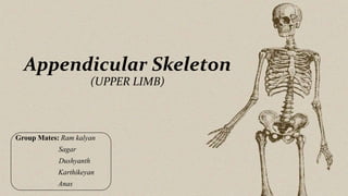
3. Introduction:
The term "appendicular" comes from the Latin
word "appendere," which means "to hang on." This
is fitting, as the bones of the appendicular skeleton
are essentially "hanging" from the axial skeleton.
The appendicular skeleton consists of 126 bones.
It allows us to move and manipulate objects.
4. Includes all bones besides axial skeleton
• The limbs
• The supportive girdles (Pectoral and Pelvic girdles)
The Pectoral girdle with the upper limbs and the
Pelvic girdle with the lower limb.
Limbs comprises of Humerus, Radius, Ulna,
Carpals, Metacarpals, Phalanges.
5. Scapula
The scapula is also known as the shoulder blade.
It articulates with the humerus at with the
glenohumeral joint, and with the clavicle at the
acromioclavicular joint. In doing so, the scapula
connects the upper limb to the trunk.
It is a triangular, flat bone, which serves as a
site for attachment for many (17) muscles.
6. Scapula
Costal Surface
The costal (anterior) surface of the scapula faces the ribcage.
It contains a large concave depression over most of its surface, known as
the subscapular fossa. The subscapularis (rotator cuff muscle) originates from
this fossa. Originating from the superolateral surface of the costal scapula is
the coracoid process. It is a hook-like projection, which lies just underneath
the clavicle.
7. Three muscles attach to the coracoid process: the pectoralis minor,
coracobrachialis, and the short head of the biceps brachii.
Lateral Surface
The lateral surface of the scapula faces the humerus. It is the site of the
glenohumeral joint, and of various muscle attachments. Its important bony
landmarks include:
•Glenoid fossa – a shallow cavity, located superiorly on the lateral border.
• It articulates with the head of the humerus to form the glenohumeral
(shoulder) joint.
8. •Supraglenoid tubercle – a roughening immediately superior to the glenoid
fossa.
• The place of attachment of the long head of the biceps brachii.
•Infraglenoid tubercle – a roughening immediately inferior to the glenoid
fossa.
• The place of attachment of the long head of the triceps brachii.
Posterior Surface
The posterior surface of the scapula faces outwards. It is a site of origin for
the majority of the rotator cuff muscles of the shoulder.
9. It is marked by:
•Spine – the most prominent feature of the posterior scapula. It runs
transversely across the scapula, dividing the surface into two.
•Acromion – projection of the spine that arches over the glenohumeral joint
and articulates with the clavicle at the acromioclavicular joint.
•Infraspinous fossa – the area below the spine of the scapula, it displays a
convex shape.
• The infraspinatus muscle originates from this area.
10. •Supraspinous fossa – the area above the spine of the scapula, it is much
smaller than the infraspinous fossa, and is more convex in shape.
• The supraspinatus muscle originates from this area.
Articulations
The scapula has two main articulations:
•Glenohumeral joint – between the glenoid fossa of the scapula and the
head of the humerus.
•Acromioclavicular joint – between the acromion of the scapula and the
clavicle.
12. Clavicle
• Also called as collarbones or beauty bones.
• It is a long, S-shaped bone which makes up the anterior portion of the
shoulder girdles.
• The clavicle (collarbone) extends between the manubrium of the
sternum and the acromion of the scapula.
13. The clavicle has three main functions:
• Attaches the upper limb to the trunk as part of the ‘shoulder girdle’.
• Protects the underlying neurovascular structures supplying the upper
limb.
• Transmits force from the upper limb to the axial skeleton.
14. Bony Landmarks and Articulations
• Facing forward, the medial aspect is convex, and the lateral aspect
concave.
• It can be divided into a sternal end, a shaft and an acromial end.
Sternal (medial) End
• The sternal end contains a large facet – for articulation with the
manubrium of the sternum at the sternoclavicular joint.
• The inferior surface of the sternal end is marked by a rough oval
depression for the costoclavicular ligament (a ligament of the SC joint).
15. Shaft
The shaft of the clavicle acts a point of origin and attachment for several
muscles – deltoid, trapezius, subclavius, pectoralis major,
sternocleidomastoid and sternohyoid.
Acromial (lateral) End
The acromial end houses a small facet for articulation with the acromion of
the scapula at the acromioclavicular joint. It also serves as an attachment
point for two ligaments:
16. • Conoid tubercle – attachment point of the conoid ligament, the medial
part of the coracoclavicular ligament.
• Trapezoid line – attachment point of the trapezoid ligament, the lateral
part of the coracoclavicular ligament.
• The coracoclavicular ligament is a very strong structure, effectively
suspending the weight of the upper limb from the clavicle.
• It is the most commonly fractured bone in the human skeleton.
• 15% of fractures occur in the lateral third.
• 80% occur in the middle third
• 5% occur in the medial third.
17. Humerus
• The humerus is a long bone of the upper limb, which extends from the
shoulder to the elbow.
• The proximal aspect of the humerus articulates with the glenoid fossa of
the scapula, forming the glenohumeral joint. Distally, at the elbow joint,
the humerus articulates with the head of the radius and trochlear notch of
the ulna.
18. Proximal Landmarks
• The proximal humerus is marked by a head, anatomical neck, surgical
neck, greater and lesser tuberosity and intertubercular sulcus.
• The upper end of the humerus consists of the head. This faces medially,
upwards and backwards and is separated from the greater and lesser
tuberosities by the anatomical neck.
• The greater tuberosity is located laterally on the humerus and
has anterior and posterior surfaces. It serves as an attachment site
for three of the rotator cuff muscles – supraspinatus,
infraspinatus and teres minor
19. • The lesser tuberosity is much smaller, and more medially located
on the bone. It only has an anterior surface. It provides attachment
for the last rotator cuff muscle – the subscapularis.
• Separating the two tuberosities is a deep groove, known as
the intertubercular sulcus.
20. • On the lateral side of the humeral shaft is a roughened surface where the
deltoid muscle attaches. This is known is as the deltoid tuberosity.
• Distally, the trochlea is located medially, and extends onto the posterior
aspect of the bone. Lateral to the trochlea is the capitulum, which
articulates with the radius.
• Also located on the distal portion of the humerus are three depressions,
known as the coronoid, radial and
olecranon fossae.
• They accommodate the forearm bones
during flexion or extension at the elbow.
21. Radius
• The radius is a long bone in the forearm. It lies laterally and parallel
to ulna.
• The radius and ulna bones articulate with each other both proximally and
distally forming proximal and distal radioulnar joints.
• Together they provide the capability of rotational movement i.e.,
pronation and supination.
• Radial tuberosity – A bony projection, which serves as the place of
attachment of the biceps brachii muscle.
22. • The proximal end of the radius articulates with the capitulum of the
humerus and the radial notch of ulna.
• The shaft of the radius is triangular in cross-section and provides
attachments for several muscles of the form arm.
• The distal end is large, with two articular surfaces: one below for the
carpals and one medial (ulnar notch) for the head of the ulna.
23. Ulna
• The ulna is a long bone in the forearm. It lies medially and parallel to
the radius.
• The ulna acts as the stabilizing bone, with the radius pivoting to produce
movement.
• Proximally, the ulna articulates with the humerus at the elbow joint.
Distally, the ulna articulates with the radius, forming the distal radio-
ulnar joint.
24. •Olecranon – a large projection of bone that extends proximally, forming
part of trochlear notch. The triceps brachii muscle attaches to its superior
surface.
•Coronoid process – this ridge of bone projects outwards anteriorly,
forming part of the trochlear notch.
•Trochlear notch – formed by the olecranon and coronoid process. It
articulates with the trochlea of the humerus.
•Radial notch – located on the lateral surface of the trochlear notch, this
area articulates with the head of the radius.
25. • Tuberosity of ulna – a roughening immediately distal to the coronoid
process. It is where the brachialis muscle attaches.
• The distal end of the ulna is much smaller in diameter than the proximal
end, with distal projection – the ulnar styloid process.
• The head articulates with the ulnar notch of the radius to form the distal
radio-ulnar joint.
26. Carpals
• The carpal bones are a group of eight, irregularly shaped bones. They are
organized into two rows: proximal and distal.
• Proximally, the scaphoid and lunate articulate with the radius to form
the wrist joint (also known as the ‘radio-carpal joint’).
• In the distal row, all of the carpal bones articulate with the metacarpals.
• The scaphoid bone of the hand is the most commonly fractured carpal
bone
28. Metacarpals
The metacarpal bones articulate proximally with the carpals, and distally
with the proximal phalanges. They are numbered, and each associated with
a digit:
•Metacarpal I – Thumb.
•Metacarpal II – Index finger.
•Metacarpal III – Middle finger.
•Metacarpal IV – Ring finger.
•Metacarpal V – Little finger.
29. • Each metacarpal consists of a base, shaft and a head. The medial and
lateral surfaces of the metacarpals are concave, allowing attachment of
the interossei muscles.
30. Phalanges
• The phalanges are the bones of the fingers. It comprises of 14 bones.
• The thumb has a proximal and distal phalanx, while the rest of the digits
have proximal, middle and distal phalanges.
• The Phalangeal formula is 2,3,3,3,3.
• The thumb is called as “Pollex”.