
Recommended
More Related Content
Similar to Anatomy Lesson_ Realistic Skeleton for Education by Slidesgo (2).pptx
Similar to Anatomy Lesson_ Realistic Skeleton for Education by Slidesgo (2).pptx (20)
Recently uploaded
Recently uploaded (20)
Anatomy Lesson_ Realistic Skeleton for Education by Slidesgo (2).pptx
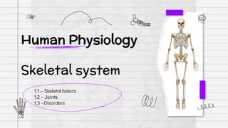
- 1. 1.1 – Skeletal basics 1.2 – Joints 1.3 - Disorders Human Physiology Skeletal system
- 2. SKELETAL SYSTEM BONES ITS FURTHER DIVIDED INTO AXIAL AND APPENDICULAR SKELETON MOVEMENT AND LOCOMOTION Hard matrix Made of Calcium salts. Pliable matrix Made of chondroitin salts 206 bones in a adult CARTILAGES
- 3. AXIAL SKELETON • 80 bones distributed among main axis • The skull, vertebral column, sternum and ribs constitute axial skeleton
- 4. Skull • The skull is composed of two sets of bones Cranial and Facial • Cranial bones are 8 in number. They form the hard protective outer covering, cranium for the brain • The facial region is made up of 14 skeletal elements which form the front part of the skull • A single U-shaped bone called hyoid is present at the base of the buccal cavity and it is also included in the skull Middle Ear Malleus Incus Stapes EAR OSSICLES (COMBINELY CALLED)
- 5. Vertebral column • Our vertebral column is formed by 26 serially arranged units called vertebrae and is dorsally placed. • Extends from the base of the skull and constitutes the main framework of the trunk • Each vertebra has a central hollow portion (neural canal) through which the spinal cord passes • First vertebra is the atlas and it articulates with the occipital condyles. • The vertebral column is differentiated into cervical (7), thoracic (12), lumbar (5), sacral (1-fused) and coccygeal (1-fused) regions starting from the skull. • The number of cervical vertebrae are seven in almost all mammals including human beings. • Sternum is a flat bone on the ventral midline of thorax. • The vertebral column protects the spinal cord, supports the head and serves as the point of attachment for the ribs and musculature of the back
- 6. RIBS • Each rib is a thin flat bone connected dorsally to the vertebral column and ventrally to the sternum. • It has two articulation surfaces on its dorsal end and is hence called bicephalic • Dorsally, they are attached to the thoracic vertebrae and ventrally connected to the sternum with the help of hyaline cartilage • The 8th, 9th and 10th pairs of ribs do not articulate directly with the sternum but join the seventh rib with the help of hyaline cartilage. • These are called vertebrochondral (false) ribs. Last 2 pairs (11th and 12th) of ribs are not connected ventrally and are therefore, called floating ribs. Thoracic vertebrae, ribs and sternum together form the rib cage
- 7. Appendicular skeleton • limbs alongwith their girdles constitute the appendicular skeleton.
- 8. LIMBS • Each limb is made of 30 bones • The bones of the hand (fore limb) are humerus, radius and ulna, carpals (wrist bones – 8 in number), metacarpals (palm bones – 5 in number) and phalanges (digits – 14 in number) • Femur (thigh bone – the longest bone), tibia and fibula, tarsals (ankle bones – 7 in number), metatarsals (5 in number) and phalanges (digits – 14 in number) are the bones of the legs (hind limb) . A cup shaped bone called patella cover the knee ventrally (knee cap).
- 9. Gridle • Pectoral and Pelvic girdle bones help in the articulation of the upper and the lower limbs respectively with the axial skeleton • Each girdle is formed of two halves. Each half of pectoral girdle consists of a clavicle and a scapula • Scapula is a large triangular flat bone situated in the dorsal part of the thorax between the second and the seventh ribs. • The dorsal, flat, triangular body of scapula has a slightly elevated ridge called the spine which projects as a flat, expanded process called the acromion • The clavicle articulates with this. Below the acromion is a depression called the glenoid cavity which articulates with the head of the humerus to form the shoulder joint • Each clavicle is a long slender bone with two curvatures. This bone is commonly called the collar bone.
- 10. Gridle • Pectoral and Pelvic girdle bones help in the articulation of the upper and the lower limbs respectively with the axial skeleton • Each girdle is formed of two halves. Each half of pectoral girdle consists of a clavicle and a scapula • Scapula is a large triangular flat bone situated in the dorsal part of the thorax between the second and the seventh ribs. • The dorsal, flat, triangular body of scapula has a slightly elevated ridge called the spine which projects as a flat, expanded process called the acromion • The clavicle articulates with this. Below the acromion is a depression called the glenoid cavity which articulates with the head of the humerus to form the shoulder joint • Each clavicle is a long slender bone with two curvatures. This bone is commonly called the collar bone.
- 11. JOINTS 02
- 12. JOINTS • Joints are points of contact between bones, or between bones and cartilages • Force generated by the muscles is used to carry out movement through joints, where the joint acts as a fulcrum. • Joints have been classified into three major structural forms, namely, fibrous, cartilaginous and synovial. Fibrous joints do not allow any movement. This type of joint is shown by the flat skull bones which fuse end-to-end with the help of dense fibrous connective tissues in the form of sutures, to form the cranium In cartilaginous joints, the bones involved are joined together with the help of cartilages. The joint between the adjacent vertebrae in the vertebral column is of this pattern and it permits limited movements. Synovial joints are characterised by the presence of a fluid filled synovial cavity between the articulating surfaces of the two bones. Such an arrangement allows considerable movement. These joints help in locomotion and many other movements. Eg: Ball and socket joint (between humerus and pectoral girdle), hinge joint (knee joint)
- 14. Myasthenia gravis Auto immune disorder affecting neuromuscular junction leading to fatigue, weakening and paralysis of skeletal muscle.
- 15. Muscular dystrophy: Progressive degeneration of skeletal muscle mostly due to genetic disorder
- 16. Tetany Rapid spasms (wild contractions) in muscle due to low Ca++ in body fluid
- 17. Arthritis : Inflammation of joints.
- 18. Osteoporosis Age-related disorder characterised by decreased bone mass and increased chances of fractures. Decreased levels of estrogen is a common cause.
- 19. Gout Inflammation of joints due to accumulation of uric acid crystals
- 20. Thanks y’all