Mand. edent. found / dental implant courses by Indian dental academy
•
2 likes•378 views

The Indian Dental Academy is the Leader in continuing dental education , training dentists in all aspects of dentistry and offering a wide range of dental certified courses in different formats.for more details please visit www.indiandentalacademy.com

Recommended
Recommended
More Related Content
What's hot
What's hot (20)
Viewers also liked
Viewers also liked (11)
Similar to Mand. edent. found / dental implant courses by Indian dental academy
Similar to Mand. edent. found / dental implant courses by Indian dental academy (20)
More from Indian dental academy
More from Indian dental academy (20)
Recently uploaded
Recently uploaded (20)
Mand. edent. found / dental implant courses by Indian dental academy
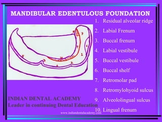
- 1. MANDIBULAR EDENTULOUS FOUNDATION 1. Residual alveolar ridge 2. Labial Frenum 3. Buccal frenum 4. Labial vestibule 5. Buccal vestibule 6. Buccal shelf 7. Retromolar pad 8. Retromylohyoid sulcus 9. Alveololingual sulcus 10. Lingual frenumwww.indiandentalacademy.com INDIAN DENTAL ACADEMY Leader in continuing Dental Education
- 2. 1. Labial Frenum: It is a fibrous band covered by mucous membrane extending from labial aspect of residual alveolar ridge to the lower lip. It has no muscle attachment and is inserted in vertical direction. It is best demonstrated by forward pull of lip. It is accommodated by a groove in the mandibular denture. www.indiandentalacademy.com
- 3. • 2. Labial Vestibule: • The portion of the oral cavity bounded on one side by the teeth, gingival and alveolar ridge (in the edentulous mouth the residual ridge) and on the other side by lips anterior to the buccal frenula. www.indiandentalacademy.com
- 4. Boundaries: 1. Labial aspect of residual alveolar ridge 2. Muco labial alveolar fold. 3. Orbicularis oris muscle (Lip) The labial flange of the mandibular denture occupies this space. www.indiandentalacademy.com
- 5. • 3. Buccal Frenum: • Single or multiple folds of mucous membrane extending from buccal mucous membrane and reflects towards the slope of the crest of alveolar mucosa distal to canine region. Reflection is in anterior posterior direction. It overlies depressor anguliorismuscle.www.indiandentalacademy.com
- 6. • 4. Buccal Vestibule: • The portion of the oral cavity bounded on one side by the teeth, gingival, and alveolar ridge (in edentulous mouth residual ridge) and on the lateral side by the cheeks posterior to the buccal frenum. It overlies fibres of buccinator muscle. Buccal flange of the mandibular denture occupies this space. www.indiandentalacademy.com
- 7. • 5. Buccal shelf area: • Bounded laterally by external oblique ridge and internally by slope of residual ridge. Bone in this area is very dense and is placed horizontal to stress land. Forces can thus be directed more nearly at right angles to buccal shelf than any other area of support. It is called as “primary Stress Bearing” area. www.indiandentalacademy.com
- 8. • 6. External oblique ridge: • It is a smooth ridge on the buccal surface of the body of the mandible that extends from the anterior border of the ramus with diminishing prominence downward and forward to the region of mental foramen. www.indiandentalacademy.com
- 9. • 7. Retromolar pad: • A mass of tissue comprised of non- keratinised mucosa located posterior to the retromolar papilla and over lying loose glandular connective tissue. This freely movable area should be differentiated from the pear shaped pad. www.indiandentalacademy.com
- 10. • Pear Shaped Pad: The most distal extension of attached keratinised mucosa overlying the mandibular ridge crest formed by the scarring pattern following extraction of the most posterior molar. It should be differentiated from retromolar area. • Retromolar pad contains fibres pterygomandibular raphae, fibres of superior pharyugeul constrictor, and buccinator muscles; fibres of temporal tendon and glandular tissue. It must be covered by the denture base to aid in stability. www.indiandentalacademy.com
- 11. • 8. Lingual frenum: • This frenum overlies genioglossus muscle, which originates from superior genial tubercule. Exhibit differing configurations in width and height. Surgical intervention is indicated when it interferes with border extension and stability of mandibular denture. Also in case of tongue tie (Restricted tongue movements).www.indiandentalacademy.com
- 12. • 9. Sub-lingual fold: • The crescent shaped area on the floor of the mouth following the inner wall of the mandible and tapering towards the molar regions. It is formed by the sub- lingual gland and Submandibular duct. • It is the fold of mucous membrane from the tongue to residual ridge www.indiandentalacademy.com
- 13. • 10. Mylohypoid muscle: • It forms muscular floor of the mouth. Arises from the mylohyoid ridge and rises progressively on the body of mandible. Mylohyoid muscle influences the mid and anterior portions of interior border of the lingual flange. Length of the flange is determined by displaceability of the floor of the mouth and movements of the tongue. www.indiandentalacademy.com
- 14. • 11. Retromylohoid space: • An anatomic area in the Alveololingual sulcus just lingual to the retromolar pad bounded anteriorly by the mylohyoid ridge, posteriorly by retromylohyoid curtain, inferiorly by the floor of the alveololingual sulcus and lingually by the anterior tonsillar pillar when the tongue is in a relaxed position. • This space lies at the distal and of alveolo-lingual sulcus and provides bracing to the mandibular denture. www.indiandentalacademy.com
- 15. Boundaries: •Anteriorly – Mylohoid ridge •Posteriorly – Retromylohoid curtain, which is formed by superior constrictor muscle. •Laterally – Mandible and pterygo mandible raphae. •Lingually – anterior tonsillar pillar. •Inferiorly – Floor of alveolo lingual sulcus.www.indiandentalacademy.com
- 16. 12. Residual ridge: The portion of the alveolar process and its soft tissue covering that remains after the removal of teeth. www.indiandentalacademy.com