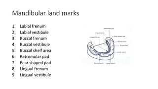
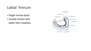
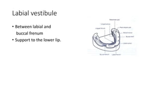
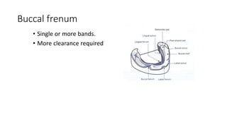
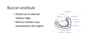
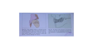
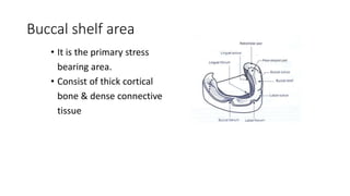
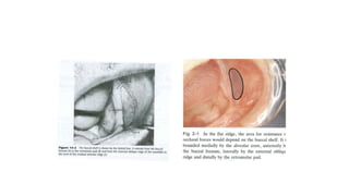
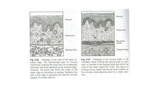
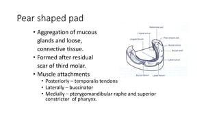
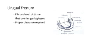
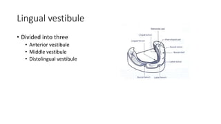
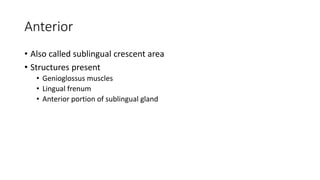
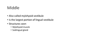
This document describes several important anatomical landmarks in the mandibular region for denture fabrication. It outlines 9 key landmarks: the labial frenum, labial vestibule, buccal frenum, buccal vestibule, buccal shelf area, retromolar pad, pear shaped pad, lingual frenum, and lingual vestibule. Each landmark is defined and its relevance to denture construction is explained. For example, the buccal shelf area is a primary stress bearing area consisting of thick cortical bone and dense connective tissue, and the retromolar pad forms the distal end of the denture and consists of loose connective tissue. Understanding these mandibular landmarks is essential
9. Buccal vestibule
• Extend up to external
oblique ridge.
• Denture borders may
overextend in this region
10.
11. Buccal shelf area
• It is the primary stress
bearing area.
• Consist of thick cortical
bone & dense connective
tissue
12.
13.
14. Retromolar pad area
• Forms the distal end of
the denture.
• Consist of loose
connective tissue
15. Pear shaped pad
• Aggregation of mucous
glands and loose,
connective tissue.
• Formed after residual
scar of third molar.
• Muscle attachments
• Posteriorly – temporalis tendons
• Laterally – buccinator
• Medially – pterygomandibular raphe and superior
constrictor of pharynx.
18. Anterior
• Also called sublingual crescent area
• Structures present
• Genioglossus muscles
• Lingual frenum
• Anterior portion of sublingual gland
19.
20. Middle
• Also called mylohyoid vestibule
• Is the largest portion of lingual vestibule
• Structures seen
• Mylohyoid muscle
• Sublingual gland
21.
22. Posterior
• Also called lateral throat form or
retromylohyoid fossa
• Boundaries
• Anteriorly – mylohyoid muscle
• Laterally – pear shaped pad
• Posteriorly – retromylohyoid curtain
• Medially - tongue
23.
24. References
• BERNARD LEWIN
• SHELDON WINKLER
• SHARRY
• Characteristics of anatomical landmarks in the mandibular
interforaminal region: A cone-beam computed tomography
study. Med oral patol cir buccal. 2012 ; 17(3): 420–5.
25. • An In-vivo Comparison of Vertical and Horizontal Distance
between Incisive Papilla and Incisal Edge of Maxillary Central
Incisors in Dentates with Different Arch Forms. JCDR. 2017; 11(4)
• Radiographic evaluation of anatomical variables in maxilla and
mandible in relation to dental implant placement. IJDR.2016; 27(4):
344-7
26. • THE SIGNIFICANCE OF ANATOMIC LANDMARKS IN COMPLETE
DENTURE SERVICE. J. Pros. Den. May-June, 1964; 14(3); 456-9
• Estimation of effectiveness of different anatomical landmarks in re-
establishing lost occlusal plane in edentates: An all-inclusive review of
literature.
International Journal of Medical and Health Research. 2018; 4(8); 52-
6