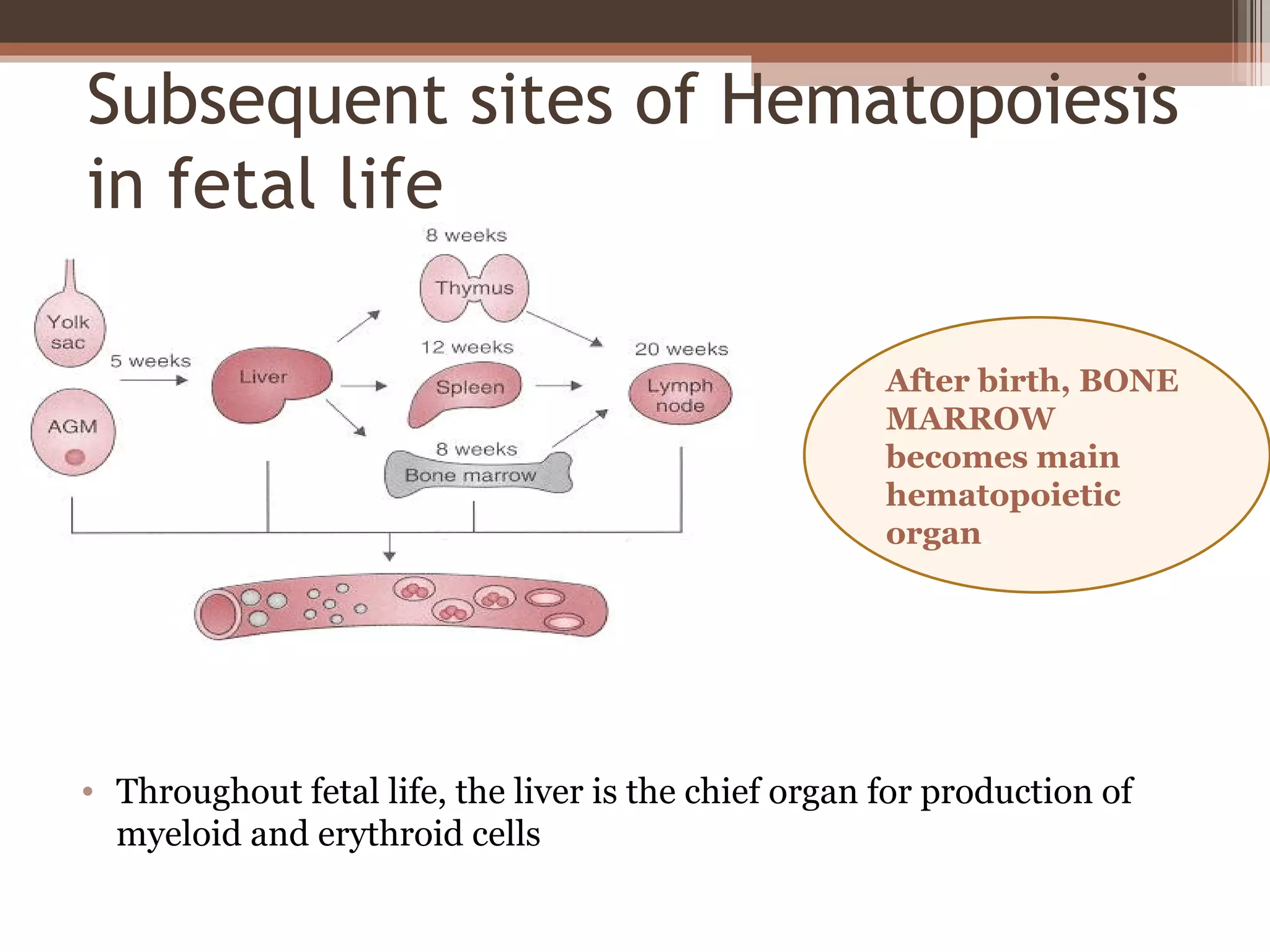
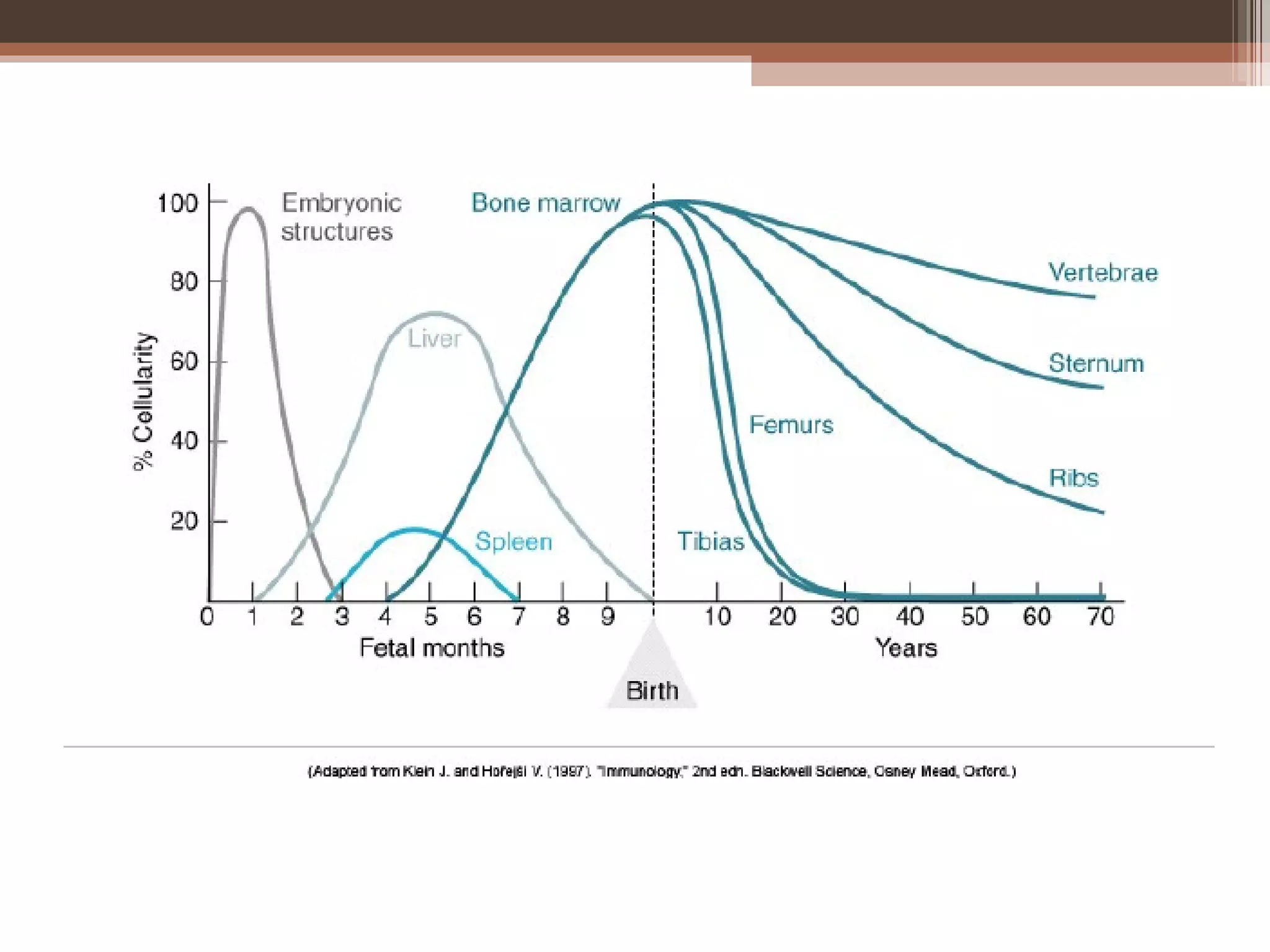
Bone marrow
Anatomy, Functions, Aspiration & Reporting


                      Dr. Narender Kumar
Anatomy..
• Two types of bone marrow:
 ▫ Medulla ossium rubra (Red Marrow -
   consisting mainly of hematopoietic tissue)
 ▫ Medulla ossium flava (Yellow Marrow -
   consisting mainly of fat cells)
• On average, bone marrow constitutes 4% of the
  total body mass of humans
• In adults weighing 65 kg (143 lbs), bone marrow
  accounts for approximately 2.6 kg (5.7 lbs)
• The hematopoeitic compartment of bone
  marrow produces approximately 500 billion
  blood cells per day
Primitive Hematopoiesis
• Derived from the extra- embryonic
  YOLK SAC;
• Consists mainly of nucleated erythroid
  cells that carry oxygen to the
  developing embryonic tissues… an early
   circulatory system.
• Probably starts ~ 4 weeks in humans
Definitive Hematopoietic System
• As the embryo’s size increases,
  primitive system superceded by
  definitive hematopoietic system,
  which originates in the embryo
  itself and continues throughout
  adult life.
• 1st definitive multipotent
  hematopoietic stem cells are
  generated within the
  embryonic AGM region of the
  para-aortic splanchnopleuric
  mesoderm (day 30-37 in human)
Subsequent sites of Hematopoiesis
in fetal life

                                                      After birth, BONE
                                                      MARROW
                                                      becomes main
                                                      hematopoietic
                                                      organ.


• Throughout fetal life, the liver is the chief organ for production of
  myeloid and erythroid cells
Pattern of distribution..
• At birth, all bone marrow is red.
• With age, more and more of it is converted to the
  yellow type; only around half of adult bone
  marrow is red
• In Adults Red marrow is found mainly in the
  Flat bones and in the epiphyseal ends of
  long bones such as the femur and humerus
• Yellow marrow is found in the medullary cavity,
  the hollow interior of the middle portion of long
  bones
Bone Marrow
• Bone marrow is specially designed to support the
  proliferation, differentiation, and maintenance of
  hematopoietic cells
• The stroma of the bone marrow is all tissue not directly
  involved in the primary function of hematopoiesis.
• Cells that constitute the bone marrow stroma are:
  • fibroblasts (reticular connective tissue)
  • macrophages
  • adipocytes
  • osteoblasts
  • osteoclasts
  • endothelial cells, which form the sinusoids.
• Bone marrow examination refers to the
  pathologic analysis of samples of bone marrow
  obtained by bone marrow aspiration and
  bone marrow biopsy (often called a trephine
  biopsy)
• The aspirate yields semi-liquid bone marrow,
  which can be examined
 •   under a light microscope as well as
 •   analyzed by flow cytometry,
 •   chromosome analysis, or
 •   polymerase chain reaction (PCR).
• Frequently, a trephine biopsy is also obtained,
  which yields a narrow, cylindrically shaped solid
  piece of bone marrow which is examined
  microscopically (sometimes with the aid of
  immunohistochemistry) for cellularity and
  infiltrative processes.
How the test is performed…
• usually performed on the back
  of the hipbone, or posterior
  iliac crest.
• However, an aspirate can also
  be obtained from the sternum
   (breastbone).
• A trephine biopsy should
  never be performed on the
  sternum, however, due to the
  risk of injury to blood vessels,
  lungs or the heart.
• Sahla Bone Marrow Needle, iliac crest, with adjustable
  stop
• 14 (2.0mm) x 50mm
• 16 (1.6mm) x 50mm
• 18 (1.2mm) x 50mm (in child)
Contraindications

• The only absolute reason to avoid is the
  presence of a severe bleeding disorder

Complications
• While mild soreness lasting 12-24 hours is
  common after a bone marrow examination,
  serious complications are extremely rare.
Erythroid series
Myeloid series
Megakaryocytic series
Monocytic series
*Proerythroblast
**Early erythroblast (Basophillic )
***Intermediate erythroblast (Polychromatic)
****Late erythroblast (Orthochromatic )
Erythroid precursors
• Normal red cells are
  produced in the bone
  marrow from erythroid
  precursors or
  erythroblasts.
• The earliest
  morphologically
  recognisable red cell
  precursor is derived from
  an erythroid progenitor
  cell which in turn is
  derived from a
  multipotent
  haemopoietic progenitor
  cell.
proerythroblast
• Normal proerythroblast
  [dark red arrow] in the
  bone marrow. This is a
  large cell with a round
  nucleus and a finely
  stippled chromatin
  pattern. Nucleoli are
  sometimes apparent.
• The cytoplasm is
  moderately to strongly
  basophilic.
Basophilic erythroblast
• Spherical nucleus, nucleoli
  not visible, basophilic
  cytoplasm
Polychromatophilic erythroblast
• smaller nucleus –condensed
  chromatin, baso- and
  eosinophilia in the cytoplasm
Orthochromatophilic erythroblast
• small nucleus with highly
  condensed chromatin, nucleus
  extruded, eosinophilic
  cytoplasm
Normal erythroblasts in the BM
Myeloid precursors


• Myeloblast promyelocyte myelocyte 
  metamyelocyte  band form  mature
  neutrophil.
credit -Dr. Lekst rom Hines, JEM 1999
Myeloid precursors
• Normal myeloblasts have no granules but
  abnormal myeloblasts may have a few granules.
• Myeloblasts undergo one cell division and
  mature into promyelocytes.
• Promyelocytes have primary or azurophilic
  granules. They have a Golgi zone a pale area
  adjacent to the nucleus that is the site of granule
  production. The chromatin pattern of a
  promyelocyte shows some condensation or
  clumping, in contrast to the diffuse chromatin
  pattern of a myeloblast, but nucleoli are still
  visible.
Normal granulocyte precursors in
  the bone marrow
 • Note the myeloblast
   [dark red arrow] with a
   high nucleocytoplasmic
   ratio, diffuse chromatin
   pattern and nucleolus.
 • There is a promyelocyte
   [green arrow] which is
   larger and has a lower
   nucleocytoplasmic ratio
   and abundant azurophilic
   granules.
Myelocytes
• Myelocytes are smaller
  than promyelocytes and
  have specific granules
  that indicate whether
  they are of neutrophil,
  eosinophil or basophil
  lineage.
• The nucleolus is no
  longer visible.
Eosinophilic myelocyte
A neutrophil metamyelocyte
• The metamyelocyte
  differs from a
  myelocyte in having
  some indentation of
  the nucleus
• It differs from a band
  form in not having
  any part of its nucleus
  with two parallel
  edges
Neutrophilic
metamyelocyte
Eosinophilic
metamyelocyte
Basophilic metamyelocyte
Band or juvenile Neutrophils

• There are smaller
  numbers of cells of
  neutrophil lineage
  with non-segmented
  nuclei. They are
  referred to as
  neutrophil band cells
  or band forms. They
  are less mature than
  segmented
  neutrophils.
Compare the different cell types:


      basophilic (myeloid)    eosinophilic (myeloid)


     neutrophilic (myeloid)        erythroid
*Megakaryoblasts
** Promegakaryocytes
***Megakaryocytes
Megakaryoblasts
• Megakaryoblasts are
  the precursors of the
  megakarycytes.
• They may show
  cytoplasmic blebbing.
Promegakaryocyte
Megakaryocyte
Monocytopoiesis

- Monoblast

- Promonocyte

- Monocyte
Promonocyte
19-
                 49

Hematopoiesis
Systemic scheme for Examining
aspirated BM films
• Low power (x10)
 ▫   Determine cellularity
 ▫   Identify megakaryocytes
 ▫   Look for clumps of abnormal cells
 ▫   Identify macrophages
Systemic scheme for Examining
aspirated BM films
• Higher power (x40, x100)
 ▫ Identify all stages of maturation of myeloid and
   erythroid cells.
 ▫ Determine the M:E ratio
 ▫ Perform a differential count
 ▫ Look for areas of BM necrosis.
 ▫ Assess the iron content.
Assessment of BM cellularity
• Cellularity cannot be assessed without knowing
  the age of a patient.
• A young child on average has about 80% of the
  intertrabecular space occupied by haemopoietic
  cells whereas in a 75-year-old the average has
  fallen to around 30%.
• 100 – Age of Patient
Comparing normo, hyper, &
hypocellular marrows
M:E ratio
• The M:E ratio is the ratio of all granulocytic plus
  monocytic cells (Myeloid) to all erythroblasts
  (Erythroid).
• For all bone marrow aspirates examined, the
  report should specify the M:E ratio and the
  percentage of lymphocytes and plasma cells.
• A differential count of at least 200-300 cells
  should be performed.
• If there is any borderline abnormality, e.g. in the
  number of blasts, lymphocytes or plasma cells, a
  500 cell differential count should be
  performed.
• Only after the bone marrow has been
  carefully assessed on low and medium power
  the X100 oil should be used to assess cellular
  detail
BM iron stores
• Once all normal and
  abnormal bone marrow
  cells have been assessed
  on a routine stain an iron
  stain should be
  examined, using a
  medium power objective
  (X 40 or X 50). Storage
  iron, which stains blue,
  should be assessed in
  bone marrow fragments.
  This image shows
  normal bone marrow
  iron.
Reporting results
• List the various descriptive comments regarding
  all the cell lines present in the BM
• Mention the striking abnormalities separately.
• Write your impression and any
  recommendations to the clinician.
Cellular constitution of the red bone
marrow parenchyma
Trephine biopsy
• Jamshidi Type Bone Marrow Biopsy Needle,
 •   8swg (4.0mm) x 100mm
 •   11swg (3.0mm) x 100mm
 •   13swg (2.3mm) x 90mm
 •   14swg (2.0mm) x 90mm (In child)
Adequacy of biopsy
• should contain at least five to six intertrabecular
  spaces and, after processing,
• should be at least 2–3 cm in length
• Others have considered that 1.5–2 cm is an
  acceptable length
Indications or areas of controversy
• Inadequate or failed aspirate.
• Need for accurate assessment of cellularity,
  whether increased or decreased.
• Suspected focal lesion (for example, suspected
  granulomatous disease or lymphoma).
• Suspected bone marrow fibrosis.
• Need to study bone marrow architecture.
• Need to study bone structure or bone marrow
  blood vessels
Bone marrow class
Bone marrow class

Bone marrow class

  • 1.
    Bone marrow Anatomy, Functions,Aspiration & Reporting Dr. Narender Kumar
  • 2.
  • 3.
    • Two typesof bone marrow: ▫ Medulla ossium rubra (Red Marrow - consisting mainly of hematopoietic tissue) ▫ Medulla ossium flava (Yellow Marrow - consisting mainly of fat cells)
  • 4.
    • On average,bone marrow constitutes 4% of the total body mass of humans • In adults weighing 65 kg (143 lbs), bone marrow accounts for approximately 2.6 kg (5.7 lbs) • The hematopoeitic compartment of bone marrow produces approximately 500 billion blood cells per day
  • 6.
    Primitive Hematopoiesis • Derivedfrom the extra- embryonic YOLK SAC; • Consists mainly of nucleated erythroid cells that carry oxygen to the developing embryonic tissues… an early circulatory system. • Probably starts ~ 4 weeks in humans
  • 7.
    Definitive Hematopoietic System •As the embryo’s size increases, primitive system superceded by definitive hematopoietic system, which originates in the embryo itself and continues throughout adult life. • 1st definitive multipotent hematopoietic stem cells are generated within the embryonic AGM region of the para-aortic splanchnopleuric mesoderm (day 30-37 in human)
  • 8.
    Subsequent sites ofHematopoiesis in fetal life After birth, BONE MARROW becomes main hematopoietic organ. • Throughout fetal life, the liver is the chief organ for production of myeloid and erythroid cells
  • 10.
    Pattern of distribution.. •At birth, all bone marrow is red. • With age, more and more of it is converted to the yellow type; only around half of adult bone marrow is red • In Adults Red marrow is found mainly in the Flat bones and in the epiphyseal ends of long bones such as the femur and humerus • Yellow marrow is found in the medullary cavity, the hollow interior of the middle portion of long bones
  • 11.
    Bone Marrow • Bonemarrow is specially designed to support the proliferation, differentiation, and maintenance of hematopoietic cells • The stroma of the bone marrow is all tissue not directly involved in the primary function of hematopoiesis. • Cells that constitute the bone marrow stroma are: • fibroblasts (reticular connective tissue) • macrophages • adipocytes • osteoblasts • osteoclasts • endothelial cells, which form the sinusoids.
  • 14.
    • Bone marrowexamination refers to the pathologic analysis of samples of bone marrow obtained by bone marrow aspiration and bone marrow biopsy (often called a trephine biopsy)
  • 15.
    • The aspirateyields semi-liquid bone marrow, which can be examined • under a light microscope as well as • analyzed by flow cytometry, • chromosome analysis, or • polymerase chain reaction (PCR). • Frequently, a trephine biopsy is also obtained, which yields a narrow, cylindrically shaped solid piece of bone marrow which is examined microscopically (sometimes with the aid of immunohistochemistry) for cellularity and infiltrative processes.
  • 16.
    How the testis performed…
  • 17.
    How the testis performed… • usually performed on the back of the hipbone, or posterior iliac crest. • However, an aspirate can also be obtained from the sternum (breastbone). • A trephine biopsy should never be performed on the sternum, however, due to the risk of injury to blood vessels, lungs or the heart.
  • 18.
    • Sahla BoneMarrow Needle, iliac crest, with adjustable stop • 14 (2.0mm) x 50mm • 16 (1.6mm) x 50mm • 18 (1.2mm) x 50mm (in child)
  • 19.
    Contraindications • The onlyabsolute reason to avoid is the presence of a severe bleeding disorder Complications • While mild soreness lasting 12-24 hours is common after a bone marrow examination, serious complications are extremely rare.
  • 20.
  • 22.
    *Proerythroblast **Early erythroblast (Basophillic) ***Intermediate erythroblast (Polychromatic) ****Late erythroblast (Orthochromatic )
  • 23.
    Erythroid precursors • Normalred cells are produced in the bone marrow from erythroid precursors or erythroblasts. • The earliest morphologically recognisable red cell precursor is derived from an erythroid progenitor cell which in turn is derived from a multipotent haemopoietic progenitor cell.
  • 24.
    proerythroblast • Normal proerythroblast [dark red arrow] in the bone marrow. This is a large cell with a round nucleus and a finely stippled chromatin pattern. Nucleoli are sometimes apparent. • The cytoplasm is moderately to strongly basophilic.
  • 25.
    Basophilic erythroblast • Sphericalnucleus, nucleoli not visible, basophilic cytoplasm
  • 26.
    Polychromatophilic erythroblast • smallernucleus –condensed chromatin, baso- and eosinophilia in the cytoplasm
  • 27.
    Orthochromatophilic erythroblast • smallnucleus with highly condensed chromatin, nucleus extruded, eosinophilic cytoplasm
  • 28.
  • 30.
    Myeloid precursors • Myeloblastpromyelocyte myelocyte  metamyelocyte  band form  mature neutrophil.
  • 31.
    credit -Dr. Lekstrom Hines, JEM 1999
  • 32.
    Myeloid precursors • Normalmyeloblasts have no granules but abnormal myeloblasts may have a few granules. • Myeloblasts undergo one cell division and mature into promyelocytes. • Promyelocytes have primary or azurophilic granules. They have a Golgi zone a pale area adjacent to the nucleus that is the site of granule production. The chromatin pattern of a promyelocyte shows some condensation or clumping, in contrast to the diffuse chromatin pattern of a myeloblast, but nucleoli are still visible.
  • 33.
    Normal granulocyte precursorsin the bone marrow • Note the myeloblast [dark red arrow] with a high nucleocytoplasmic ratio, diffuse chromatin pattern and nucleolus. • There is a promyelocyte [green arrow] which is larger and has a lower nucleocytoplasmic ratio and abundant azurophilic granules.
  • 34.
    Myelocytes • Myelocytes aresmaller than promyelocytes and have specific granules that indicate whether they are of neutrophil, eosinophil or basophil lineage. • The nucleolus is no longer visible.
  • 35.
  • 36.
    A neutrophil metamyelocyte •The metamyelocyte differs from a myelocyte in having some indentation of the nucleus • It differs from a band form in not having any part of its nucleus with two parallel edges
  • 37.
  • 38.
  • 39.
  • 40.
    Band or juvenileNeutrophils • There are smaller numbers of cells of neutrophil lineage with non-segmented nuclei. They are referred to as neutrophil band cells or band forms. They are less mature than segmented neutrophils.
  • 41.
    Compare the differentcell types: basophilic (myeloid) eosinophilic (myeloid) neutrophilic (myeloid) erythroid
  • 42.
  • 43.
    Megakaryoblasts • Megakaryoblasts are the precursors of the megakarycytes. • They may show cytoplasmic blebbing.
  • 44.
  • 45.
  • 46.
  • 47.
  • 49.
    19- 49 Hematopoiesis
  • 50.
    Systemic scheme forExamining aspirated BM films • Low power (x10) ▫ Determine cellularity ▫ Identify megakaryocytes ▫ Look for clumps of abnormal cells ▫ Identify macrophages
  • 51.
    Systemic scheme forExamining aspirated BM films • Higher power (x40, x100) ▫ Identify all stages of maturation of myeloid and erythroid cells. ▫ Determine the M:E ratio ▫ Perform a differential count ▫ Look for areas of BM necrosis. ▫ Assess the iron content.
  • 52.
    Assessment of BMcellularity • Cellularity cannot be assessed without knowing the age of a patient. • A young child on average has about 80% of the intertrabecular space occupied by haemopoietic cells whereas in a 75-year-old the average has fallen to around 30%. • 100 – Age of Patient
  • 53.
    Comparing normo, hyper,& hypocellular marrows
  • 54.
    M:E ratio • TheM:E ratio is the ratio of all granulocytic plus monocytic cells (Myeloid) to all erythroblasts (Erythroid). • For all bone marrow aspirates examined, the report should specify the M:E ratio and the percentage of lymphocytes and plasma cells. • A differential count of at least 200-300 cells should be performed. • If there is any borderline abnormality, e.g. in the number of blasts, lymphocytes or plasma cells, a 500 cell differential count should be performed.
  • 55.
    • Only afterthe bone marrow has been carefully assessed on low and medium power the X100 oil should be used to assess cellular detail
  • 56.
    BM iron stores •Once all normal and abnormal bone marrow cells have been assessed on a routine stain an iron stain should be examined, using a medium power objective (X 40 or X 50). Storage iron, which stains blue, should be assessed in bone marrow fragments. This image shows normal bone marrow iron.
  • 57.
    Reporting results • Listthe various descriptive comments regarding all the cell lines present in the BM • Mention the striking abnormalities separately. • Write your impression and any recommendations to the clinician.
  • 58.
    Cellular constitution ofthe red bone marrow parenchyma
  • 59.
  • 60.
    • Jamshidi TypeBone Marrow Biopsy Needle, • 8swg (4.0mm) x 100mm • 11swg (3.0mm) x 100mm • 13swg (2.3mm) x 90mm • 14swg (2.0mm) x 90mm (In child)
  • 62.
    Adequacy of biopsy •should contain at least five to six intertrabecular spaces and, after processing, • should be at least 2–3 cm in length • Others have considered that 1.5–2 cm is an acceptable length
  • 64.
    Indications or areasof controversy • Inadequate or failed aspirate. • Need for accurate assessment of cellularity, whether increased or decreased. • Suspected focal lesion (for example, suspected granulomatous disease or lymphoma). • Suspected bone marrow fibrosis. • Need to study bone marrow architecture. • Need to study bone structure or bone marrow blood vessels

Editor's Notes

  • #9 After about day 37 (5 weeks), HSCs from the AGM begin to colonize the fetal liver, and at 6 weeks hematopoiesis (definitive) takes place in the fetal liver until bone marrow is formed