SlideShare a Scribd company logo
1 of 67
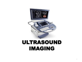
ULTRASOUND
IMAGING
1
Learning objectives
• Present an introduction to medical
ultrasonography at the level of a third or
fourth year medical student.
• After the brief course, the student should be
able to:
– Understand basic physics and terminology of
ultrasound
– State the advantages and disadvantages of
ultrasound
– Understand how the images are obtained
– Begin to interpret simple ultrasound images.
Outline
• Brief history of ultrasound technology
• Advantages, disadvantages
• What is ultrasound?
• Physics
• Probes, controls, machine, Doppler
• Terminology
• What structures look like
• Abdominal ultrasound
• Example of liver/gallbladder
• Quick review questions
History
• Medical use – 1940s
• Developing technology – 1950s
• Advancements – 1960s/1970s
• Real time ultrasound – 1980s
• 3D and 4D images – 1990s
Advantages
• Lack of radiation
• Quick, adaptable
• Looking at different layers/planes
• High resolution in low fat areas
• Portable
• Less expensive
Disadvantages
• Operator-dependent
• Depends on area to view
• Depends on body habitus of patient
• Artifact
• Location
What is ultrasound?
• Sound waves
• Waves have amplitude and frequency
• Frequency – measured in Hz
• Pulse-echo principle – crystals respond to
sound waves
• Ultrasound waves – transmitted well through
fluids and poorly through gases
What is Ultrasound?
• Ultrasound or ultrasonography is a medical
imaging technique that uses high frequency
sound waves and their echoes. The technique
is similar to the echolocation used by bats,
whales and dolphins, as well as SONAR used
by submarines.
In ultrasound, the following events
happen:
• The ultrasound machine transmits high-frequency (1 to 5 megahertz)
sound pulses into your body using a probe.
• The sound waves travel into your body and hit a boundary between
tissues (e.g. between fluid and soft tissue, soft tissue and bone).
• Some of the sound waves get reflected back to the probe, while some
travel on further until they reach another boundary and get reflected.
• The reflected waves are picked up by the probe and relayed to the
machine.
• The machine calculates the distance from the probe to the tissue or organ
(boundaries) using the speed of sound in tissue (5,005 ft/s or1,540 m/s)
and the time of the each echo's return (usually on the order of millionths
of a second).
• The machine displays the distances and intensities of the echoes on the
screen, forming a two dimensional image like the one shown below.
The Ultrasound Machine
• Transducer probe - probe that sends and receives the sound waves
• Central processing unit (CPU) - computer that does all of the calculations
and contains the electrical power supplies for itself and the transducer
probe
• Transducer pulse controls - changes the amplitude, frequency and
duration of the pulses emitted from the transducer probe
• Display - displays the image from the ultrasound data processed by the
CPU
• Keyboard/cursor - inputs data and takes measurements from the display
• Disk storage device (hard, floppy, CD) - stores the acquired images
• Printer - prints the image from the displayed data
Transducer probe
• Is the main part of the ultrasound machine.
• The transducer probe makes the sound waves and receives the echoes.
• The mouth and ears of the ultrasound machine. The transducer probe
generates and receives sound waves using a principle called the
piezoelectric (pressure electricity) effect, which was discovered by Pierre
and Jacques Curie in 1880.
• In the probe, there are one or more quartz crystals called piezoelectric
crystals.
• When an electric current is applied to these crystals, they change shape
rapidly. The rapid shape changes, or vibrations, of the crystals produce
sound waves that travel outward. Conversely, when sound or pressure
waves hit the crystals, they emit electrical currents.
• Therefore, the same crystals can be used to send and receive sound
waves. The probe also has a sound absorbing substance to eliminate back
reflections from the probe itself, and an acoustic lens to help focus the
emitted sound waves.
Physics
• Speed of ultrasound – average 1,540
m/second, but depends on tissue
• Acoustic impedance – density of tissue
multiplied by velocity of sound; harder
substances (like bone) usually have higher
acoustic impedance (than substances like
water or fat)
Terminology
• Echogenicity – measure of jumps in
impedance of an organ (NOT density)
• Hyperechoic – bright; many echos
• Hypoechoic – dark; few echos
• Anechoic – black; no echoes
• Shadowing
Frequencies
• Higher frequencies – better resolution but less
penetration [useful for thyroid, testes,
superficial blood vessels]
• Lower frequencies – better penetration but
worse resolution [useful for the abdomen,
aorta]
Orientation of probe
Head Feet
Transverse/axial
Head
Feet
Feet
Head
Coronal Longitudinal
Different probes
Linear probes Convex probes
Marker on right side of screen used to show scale
Ultrasound probe
Where pizoelectric
crystal is internally
Connects probe to cable,
which is connected to
ultrasound machine
How to hold an ultrasound probe
Types of probes – linear
Linear probes
Probe
Direction of waves
Linear
Image from UCMC
Types of probes – convex
Convex probes (also known as
‘curved’)
Direction of waves
Probe
Large convex
Large curved probe
Images from UCMC
Small convex
Small curved probe – similar to sector probe; used to picture a baby’s head
Images from UCMC
Ultrasound machine
Ultrasound machine - portable
4 main controls
Doppler
Freeze
Depth
Gain
How to operate machine
Doppler
Images from UCMC
Color Doppler – blue and red show
flow of blood
Spectral Doppler – waveform showing
flow (same as color, but quantitative)
• Tells you direction of flow, and its
magnitude
Doppler
Images from UCMC
Color Doppler – kidney , showing
segmental arteries and veins
Spectral Doppler – right testicle with
low impedance arterial waveform
• A little more detail
What structures look like. . .
Pleural effusion - anechoic
Image from UCMC
Pleural effusion - anechoic
Image from UCMC
Pleural
effusion
Liver
Skin/abdo
minal wall
Coronal view neonatal brain
hydrocephalus – anechoic
Image from UCMC
Coronal view neonatal brain
hydrocephalus – anechoic
Image from UCMC
Mid-line
Lateral ventricles
Third ventricle
Posterior lateral
horns
Skull
Sylvian fissure
Conus medullaris – hypoechoic
Image from UCMC
Conus medullaris – hypoechoic
Image from UCMC
Vertebral body
Spinal cord in spinal canal
Posterior process
Kidneys – parenchyma hypoechoic
to liver
Image from UCMC
Kidneys – parenchyma hypoechoic
to liver
Image from UCMC
Liver
Kidney
Liver-
kidney
interface
Calyx
Cortex
Fat
Gallstones – hyperechoic and
shadowing underneath
Image from UCMC
Gallstones – hyperechoic and
shadowing underneath
Image from UCMC
Kidney
Gallstone
Gallbladder
Shadowing
Liver
Liver/gallbladder example
• Now – try to think of an ultrasound from start
to finish; we will take the example of a
liver/gallbladder ultrasound
• Liver parenchyma
• Ducts
• Vascular system
• Gallbladder, stones
Indications
• Hernia
• Tumors/cancers/metastasis
• Ascites
• Organomegaly
• Free peritoneal fluid s/p trauma
• Gallbladder or kidney stones
• Evaluation of liver anatomy and ducts
• Pancreatitis
• Abscess
• Appendicitis
• Ultrasound guided biopsy
Liver/gallbladder example
• NPO 4-6 hours – gallbladder distended and
bowel gas minimal
• Longitudinal, transverse, and coronal scans in
supine and left posterior oblique positions
• Subcostal approach good for most of liver, but
may need intercostal approach for most
superior part
• Use Doppler to distinguish blood vessels and
ducts
Abdominal ultrasound
• Layperson description (what your patient gets
told):
http://www.nlm.nih.gov/medlineplus/ency/ar
ticle/003777.htm
• Example of how to do an abdominal
ultrasound:
http://www.youtube.com/watch?v=7Y6wFXf
muvg
Liver/gallbladder anatomy
Middle hepatic vein
Gallbladder
Small bowel with
contrast
Distended bladder
Stomach
Image from UCMC
Liver/gallbladder anatomy
IVC coursing
through liver
Aorta passing
anterior to IVC
and bifurcating
into right and left
common iliac
arteries
Portal vein
formed by
SMV and
splenic
vein, going
to liver
Branches of SMA
and SMV
Image from UCMC
Liver/gallbladder anatomy
Spleen
Bowel
Liver
Right kidney
Left kidney
Image from UCMC
Liver/gallbladder
CT – sagittal cut U/S – longitudinal orientation
Liver Gallbladder Right kidney Liver Right kidney
Images from UCMC
Liver/gallbladder
CT – axial cut
Right, middle, and left hepatic veins
Draining into the IVC
U/S – transverse orientation
Images from UCMC
Liver/gallbladder
Liver
CT – axial cut
Kidneys
Gallbladder
U/S – transverse orientation
Liver Right kidney
Gallbladder
Splenic vein
Images from UCMC
Liver/gallbladder
U/S – transverse orientation
IVC
Pancreas
Left lobe of liver
Left kidney
Spleen
Splenic vein
CT – axial cut
Right portal vein
IVC
Aorta
Images from UCMC
Summary
• You should now understand ultrasound basics
• Physics, orientation, probes
• Terms, anatomy
• Much more to learn
Quick review questions
1. What is ultrasound? What types of tissues
does it travel well through? Poorly through?
Quick review questions
2. How does the frequency of the ultrasound
affect resolution and penetration?
Quick review questions
3. Name some types of hyperechoic,
hypoechoic, and anechoic tissues. Can you
picture them as they would appear on an
ultrasound image?
Quick review questions
4. What are some pros and cons of using
ultrasound?
Quick review questions
5. What shapes do the probes come in? Can you
describe a few of the differences of the probe
types?
Quick review questions
6. What are the three main ways in which the
ultrasound probe can be held (i.e. the
orientations of the probes)?
Quick review questions
7. Name at least three indications for an
abdominal ultrasound.
What are some of the things you could tell a
patient when they are preparing for an
abdominal ultrasound and orders you might
need to write?
Quick review questions
8. (Difficult) What are these ultrasound images
of? What do they show?
References
• Pediatric Sonography, by Siegel, 4th edition
• Ultrasound Teaching Manual, by Hofer: Thieme medical
publishers
• Abdominal Ultrasound: Step by Step, by Block, Thieme
medical publishers
• ACS Ultrasound for Surgeons, 1998
• http://www.ultrasound-images.com/liver.htm
• http://www.ultrasoundcases.info/
• http://www.genesis.net.au/~ajs/projects/medical_physics/ult
rasound/index.html
• http://www.radiologytoday.net/archive/rt_120108p28.shtml
• http://www.ultrasoundpaedia.com/
Questions?

More Related Content

What's hot

Ultrasound Physics
Ultrasound PhysicsUltrasound Physics
Ultrasound Physicsu.surgery
 
Physical Principles Of Ultrasound
Physical Principles Of UltrasoundPhysical Principles Of Ultrasound
Physical Principles Of Ultrasoundu.surgery
 
Ultrasound artifacts
Ultrasound artifactsUltrasound artifacts
Ultrasound artifactsansaripv
 
Encoding and image formation
Encoding and image formationEncoding and image formation
Encoding and image formationAnkit Mishra
 
ULTRASOUND IMAGING PRINCIPLES
ULTRASOUND IMAGING PRINCIPLESULTRASOUND IMAGING PRINCIPLES
ULTRASOUND IMAGING PRINCIPLESINDIA ULTRASOUND
 
Presentation1, basic principle of ultrasound.
Presentation1, basic principle of ultrasound.Presentation1, basic principle of ultrasound.
Presentation1, basic principle of ultrasound.Abdellah Nazeer
 
Doppler Ultrasonography And Advancements in USG
Doppler Ultrasonography And Advancements in USGDoppler Ultrasonography And Advancements in USG
Doppler Ultrasonography And Advancements in USGSudil Paudyal
 
Types of Ultrasound Probes
Types of Ultrasound ProbesTypes of Ultrasound Probes
Types of Ultrasound Probeshafsatalat1
 
Ultrasound physics and image optimization1 (1)
Ultrasound physics and image optimization1 (1)Ultrasound physics and image optimization1 (1)
Ultrasound physics and image optimization1 (1)Prajwith Rai
 
Ultrasound imaging
Ultrasound imagingUltrasound imaging
Ultrasound imagingAbid Hussain
 
MRI spin echo pulse sequences
MRI spin echo pulse sequencesMRI spin echo pulse sequences
MRI spin echo pulse sequencesShiva Prakash
 
Ultrasound machine
Ultrasound machineUltrasound machine
Ultrasound machineNamra Afzal
 
ULTRASOUND PHYSICS
ULTRASOUND PHYSICSULTRASOUND PHYSICS
ULTRASOUND PHYSICSNavni Garg
 
Basic physics of ultrasound.JH
Basic physics of ultrasound.JHBasic physics of ultrasound.JH
Basic physics of ultrasound.JHhari baskar
 
Basic principle of ct and ct generations
Basic principle of ct and ct generationsBasic principle of ct and ct generations
Basic principle of ct and ct generationsTarun Goyal
 

What's hot (20)

Ultrasound Physics
Ultrasound PhysicsUltrasound Physics
Ultrasound Physics
 
Physical Principles Of Ultrasound
Physical Principles Of UltrasoundPhysical Principles Of Ultrasound
Physical Principles Of Ultrasound
 
Ultrasound artifacts
Ultrasound artifactsUltrasound artifacts
Ultrasound artifacts
 
Encoding and image formation
Encoding and image formationEncoding and image formation
Encoding and image formation
 
ULTRASOUND IMAGING PRINCIPLES
ULTRASOUND IMAGING PRINCIPLESULTRASOUND IMAGING PRINCIPLES
ULTRASOUND IMAGING PRINCIPLES
 
Presentation1, basic principle of ultrasound.
Presentation1, basic principle of ultrasound.Presentation1, basic principle of ultrasound.
Presentation1, basic principle of ultrasound.
 
Doppler Ultrasonography And Advancements in USG
Doppler Ultrasonography And Advancements in USGDoppler Ultrasonography And Advancements in USG
Doppler Ultrasonography And Advancements in USG
 
Types of Ultrasound Probes
Types of Ultrasound ProbesTypes of Ultrasound Probes
Types of Ultrasound Probes
 
Basics in ct
Basics in ctBasics in ct
Basics in ct
 
Ct Detectors
Ct DetectorsCt Detectors
Ct Detectors
 
Ultrasound and imaging
Ultrasound and imaging Ultrasound and imaging
Ultrasound and imaging
 
Ultrasound physics and image optimization1 (1)
Ultrasound physics and image optimization1 (1)Ultrasound physics and image optimization1 (1)
Ultrasound physics and image optimization1 (1)
 
Ultrasound imaging
Ultrasound imagingUltrasound imaging
Ultrasound imaging
 
Introduction to ultrasound
Introduction to ultrasoundIntroduction to ultrasound
Introduction to ultrasound
 
MRI spin echo pulse sequences
MRI spin echo pulse sequencesMRI spin echo pulse sequences
MRI spin echo pulse sequences
 
Ultrasound machine
Ultrasound machineUltrasound machine
Ultrasound machine
 
ULTRASOUND PHYSICS
ULTRASOUND PHYSICSULTRASOUND PHYSICS
ULTRASOUND PHYSICS
 
Basic principles of CT scanning
Basic principles of CT scanningBasic principles of CT scanning
Basic principles of CT scanning
 
Basic physics of ultrasound.JH
Basic physics of ultrasound.JHBasic physics of ultrasound.JH
Basic physics of ultrasound.JH
 
Basic principle of ct and ct generations
Basic principle of ct and ct generationsBasic principle of ct and ct generations
Basic principle of ct and ct generations
 

Similar to Ultrasound Basics.ppt

Ultrasound-1.pptx
Ultrasound-1.pptxUltrasound-1.pptx
Ultrasound-1.pptxcutefairy5
 
Ultrasonography
UltrasonographyUltrasonography
UltrasonographySai Ram
 
Ultrasound imaging
Ultrasound imagingUltrasound imaging
Ultrasound imagingAnjan Dangal
 
Spom Imaging
Spom ImagingSpom Imaging
Spom ImagingClaraha
 
bams-4th-year-shalya-ultrasound-05-05-2020.pptx
bams-4th-year-shalya-ultrasound-05-05-2020.pptxbams-4th-year-shalya-ultrasound-05-05-2020.pptx
bams-4th-year-shalya-ultrasound-05-05-2020.pptxsaid462049
 
Ultrasonography in Animals.pptxblba jhaha
Ultrasonography in Animals.pptxblba jhahaUltrasonography in Animals.pptxblba jhaha
Ultrasonography in Animals.pptxblba jhahaIzzatAftab
 
BASICS OF US & DUPLEX (NEW).ppt
BASICS OF US & DUPLEX (NEW).pptBASICS OF US & DUPLEX (NEW).ppt
BASICS OF US & DUPLEX (NEW).pptmohamedabushanab1
 
basicechocardiography-170913182344 (2).pdf
basicechocardiography-170913182344 (2).pdfbasicechocardiography-170913182344 (2).pdf
basicechocardiography-170913182344 (2).pdflittlealphonsa
 
первая лекция иностр.pptx
 первая лекция иностр.pptx первая лекция иностр.pptx
первая лекция иностр.pptxssuser13730b1
 
Introduction to echocardiography
Introduction to echocardiography Introduction to echocardiography
Introduction to echocardiography Bibini Bab
 

Similar to Ultrasound Basics.ppt (20)

Ultrasound-1.pptx
Ultrasound-1.pptxUltrasound-1.pptx
Ultrasound-1.pptx
 
Basics of Ultrasound
Basics of UltrasoundBasics of Ultrasound
Basics of Ultrasound
 
Ultrasonography
UltrasonographyUltrasonography
Ultrasonography
 
Ultrasound
UltrasoundUltrasound
Ultrasound
 
Ultrasound imaging
Ultrasound imagingUltrasound imaging
Ultrasound imaging
 
Spom Imaging
Spom ImagingSpom Imaging
Spom Imaging
 
bams-4th-year-shalya-ultrasound-05-05-2020.pptx
bams-4th-year-shalya-ultrasound-05-05-2020.pptxbams-4th-year-shalya-ultrasound-05-05-2020.pptx
bams-4th-year-shalya-ultrasound-05-05-2020.pptx
 
ultrasound JP.pptx
ultrasound JP.pptxultrasound JP.pptx
ultrasound JP.pptx
 
Us imaging
Us imagingUs imaging
Us imaging
 
ultrasonography.pptx
ultrasonography.pptxultrasonography.pptx
ultrasonography.pptx
 
Ultrasonography in Animals.pptxblba jhaha
Ultrasonography in Animals.pptxblba jhahaUltrasonography in Animals.pptxblba jhaha
Ultrasonography in Animals.pptxblba jhaha
 
BASICS OF US & DUPLEX (NEW).ppt
BASICS OF US & DUPLEX (NEW).pptBASICS OF US & DUPLEX (NEW).ppt
BASICS OF US & DUPLEX (NEW).ppt
 
Ultrasound
UltrasoundUltrasound
Ultrasound
 
Ultrasound final.pptx
Ultrasound final.pptxUltrasound final.pptx
Ultrasound final.pptx
 
Basic echocardiography
Basic echocardiographyBasic echocardiography
Basic echocardiography
 
basicechocardiography-170913182344 (2).pdf
basicechocardiography-170913182344 (2).pdfbasicechocardiography-170913182344 (2).pdf
basicechocardiography-170913182344 (2).pdf
 
первая лекция иностр.pptx
 первая лекция иностр.pptx первая лекция иностр.pptx
первая лекция иностр.pptx
 
Introduction to echocardiography
Introduction to echocardiography Introduction to echocardiography
Introduction to echocardiography
 
Us physics 3
Us physics 3Us physics 3
Us physics 3
 
Ultasound
UltasoundUltasound
Ultasound
 

Recently uploaded

Final demo Grade 9 for demo Plan dessert.pptx
Final demo Grade 9 for demo Plan dessert.pptxFinal demo Grade 9 for demo Plan dessert.pptx
Final demo Grade 9 for demo Plan dessert.pptxAvyJaneVismanos
 
“Oh GOSH! Reflecting on Hackteria's Collaborative Practices in a Global Do-It...
“Oh GOSH! Reflecting on Hackteria's Collaborative Practices in a Global Do-It...“Oh GOSH! Reflecting on Hackteria's Collaborative Practices in a Global Do-It...
“Oh GOSH! Reflecting on Hackteria's Collaborative Practices in a Global Do-It...Marc Dusseiller Dusjagr
 
Capitol Tech U Doctoral Presentation - April 2024.pptx
Capitol Tech U Doctoral Presentation - April 2024.pptxCapitol Tech U Doctoral Presentation - April 2024.pptx
Capitol Tech U Doctoral Presentation - April 2024.pptxCapitolTechU
 
18-04-UA_REPORT_MEDIALITERAСY_INDEX-DM_23-1-final-eng.pdf
18-04-UA_REPORT_MEDIALITERAСY_INDEX-DM_23-1-final-eng.pdf18-04-UA_REPORT_MEDIALITERAСY_INDEX-DM_23-1-final-eng.pdf
18-04-UA_REPORT_MEDIALITERAСY_INDEX-DM_23-1-final-eng.pdfssuser54595a
 
Roles & Responsibilities in Pharmacovigilance
Roles & Responsibilities in PharmacovigilanceRoles & Responsibilities in Pharmacovigilance
Roles & Responsibilities in PharmacovigilanceSamikshaHamane
 
ECONOMIC CONTEXT - LONG FORM TV DRAMA - PPT
ECONOMIC CONTEXT - LONG FORM TV DRAMA - PPTECONOMIC CONTEXT - LONG FORM TV DRAMA - PPT
ECONOMIC CONTEXT - LONG FORM TV DRAMA - PPTiammrhaywood
 
Earth Day Presentation wow hello nice great
Earth Day Presentation wow hello nice greatEarth Day Presentation wow hello nice great
Earth Day Presentation wow hello nice greatYousafMalik24
 
Crayon Activity Handout For the Crayon A
Crayon Activity Handout For the Crayon ACrayon Activity Handout For the Crayon A
Crayon Activity Handout For the Crayon AUnboundStockton
 
Difference Between Search & Browse Methods in Odoo 17
Difference Between Search & Browse Methods in Odoo 17Difference Between Search & Browse Methods in Odoo 17
Difference Between Search & Browse Methods in Odoo 17Celine George
 
Proudly South Africa powerpoint Thorisha.pptx
Proudly South Africa powerpoint Thorisha.pptxProudly South Africa powerpoint Thorisha.pptx
Proudly South Africa powerpoint Thorisha.pptxthorishapillay1
 
ENGLISH 7_Q4_LESSON 2_ Employing a Variety of Strategies for Effective Interp...
ENGLISH 7_Q4_LESSON 2_ Employing a Variety of Strategies for Effective Interp...ENGLISH 7_Q4_LESSON 2_ Employing a Variety of Strategies for Effective Interp...
ENGLISH 7_Q4_LESSON 2_ Employing a Variety of Strategies for Effective Interp...JhezDiaz1
 
MICROBIOLOGY biochemical test detailed.pptx
MICROBIOLOGY biochemical test detailed.pptxMICROBIOLOGY biochemical test detailed.pptx
MICROBIOLOGY biochemical test detailed.pptxabhijeetpadhi001
 
AmericanHighSchoolsprezentacijaoskolama.
AmericanHighSchoolsprezentacijaoskolama.AmericanHighSchoolsprezentacijaoskolama.
AmericanHighSchoolsprezentacijaoskolama.arsicmarija21
 
MARGINALIZATION (Different learners in Marginalized Group
MARGINALIZATION (Different learners in Marginalized GroupMARGINALIZATION (Different learners in Marginalized Group
MARGINALIZATION (Different learners in Marginalized GroupJonathanParaisoCruz
 
Employee wellbeing at the workplace.pptx
Employee wellbeing at the workplace.pptxEmployee wellbeing at the workplace.pptx
Employee wellbeing at the workplace.pptxNirmalaLoungPoorunde1
 
Like-prefer-love -hate+verb+ing & silent letters & citizenship text.pdf
Like-prefer-love -hate+verb+ing & silent letters & citizenship text.pdfLike-prefer-love -hate+verb+ing & silent letters & citizenship text.pdf
Like-prefer-love -hate+verb+ing & silent letters & citizenship text.pdfMr Bounab Samir
 
Introduction to AI in Higher Education_draft.pptx
Introduction to AI in Higher Education_draft.pptxIntroduction to AI in Higher Education_draft.pptx
Introduction to AI in Higher Education_draft.pptxpboyjonauth
 

Recently uploaded (20)

Final demo Grade 9 for demo Plan dessert.pptx
Final demo Grade 9 for demo Plan dessert.pptxFinal demo Grade 9 for demo Plan dessert.pptx
Final demo Grade 9 for demo Plan dessert.pptx
 
“Oh GOSH! Reflecting on Hackteria's Collaborative Practices in a Global Do-It...
“Oh GOSH! Reflecting on Hackteria's Collaborative Practices in a Global Do-It...“Oh GOSH! Reflecting on Hackteria's Collaborative Practices in a Global Do-It...
“Oh GOSH! Reflecting on Hackteria's Collaborative Practices in a Global Do-It...
 
Capitol Tech U Doctoral Presentation - April 2024.pptx
Capitol Tech U Doctoral Presentation - April 2024.pptxCapitol Tech U Doctoral Presentation - April 2024.pptx
Capitol Tech U Doctoral Presentation - April 2024.pptx
 
18-04-UA_REPORT_MEDIALITERAСY_INDEX-DM_23-1-final-eng.pdf
18-04-UA_REPORT_MEDIALITERAСY_INDEX-DM_23-1-final-eng.pdf18-04-UA_REPORT_MEDIALITERAСY_INDEX-DM_23-1-final-eng.pdf
18-04-UA_REPORT_MEDIALITERAСY_INDEX-DM_23-1-final-eng.pdf
 
Roles & Responsibilities in Pharmacovigilance
Roles & Responsibilities in PharmacovigilanceRoles & Responsibilities in Pharmacovigilance
Roles & Responsibilities in Pharmacovigilance
 
ECONOMIC CONTEXT - LONG FORM TV DRAMA - PPT
ECONOMIC CONTEXT - LONG FORM TV DRAMA - PPTECONOMIC CONTEXT - LONG FORM TV DRAMA - PPT
ECONOMIC CONTEXT - LONG FORM TV DRAMA - PPT
 
Earth Day Presentation wow hello nice great
Earth Day Presentation wow hello nice greatEarth Day Presentation wow hello nice great
Earth Day Presentation wow hello nice great
 
ESSENTIAL of (CS/IT/IS) class 06 (database)
ESSENTIAL of (CS/IT/IS) class 06 (database)ESSENTIAL of (CS/IT/IS) class 06 (database)
ESSENTIAL of (CS/IT/IS) class 06 (database)
 
Crayon Activity Handout For the Crayon A
Crayon Activity Handout For the Crayon ACrayon Activity Handout For the Crayon A
Crayon Activity Handout For the Crayon A
 
Difference Between Search & Browse Methods in Odoo 17
Difference Between Search & Browse Methods in Odoo 17Difference Between Search & Browse Methods in Odoo 17
Difference Between Search & Browse Methods in Odoo 17
 
Proudly South Africa powerpoint Thorisha.pptx
Proudly South Africa powerpoint Thorisha.pptxProudly South Africa powerpoint Thorisha.pptx
Proudly South Africa powerpoint Thorisha.pptx
 
ENGLISH 7_Q4_LESSON 2_ Employing a Variety of Strategies for Effective Interp...
ENGLISH 7_Q4_LESSON 2_ Employing a Variety of Strategies for Effective Interp...ENGLISH 7_Q4_LESSON 2_ Employing a Variety of Strategies for Effective Interp...
ENGLISH 7_Q4_LESSON 2_ Employing a Variety of Strategies for Effective Interp...
 
MICROBIOLOGY biochemical test detailed.pptx
MICROBIOLOGY biochemical test detailed.pptxMICROBIOLOGY biochemical test detailed.pptx
MICROBIOLOGY biochemical test detailed.pptx
 
AmericanHighSchoolsprezentacijaoskolama.
AmericanHighSchoolsprezentacijaoskolama.AmericanHighSchoolsprezentacijaoskolama.
AmericanHighSchoolsprezentacijaoskolama.
 
MARGINALIZATION (Different learners in Marginalized Group
MARGINALIZATION (Different learners in Marginalized GroupMARGINALIZATION (Different learners in Marginalized Group
MARGINALIZATION (Different learners in Marginalized Group
 
Employee wellbeing at the workplace.pptx
Employee wellbeing at the workplace.pptxEmployee wellbeing at the workplace.pptx
Employee wellbeing at the workplace.pptx
 
9953330565 Low Rate Call Girls In Rohini Delhi NCR
9953330565 Low Rate Call Girls In Rohini  Delhi NCR9953330565 Low Rate Call Girls In Rohini  Delhi NCR
9953330565 Low Rate Call Girls In Rohini Delhi NCR
 
Like-prefer-love -hate+verb+ing & silent letters & citizenship text.pdf
Like-prefer-love -hate+verb+ing & silent letters & citizenship text.pdfLike-prefer-love -hate+verb+ing & silent letters & citizenship text.pdf
Like-prefer-love -hate+verb+ing & silent letters & citizenship text.pdf
 
Introduction to AI in Higher Education_draft.pptx
Introduction to AI in Higher Education_draft.pptxIntroduction to AI in Higher Education_draft.pptx
Introduction to AI in Higher Education_draft.pptx
 
OS-operating systems- ch04 (Threads) ...
OS-operating systems- ch04 (Threads) ...OS-operating systems- ch04 (Threads) ...
OS-operating systems- ch04 (Threads) ...
 

Ultrasound Basics.ppt

  • 2. Learning objectives • Present an introduction to medical ultrasonography at the level of a third or fourth year medical student. • After the brief course, the student should be able to: – Understand basic physics and terminology of ultrasound – State the advantages and disadvantages of ultrasound – Understand how the images are obtained – Begin to interpret simple ultrasound images.
  • 3. Outline • Brief history of ultrasound technology • Advantages, disadvantages • What is ultrasound? • Physics • Probes, controls, machine, Doppler • Terminology • What structures look like • Abdominal ultrasound • Example of liver/gallbladder • Quick review questions
  • 4. History • Medical use – 1940s • Developing technology – 1950s • Advancements – 1960s/1970s • Real time ultrasound – 1980s • 3D and 4D images – 1990s
  • 5. Advantages • Lack of radiation • Quick, adaptable • Looking at different layers/planes • High resolution in low fat areas • Portable • Less expensive
  • 6. Disadvantages • Operator-dependent • Depends on area to view • Depends on body habitus of patient • Artifact • Location
  • 7. What is ultrasound? • Sound waves • Waves have amplitude and frequency • Frequency – measured in Hz • Pulse-echo principle – crystals respond to sound waves • Ultrasound waves – transmitted well through fluids and poorly through gases
  • 8. What is Ultrasound? • Ultrasound or ultrasonography is a medical imaging technique that uses high frequency sound waves and their echoes. The technique is similar to the echolocation used by bats, whales and dolphins, as well as SONAR used by submarines.
  • 9. In ultrasound, the following events happen: • The ultrasound machine transmits high-frequency (1 to 5 megahertz) sound pulses into your body using a probe. • The sound waves travel into your body and hit a boundary between tissues (e.g. between fluid and soft tissue, soft tissue and bone). • Some of the sound waves get reflected back to the probe, while some travel on further until they reach another boundary and get reflected. • The reflected waves are picked up by the probe and relayed to the machine. • The machine calculates the distance from the probe to the tissue or organ (boundaries) using the speed of sound in tissue (5,005 ft/s or1,540 m/s) and the time of the each echo's return (usually on the order of millionths of a second). • The machine displays the distances and intensities of the echoes on the screen, forming a two dimensional image like the one shown below.
  • 10. The Ultrasound Machine • Transducer probe - probe that sends and receives the sound waves • Central processing unit (CPU) - computer that does all of the calculations and contains the electrical power supplies for itself and the transducer probe • Transducer pulse controls - changes the amplitude, frequency and duration of the pulses emitted from the transducer probe • Display - displays the image from the ultrasound data processed by the CPU • Keyboard/cursor - inputs data and takes measurements from the display • Disk storage device (hard, floppy, CD) - stores the acquired images • Printer - prints the image from the displayed data
  • 11. Transducer probe • Is the main part of the ultrasound machine. • The transducer probe makes the sound waves and receives the echoes. • The mouth and ears of the ultrasound machine. The transducer probe generates and receives sound waves using a principle called the piezoelectric (pressure electricity) effect, which was discovered by Pierre and Jacques Curie in 1880. • In the probe, there are one or more quartz crystals called piezoelectric crystals. • When an electric current is applied to these crystals, they change shape rapidly. The rapid shape changes, or vibrations, of the crystals produce sound waves that travel outward. Conversely, when sound or pressure waves hit the crystals, they emit electrical currents. • Therefore, the same crystals can be used to send and receive sound waves. The probe also has a sound absorbing substance to eliminate back reflections from the probe itself, and an acoustic lens to help focus the emitted sound waves.
  • 12.
  • 13.
  • 14.
  • 15. Physics • Speed of ultrasound – average 1,540 m/second, but depends on tissue • Acoustic impedance – density of tissue multiplied by velocity of sound; harder substances (like bone) usually have higher acoustic impedance (than substances like water or fat)
  • 16. Terminology • Echogenicity – measure of jumps in impedance of an organ (NOT density) • Hyperechoic – bright; many echos • Hypoechoic – dark; few echos • Anechoic – black; no echoes • Shadowing
  • 17. Frequencies • Higher frequencies – better resolution but less penetration [useful for thyroid, testes, superficial blood vessels] • Lower frequencies – better penetration but worse resolution [useful for the abdomen, aorta]
  • 18. Orientation of probe Head Feet Transverse/axial Head Feet Feet Head Coronal Longitudinal
  • 19. Different probes Linear probes Convex probes Marker on right side of screen used to show scale
  • 20. Ultrasound probe Where pizoelectric crystal is internally Connects probe to cable, which is connected to ultrasound machine
  • 21. How to hold an ultrasound probe
  • 22. Types of probes – linear
  • 25. Types of probes – convex
  • 26. Convex probes (also known as ‘curved’) Direction of waves Probe
  • 27. Large convex Large curved probe Images from UCMC
  • 28. Small convex Small curved probe – similar to sector probe; used to picture a baby’s head Images from UCMC
  • 32. How to operate machine
  • 33. Doppler Images from UCMC Color Doppler – blue and red show flow of blood Spectral Doppler – waveform showing flow (same as color, but quantitative) • Tells you direction of flow, and its magnitude
  • 34. Doppler Images from UCMC Color Doppler – kidney , showing segmental arteries and veins Spectral Doppler – right testicle with low impedance arterial waveform • A little more detail
  • 35. What structures look like. . .
  • 36. Pleural effusion - anechoic Image from UCMC
  • 37. Pleural effusion - anechoic Image from UCMC Pleural effusion Liver Skin/abdo minal wall
  • 38. Coronal view neonatal brain hydrocephalus – anechoic Image from UCMC
  • 39. Coronal view neonatal brain hydrocephalus – anechoic Image from UCMC Mid-line Lateral ventricles Third ventricle Posterior lateral horns Skull Sylvian fissure
  • 40. Conus medullaris – hypoechoic Image from UCMC
  • 41. Conus medullaris – hypoechoic Image from UCMC Vertebral body Spinal cord in spinal canal Posterior process
  • 42. Kidneys – parenchyma hypoechoic to liver Image from UCMC
  • 43. Kidneys – parenchyma hypoechoic to liver Image from UCMC Liver Kidney Liver- kidney interface Calyx Cortex Fat
  • 44. Gallstones – hyperechoic and shadowing underneath Image from UCMC
  • 45. Gallstones – hyperechoic and shadowing underneath Image from UCMC Kidney Gallstone Gallbladder Shadowing Liver
  • 46. Liver/gallbladder example • Now – try to think of an ultrasound from start to finish; we will take the example of a liver/gallbladder ultrasound • Liver parenchyma • Ducts • Vascular system • Gallbladder, stones
  • 47. Indications • Hernia • Tumors/cancers/metastasis • Ascites • Organomegaly • Free peritoneal fluid s/p trauma • Gallbladder or kidney stones • Evaluation of liver anatomy and ducts • Pancreatitis • Abscess • Appendicitis • Ultrasound guided biopsy
  • 48. Liver/gallbladder example • NPO 4-6 hours – gallbladder distended and bowel gas minimal • Longitudinal, transverse, and coronal scans in supine and left posterior oblique positions • Subcostal approach good for most of liver, but may need intercostal approach for most superior part • Use Doppler to distinguish blood vessels and ducts
  • 49. Abdominal ultrasound • Layperson description (what your patient gets told): http://www.nlm.nih.gov/medlineplus/ency/ar ticle/003777.htm • Example of how to do an abdominal ultrasound: http://www.youtube.com/watch?v=7Y6wFXf muvg
  • 50. Liver/gallbladder anatomy Middle hepatic vein Gallbladder Small bowel with contrast Distended bladder Stomach Image from UCMC
  • 51. Liver/gallbladder anatomy IVC coursing through liver Aorta passing anterior to IVC and bifurcating into right and left common iliac arteries Portal vein formed by SMV and splenic vein, going to liver Branches of SMA and SMV Image from UCMC
  • 53. Liver/gallbladder CT – sagittal cut U/S – longitudinal orientation Liver Gallbladder Right kidney Liver Right kidney Images from UCMC
  • 54. Liver/gallbladder CT – axial cut Right, middle, and left hepatic veins Right, middle, and left hepatic veins Draining into the IVC Draining into the IVC U/S – transverse orientation Images from UCMC
  • 55. Liver/gallbladder Liver CT – axial cut Kidneys Gallbladder U/S – transverse orientation Liver Right kidney Gallbladder Splenic vein Images from UCMC
  • 56. Liver/gallbladder U/S – transverse orientation IVC Pancreas Left lobe of liver Left kidney Spleen Splenic vein CT – axial cut Right portal vein IVC Aorta Images from UCMC
  • 57. Summary • You should now understand ultrasound basics • Physics, orientation, probes • Terms, anatomy • Much more to learn
  • 58. Quick review questions 1. What is ultrasound? What types of tissues does it travel well through? Poorly through?
  • 59. Quick review questions 2. How does the frequency of the ultrasound affect resolution and penetration?
  • 60. Quick review questions 3. Name some types of hyperechoic, hypoechoic, and anechoic tissues. Can you picture them as they would appear on an ultrasound image?
  • 61. Quick review questions 4. What are some pros and cons of using ultrasound?
  • 62. Quick review questions 5. What shapes do the probes come in? Can you describe a few of the differences of the probe types?
  • 63. Quick review questions 6. What are the three main ways in which the ultrasound probe can be held (i.e. the orientations of the probes)?
  • 64. Quick review questions 7. Name at least three indications for an abdominal ultrasound. What are some of the things you could tell a patient when they are preparing for an abdominal ultrasound and orders you might need to write?
  • 65. Quick review questions 8. (Difficult) What are these ultrasound images of? What do they show?
  • 66. References • Pediatric Sonography, by Siegel, 4th edition • Ultrasound Teaching Manual, by Hofer: Thieme medical publishers • Abdominal Ultrasound: Step by Step, by Block, Thieme medical publishers • ACS Ultrasound for Surgeons, 1998 • http://www.ultrasound-images.com/liver.htm • http://www.ultrasoundcases.info/ • http://www.genesis.net.au/~ajs/projects/medical_physics/ult rasound/index.html • http://www.radiologytoday.net/archive/rt_120108p28.shtml • http://www.ultrasoundpaedia.com/