
Recommended
More Related Content
What's hot
What's hot (20)
Similar to Lecture 6 superfamily strongyloidae
Similar to Lecture 6 superfamily strongyloidae (20)
More from farhab dvm
More from farhab dvm (20)
Recently uploaded
Recently uploaded (20)
Lecture 6 superfamily strongyloidae
- 2. Strongyloidea • There are several important parasites of domestic mammals and birds in this superfamily of bursate nematodes. • Most are characterized by a large buccal capsule which often contains teeths or cutting plates and in some there are prominent leaf crowns surrounding the mouth opening. • The adults occur on mucosal surfaces of the gastrointestinal and respiratory tracts and feeding is generally by ingestion of plugs of mucosa.
- 3. Strongyloidea With the excetion of three genera, syngamus and mammonogamus, which are parasitic in the trachea and bronchi, and stephanurus found in the perirenal area, all other genera of veterinary importance in this superfamily are found in the intestine and conveniently can be divided into two groups • Strongyles • Hook worms
- 4. Strongyloidea • The strongyles are parasitic in the large intestine and the important genera are strongylus, Triodontophorous, Trichonema (cyathostomes), Chabertia and oesophagostomum. • Hook worms are parasites of small intestine and the three genera of veterinary importance are Ancylostoma, Uncinaria and Bunostomum.
- 5. Strongyles of Horses Strongylus • Members of this genus live in the large intestine of horses and donkeys and, with Triodontophorous, are commonly known as large strongyles. Hosts • Horses and donkeys Site • Caecum and colon
- 6. Species • S. vulgaris • S. edentatus • S. equines Distribution • Worldwide
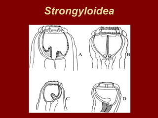
- 7. Identification Gross – Dark red worms which are easily seen against the intestinal mucosa. – The well developed buccal capsule of the adult parasite is prominent as is the bursa of the male. Microscopic – Species differentiation is based on the size and the presence and the shape of the teeth in the base of the buccal capsule.
- 8. Identification S. vulgaris 1.5-2 cm, two ear shaped rounded teeth are present S. edentatus 2.5-4.5 cm, No teeth S. equines 2.5-5 cm, three conical teeth are present. One is situated dorsally and I larger than the others.
- 10. Life cycle • The adult parasites live in the caecum and colon. • Eggs are passed in the feces and development from egg to the L3 requires about two weeks (moist and temperate). • Infection is by ingestion of L3. • Parasitic larval development of the three species differs with each other.
- 11. S. vulgaris • The L3 penetrate the intestinal mucosa and moult to L4 in the submucosa. • These then enter small arteries and migrate on the endothelium to their predilection site in the cranial mesenteric artery and its main branches. • After a period of several months the larvae moult to L5 and return to the intestinal wall via the arterial lumina.
- 12. S. vulgaris • Nodules are formed around the larvae mainly in the wall of caecum and colon, when, due to their size, they can travel no further within the arteries and subsequent rupture of these nodules releases the young adult parasites into the lumen of intestine. • The prepatent period is 6-7 months.
- 13. Strongylus equinus - life cycle • The preparasitic phase of this life cycle is virtually identical to that of Strongylus vulgaris. • Of the three large strongyles, we know the least about the parasitic phase of the life cycle of Strongylus equinus because it is less common than the others and so has been little studied. Infection is by ingestion of third stage larvae and exsheathment occurs in the small intestine.
- 14. life cycle • Exhseathed L3s invade the wall of the small intestine, cecum and colon and become encapsulated in nodules in which they molt to L4s by 12-14 days after infection. • L4s leave their nodules and cross the peritoneal cavity to the liver, the right lobe of which is in close aposition to the cecum. Most L4s will have reached the liver by 19-20 days after infection and will remain there for at least 12 weeks.
- 15. life cycle • Finally, they begin to migrate back to the large intestine by leaving the liver and crossing the abdominal cavity directly or by first passing through the pancreas ( which is closely associated with the right lobe of the liver) and then the abdominal cavity. • The final molt to immature adults (L5s) occurs about 15 weeks after infection during their migration back to the large intestine.
- 16. life cycle L5s penetrate the gut wall and enter the lumen of the large intestine via the formation of nodules. Reproduction takes place and the prepatent period is approximately 9 months.
- 17. S. edentatus- Life cycle • Freeliving stages similar to S. vulgaris • L 3 exsheaths in small intestine, penetrates gut wall to subserosa, and molts to L 4 ; remain here (usually in nodules) for 3 months • L 4 migrate to root of mesentery, some going to liver and lungs and then to wall of cecum and right ventral colon • Immature adults then appear in lumen of these portions of the large intestine • Prepatent period - 11 months
- 18. Pathogenesis Pathogenesis of migrating stages • Migrating larvae of Strongylus equinus and edentatus do not appear to cause pathological changes of enough severity to translate into recognizable clinical signs. • However, migrations of larvae through the liver may produce nodules and formation of fibrous tissue that are readily seen at necropsies of infected horses. • The early migrations of Strongylus equinus larvae into the intestinal mucosa produce hemorrhagic nodules that are easily recognized at necropsy or during abdominal surgery in horses with colic.
- 19. Pathogenesis of migrating stages • Whether or not these nodules may themselves produce colic in horses is debatable. • However, their presence certainly indicates recent infections and it is safe to assume that such horses will also be infected with other large and small strongyles and these may be primary contributors to the observed signs of colic.
- 20. Pathogenesis of migrating stages • The life cycle of Strongylus edentatus includes migration to the flanks where immature adults and L4s may be found at necropsy enclosed in subperitoneal cysts. • Although these lesions have no clinical significance, they do indicate the presence of strongyle infections and a poor parasite control program in the farm of origin.
- 21. Pathogenesis of adult worms • The pathogenic effects due to large strongyle infections in horses can be divided into those caused by adult worms in the large intestine and those caused by larvae and immature adults during their extensive migrations in other organs. • The pathogenic effects due to adults in the cecum and colon of infected horses are similar in all three species of large strongyles because they all provoke the formation of nodules in the gut wall as they pass through in completing their parasitic migrations and their methods of feeding are similar.
- 22. Pathogenesis of adult worms • All three strongylus species have large buccal cavities and are aggressive feeders. • They attach to the mucosa of the large intestine by their mouth openings and draw a plug of mucosa into the buccal cavity where it is ground up by teeth, if present, digested by secreted enzymes then drawn into the intestine by the sucking action of the muscular esophagus. • Adult worms then move to a fresh site leaving behind small bleeding ulcers that may be seen as red spots at necropsy.
- 23. Pathogenesis of adult worms • Although these worms do not appear to be actual blood suckers, damage to mucosal blood vessels during feeding may cause significant blood loss particularly if the damage extends to the level of the muscularis mucosa. • Pathophysiological studies have shown that 30ml of blood may be lost per day by infection with 75-100 Strongylus vulgaris adults. These ulcers appear to heal readily after the feeding worms move on.
- 24. Pathogenesis of adult worms However, if the damage is deep into the muscularis, as is often the case with the largest of the three strongyles (Strongylus edentatus), granulation tissue will be formed and healing will result in a circular scar at each feeding site.