
Recommended
More Related Content
What's hot
What's hot (20)
Similar to Lecture 1
Similar to Lecture 1 (20)
More from farhab dvm
More from farhab dvm (20)
Recently uploaded
Recently uploaded (20)
Lecture 1
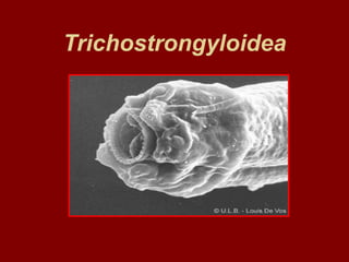
- 2. Characteristics The trichostrongyloides are small, often hair like worms which, with the exception of lung worm Dictyocaullus, parasitize the alimentary tract of animals and birds. The males have a well developed bursa and two spicules.
- 3. Characteristics The life cycle is direct and usually non migratory and the L3 is the infective stage. The trichostrongyloids are responsible for considerable mortality and widespread morbidity, especially in ruminants.
- 4. Genera The most important alimentary genera Ostertagia Haemonchus Trichostrongylus Cooperia Nematodirus Hyostrongylus Marshallgia Mecistocirrus
- 5. Ostertagia The genus is the major cause of parasitic gastritis in ruminants in all over the world. Host Ruminants Site Abomasum
- 6. Species Ostertagia ostertagi Cattle Ostertagia circumcincta Sheep and goats Ostertagia trifurcata Sheep and goats
- 7. Ostertagia ostertagi Adult worms are small (approximately 1 cm long) and brownish in color. They are difficult to see unless they are present in large numbers (thousands). All parasitic larval stages are found in the gastric glands of the abomasum.
- 8. Ostertagia ostertagi Gastric glands must be digested in order to release the larvae for observation with a microscope. Species differentiation is based on the structure of the spicules.
- 9. Life cycle • O.ostertagi has a direct life cycle. • Adults in the abomasum lay eggs that pass in feces. • Once hatched, larvae undergo two molts to become infective third-stage larvae, which migrate onto herbage and are ingested by grazing cattle. • Once ingested, these parasitic larvae grow and molt twice more to become egg-laying adults.
- 10. Life cycle • Environmental conditions of cold or excessive dryness may trigger a condition known as hypobiosis, in which larval development is arrested so that maturation may take several months. • Prepatent period is 17 to 21 days.
- 11. Effect on Host Ostertagiasis occurs in two forms • Type I disease • Type II disease.
- 12. Type 1 Disease Type I form occurs in calves during their first grazing season as a result of maturation of ingested larvae in the abomasum. This normally occurs from mid July onwards.
- 13. Type II Disease Type II disease occurs as a result of resumed development of larvae which have undergone arrested development at the early fourth stage, or hypobiosis. This usually occurs in late winter or spring and results from the maturation of larvae ingested during the previous autumn.
- 14. Effect on Host • Ingested larvae enter the glands of the abomasum, where they grow and molt to become adults (17 to 21 days), at the same time causing erosion of the cells. • Cells damaged by the larvae are replaced by rapidly dividing cells that lack the function of producing hydrochloric acid and the enzyme pepsinogen.
- 15. Effect on Host • In a normal abomasum, the pH of the contents is maintained at 2 to 2.5. When large numbers of the cells are lost, the pH of the abomasal contents may rise to 7. Consequences of this are • (a) pepsinogen is not activated to its active form, pepsin, • (b) proteins are not denatured and digested, • (c) there is an increase in the number of bacteria in the abomasum.
- 16. Effect on Host • Another important result of the cell damage is leakage of blood proteins into the gut. • Dietary energy and protein which would otherwise be used for growth must be used to replace these proteins. Weight loss is the result. • Albumin in the intestine also inhibits fluid absorption by the gut, causing diarrhea. Severe infections may cause death.
- 17. Effect on Host • The loss of albumin also causes body fluids to collect in lower parts of the body such as under the jaw (bottle jaw) or in the abdomen (ascites). • The disease condition produced by these worms is known as ostertagiasis and is characterized by severe diarrhea, edema, and weight loss leading to emaciation.
- 18. Diagnostic Information The clinical signs of inappetance, weight loss and diarrhoea The season. For example, Type I occurs from July until September and Type II from March to May Faecal egg counts. In Type I disease these can be more than 1,000 eggs per gram; in Type II counts are highly variable, may even be negative and are of limited value.
- 19. Diagnostic Information • Plasma pepsinogen levels. In clinically affected animals up to two years old these are usually raised. • Post-mortem examination • Strongyle-type eggs appear in the feces • Larvae cultured from them may be identified as Ostertagia.
- 20. TREATMENT • Type I disease responds well to treatment at the standard dosage rates with any of the benzimidazoles (albendazole, fenbendazole or oxfendazole), the probenzimidazoles, levamisole, or ivermectin. • All of these drugs are effective against developing larvae and adult stages. • Following treatment calves should be moved to pasture which has not been grazed by cattle in the same year. The field where the outbreak has originated may be grazed by sheep or rested until the following June.
- 21. TREATMENT • For the successful treatment of Type II disease it is necessary to use drugs which are effective against inhibited larvae as well as developing larvae and adult stages. • Only the benzimidazoles or ivermectin are useful in the treatment of Type II disease when used at standard dosage levels although the probenzimidazoles are also effective at higher dose rates.
- 22. Control • Avoid overstocking • Use pasture management to avoid the accumulation of infective larvae on herbage • Treat regularly with anthelmintics