
Recommended
More Related Content
What's hot
What's hot (20)
Similar to Lecture 7 triodonto, trichonema, oesophago, stephnurus
Similar to Lecture 7 triodonto, trichonema, oesophago, stephnurus (20)
More from farhab dvm
More from farhab dvm (20)
Recently uploaded
Recently uploaded (20)
Lecture 7 triodonto, trichonema, oesophago, stephnurus
- 2. Triodontophorous Members of this genus are non migratory large strongyles frequently occur in the large numbers in the colon and contributes to the deleterious effect of mixed strongyle infection. Species T. serratus T. tenuicollis T. brevicauda T. minor
- 3. Trichonema/Cyathostomes • This genus contains more than 40 species, popularly known as trichonemes, cyathostomes or small strongyles. • These parasites are present in the large intestine of horses. • Their effects on the host range from poor performance to clinical signs of severe entritis.
- 4. Trichonema Definitive Host Spectrum • 13 genera and approximately 44 species in the horse; 3 genera and 30 species in elephants Intermediate Hosts • None Geographic Distribution • Worldwide
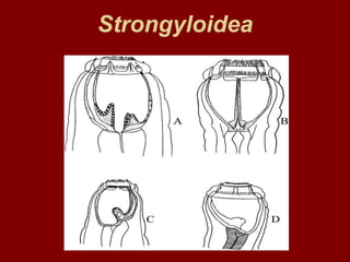
- 5. Morphology • Adults - mouth directed forward ; short, thick-walled, cylindrical buccal capsule. • Body lengths vary per species but about 1/4 to 1/2 inch long; males bursate with spicules equal. • Eggs - similar to large strongyle
- 6. Life Cycle (Stages) • Free living stages same as large strongyles • L 3 infective; exsheath in stomach, small intestine but do not migrate - only forms nodules • L 3 enter walls of cecum, colon; develop and molt; return to gut lumen as early L 5 • Prepatent period - 4 to six weeks up to 3 to 4 months
- 7. Pathogenesis Sites of Infection – Cecum, right ventral colon Pathogenesis/Clinical Signs – Infection with a large number of "arrested" cyathostomin larvae causes a clinical disease – Affected horses with persistent diarrhea, emaciation, hypoalbuminemia
- 8. Pathogenesis – Granulomatous colitis; masses of cyathostomin larvae embedded in mucosa = bright red L 4 – May be large numbers of L 4 , early L 5 in feces with watery diarrhea
- 9. Diagnosis & Treatment Diagnosis • Eggs identify family - Cyathostome eggs may be the majority found • Larval culture necessary for species identification - number of gut cells are a factor in identification Treatment • Benzimidazoles - BZ resistance shown by several spp; Moxidectin (best to date) – approved for horses Ivermectin or other avermectins • Pyrantel salt - probably some resistance
- 10. Other Control Measures • Clean bedding in stalls • Not running young foals with older, more immune horses • Rotation onto "clean" pastures • Strategic, regular treatment programs • Alternate dewormer types annually (one type each year, using the same type each time a dewormer is administered in one year) • Pasture contamination due to large number of parasites and eggs produced makes management difficult - it is practically impossible to remove all parasites from a horse's gut
- 11. Oesophagostomum spp. (NODULAR WORM) Definitive Host Spectrum • Cattle, sheep, goats, wild ruminants, pigs, primates (host specificity/parasite species) Intermediate Host • None Geographic Distribution • Worldwide
- 12. Species O. columbianum sheep and goat O. venulosum sheep and goat O. radiatum cattle and buffalo O. dentatum pig O. quadrispinulatum pig
- 13. Morphology Gross Stout white worm 1.0-2.0 cm long. Microscopically • The buccal capsule is small. • In many species, it is surrounded by leaf crowns. • Around the anterior oesophagus, there is a cuticular cephalic vesicle. This terminates in a cervical groove.
- 15. life cycle – Eggs are passed in feces, develop, L 1 hatch – L 1 develop to infective L 3 in the environment in 6-7 days – L 3 are ingested by DH – Larvae exsheath and penetrate the wall of the intestine (anywhere from pylorus to rectum) and become enclosed in nodules - over time these can become caseous and fibrotic – Larvae (now L 4 ) leave the nodules and begin maturation – Prepatent period - 30 to 50 days
- 16. Pathogenesis • Sites of Infection – Large intestine adults • Pylorus to rectum (in nodules) - larvae Pathogenesis/Clinical Signs • Cattle • Resistance develops by 2 years, so disease is seen only in young stock • Nodules may be 1-5 mm in diameter, contain caseous material and may become calcified • Clinical signs - anorexia, emaciation, profuse dark green diarrhea
- 18. Pathogenesis in sheep • Nodules do not appear unless there has been prior exposure and sensitization • Nodules consist of the larva, leucocytes and foreign body giant cells surrounded by fibroblasts • Nodules, when extensive, interfere with digestion, absorption and bowel movement
- 19. Pathogenesis in sheep • Suppurative nodules may rupture and cause peritonitis. • Adults are plug feeders and cause thickening of the bowel wall and excess mucus • Clinical signs - anorexia, weight loss, emaciation and dark green mucoid, sometimes bloody, diarrhea; perhaps death
- 20. Pathogenesis Pigs • Nodule formation causes enteritis, anorexia and bloody feces • In severe infections death is possible
- 21. Diagnosis & Treatment Diagnosis – Because most of the clinical effects are due to the nodules, necropsy may be necessary for confirmation – Eggs in feces cannot be distinguished from other strongylid- type eggs and need to be cultured to the L 3 stage to confirm Oesophagostomum Treatment – Benzimidazoles (e.g. fenbendazole); BZ-resistance reported – Levamisole (ruminants) – Morantel (cattle) – Avermectins are particularly effective: Ivermectin, doramectin Other Control Measures – Good management coupled with routine anthelmintic treatment is the best control
- 22. Stephanurus dentatus Kidney worm of pig Definitive Host Spectrum • Pigs, rarely and accidentally cattle, donkeys Intermediate Host • Earthworms may act as paratenic hosts Geographic Distribution • Worldwide, except northern Europe
- 23. Morphology A large stout worm upto 4.5 cm long, with a prominent buccal capsule and transparent cuticle through which the internal organs may be seen. The colour is usually pinkish. The size and site are diagnostic.
- 25. Life Cycle (Stages) • Eggs are passed in urine • Eggs hatch, larvae molt twice • Infection of DH may occur 4 ways • Ingestion of L 3 • Skin penetration by L 3 • Ingestion of earthworm containing L 3 • Transplacentally
- 26. Life Cycle (Stages) • Ingested larvae arrive in the liver via the portal circulation; percutaneous larvae arrive in the liver via the lungs and the systemic circulation • Migrate in liver 3 or more months - extensive migration of L4 may cause great damage in the form of abscesses and secondary bacterial infections • Larvae then penetrate through the liver capsule and migrate to the perirenal tissues, ureters, etc. • Prepatent period - 6 to 16 months
- 27. Pathogenesis/Clinical Signs Sites of Infection Kidneys and perirenal tissues Pathogenesis Most pathology is caused by the larvae • Skin penetration causes the formation of nodules and enlargement of superficial lymph nodes • Liver migration causes fibrosis, abscess formation, cirrhosis, adhesions • Economic loss from liver condemnation Adults may cause thickening of the ureters and cystitis Clinical signs - decreased weight gain, weight loss, stiffness of the hind legs
- 28. Diagnosis & Treatment Diagnosis • Eggs in urine - highest concentration of eggs in the last part of the first micturition in the morning • Necropsy Treatment • Levamisole - kills adults only • Ivermectin & fenbendazole - kill adults and immature stages