170male reproductive systemmale reproductive system
xtestis is covered by three layers (from
outside to inwards):
̞visceral layer of tunica vaginalis:
̎it is lined by flat mesothelial cells.ons of seminiferous tubules lined by spermatogonia, primary and secondary
xThese tight junctions form the blood–testis
barrier.
xThe tight junction divides the intercellular
compartment between the Sertoli cells
into basal and luminal compartment.
xBasal compartment contains spermato
gonia and primary spermatocytes.
xLuminal compartment contains secondary
spermatocytes and spermatids (Fig. 19.5).
Functions of Sertoli Cells
xSertoli cells provide support and nutrition
to spermatogenic cells.
xThe bloodtestis barrier protects the
spermatogenic cells from the harmful
substances (antigens) of blood.
xThey phagocytose the residual bodies.
xSertoli cells secrete androgen-binding
protein (ABP), which concentrates the
testosterone.
xIn fetal testis, Sertoli cells produce anti
mullerian hormone, which inhibits the
development of mullerian duct.
xSertoli cells are nondividing cells, highly
resistant to infection, malnutrition, and
radiation.
xThese produce inhibin, which inhibits the
secretion of follicle-stimulating hormone
(FSH).
Interstitial Cells of Leydig
xThese are large polyhedral cells lying in
the connective tissue between seminif
erous tubules.
xThese are pale staining cells with eccen
tric nucleus and cytoplasm shows unique
needleshaped crystalline inclusion
(Reinke’s crystal).
spermatocytes, spermatids, and sperms are seen.
2.Sertoli cells are seen in between the spermatogenic cells.
3.Interstitial ces of Leydig are seen in between the seminiferous tubules.
xThey secrete testoster
̞tunica albuginea:
̎it is a thin layer of connective tissue
containing collagen, blood vessels,
and lymphatics.
̎along the posterior border, tunica
albuginea is thickened to form medi
astinum testis.
̎septa arising from the mediastinum
testis divide the substance of the
testis into 200 to 300 lobules.
̎each lobule contains one to four
seminiferous tubules.
̎seminiferous tubules contain coiled
part in the front and straight part
behind.
̎straight part enters the medi
astinum testis where it joins and
forms a network called as rete testis.
̎from the upper end of rete testis
12 to 14 efferent ductules arise and
enter the epididymis.
̞tunica vasculosa:
̎highly vascularized connective
tissue which covers the individual
lobule.
microscopic structure
oftestis
seminiferous tubule
xthere are 400 to 600 seminiferous tubules
in each testis.
xeach tubule is surrounded by a basal
lamina supported by connective tissue
which contains muscle-like myoid cells.
xcontraction of myoid cells helps to move
the spermatozoa along the tubule.
xeach seminiferous tubule is lined by
stratified seminiferous epithelium which
contains spermatogenic cells and sertoli
cells(figs. 19.2and19.3).
fig. 19.2diagram of testis (h&e pencil). h&e, hematoxylin and eosin. 3.Interstitia
Berhampur CALL GIRL❤7091819311❤CALL GIRLS IN ESCORT SERVICE WE ARE PROVIDING
Lecture 2 MICROSCOPY.pptx
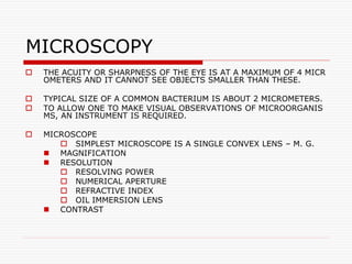
1. MICROSCOPY
THE ACUITY OR SHARPNESS OF THE EYE IS AT A MAXIMUM OF 4 MICR
OMETERS AND IT CANNOT SEE OBJECTS SMALLER THAN THESE.
TYPICAL SIZE OF A COMMON BACTERIUM IS ABOUT 2 MICROMETERS.
TO ALLOW ONE TO MAKE VISUAL OBSERVATIONS OF MICROORGANIS
MS, AN INSTRUMENT IS REQUIRED.
MICROSCOPE
SIMPLEST MICROSCOPE IS A SINGLE CONVEX LENS – M. G.
MAGNIFICATION
RESOLUTION
RESOLVING POWER
NUMERICAL APERTURE
REFRACTIVE INDEX
OIL IMMERSION LENS
CONTRAST
2. Types of microscopes
Light Microscopy
The light microscope uses visible light to detect s
mall objects
The following challenges are often encountered
when using a light microscope
obtaining sufficient contrast
finding the focal plane
obtaining good resolution
recognizing the specimen when one sees it
3. Bright Field Microscopy
Light from an incandescent source is aimed toward a lens b
eneath the stage called the condenser
This passes through the specimen, through an objective len
s, and to the eye through a second magnifying lens, the ocu
lar or eyepiece
Objects are seen in the light path because natural pigmenta
tion or stains absorb light differentially, or because they are
thick enough to absorb a significant amount of light despite
being colorless
4. A Paramecium should show up fairly well in a bright field micros
cope
However, it will not be easy to see cilia or most organelles
The condenser is used to focus light on the specimen th
rough an opening in the stage
After passing through the specimen, the light is display
ed to the eye with an apparent field that is much larger
than the area illuminated
The magnification of the image is simply the objective l
ens magnification times the ocular magnification
5. Some condensers are fixed in position, others are focusable, so
that the quality of light can be adjusted
Usually the best position for a focusable condenser is as close t
o the stage as possible
The bright field condenser usually contains an aperture diaphra
gm
This is a device that controls the diameter of the light beam co
ming up through the condenser
When the diaphragm is stopped down (nearly closed) the light
comes straight up through the center of the condenser lens an
d contrast is high
6. When the diaphragm is wide open the im
age is brighter and contrast is low
A disadvantage of having to rely solely o
n an aperture diaphragm for contrast is t
hat beyond an optimum point the more c
ontrast you produce the more you distort
the image
With a small, unstained, unpigmented sp
7. Brightfield Microscope
Study size, shape & arrangement of microbia
l cells, little information about internal
cell structure.
Tips:
Molds & large protozoa-10x, 40x
Bacteria,yeasts,small protozoa-100x oil
immersion objective
Increase amount of light when using th
e 100x oil immersion objective lens
8. BRIGHTFIELD MICROSCOPE
Allows light rays to pass directl
y through the eye without bein
g deflected by intervening opa
que plate in the condenser.
9.
10. Dark Field Viewing
Dark-field Microscope (=Ultra-microscope)
It was invented by Zsigmondy (1905)
A special condenser lens is used with an opaque disc at the
centre, so that direct rays don’t enter the objective lens
Only light scattered by the specimen enter the objective len
s to form a bright image against dark background
Dark field microscope does not have a good resolution
It is commonly used in microbiology
11.
12. Darkfield Microscope
Designed to eliminate the need
for staining to achieve contrast
between the specimen and the
background.
Condenser lens- focuses light o
n the specimen at an oblique a
ngle.
Microorganisms appear very br
ight on a dark background
13.
14. Fluorescence Microscopy
Allows the detection of molecules and ions within cells
Fluorescent dyes absorb short wavelengths of light and emit longe
r wavelengths
Barrier filters and a dichroic prism select the excitation wavelengt
h that strikes the specimen and exclude the excitation wavelength
from the detector
This allows only emitted light to reach the detector (oculars)
uses uv light source = mercury or xenon arc lamp.
- high contrast, high resolution image
15. special fluorescent dyes used to locate “mole
cules” in a specimen
- black background, bright-stained specimen
- no condenser required, light comes from ab
ove (“epi”) specimen
- multiple fluorescent probes available
- detects small quantities, molecules; can use
antibody staining techniques
16. Fluorescence microscope
Specimen is illuminated at one wa
velength of light and observed by a
light emittted at a different wavele
ngth
Fluorexcein isothiocyanate
Excitation wavelength and emissio
n wavelength
Excitation filter and barrier filter
17.
18.
19.
20. Fixed (Preserved) Specimens for Hist
ology--the Study of Tissue
1) Specimens may be preserved using chemicals suc
h as formalin, acetic acid, ethanol, and methanol
Fixation immobilizes molecules such as proteins and
lipids
2) Fixed specimens are dehydrated by serial transfer
through an ascending alcohol series, to 100% alcoho
l
3) Specimens are infiltrated with melted paraffin, pa
raffin substitute, or plastic and placed in a mold to h
arden
21. 4) Specimens are cut into 5-10 um thick sect
ions using a steel knife or razor on an instru
ment called a microtome
5) Sections are then mounted on slides,
6) Stained to achieve contrast or identify cell
structures or components, and
7) Viewed microscopically
22.
23. Many variations in technique are used to pre
pare specimens for light microscopy
Some omit the dehydration, infiltration, emb
edding and sectioning steps and use aqueous
staining systems for viewing whole mounts (
unsectioned tissues or cells)
Freezing may be used instead of chemicals to
fix tissues that need to be examined quickly
or that have components damaged by the ch
emicals
Examples are frozen biopsies and tissues in
which heat-labile structures are to be stained
24. Frozen specimens are sectioned using a cryot
ome, a microtome encased in a freezing cha
mber
Permanent slides may be made from paraffin
sections but not from frozen sections
25. Transmission
Electron Microscopes
TEM makes high-resolution (
± 1 nm) views of the inner si
de of objects. Mostly TEM is
applied on material (e.g. cell
s) that has been previously '
stained' and cut into ultrathi
n sections, but sometimes al
so intact objects < 1 µm, like
viruses and aggregates of m
acromolecules, are visualize
d.
26. Principle
Fixed, dehydrated specimens are embedded in a resin, hardened,
sectioned, stained with heavy metals such as uranium and lead, a
nd inserted into the electron column in the microscope
The electron beam is absorbed or deflected by the heavy metal st
ains and shadows are cast onto film or a phosphorescent plate at
the bottom of the column
- 2-D image
- reveals internal cell structure
- high resolution, high magnification
- electron beam is focused by magnetic field
28. Scanning Electron Microscope
In SEM the image primary el
ectrons from the source bo
mbard the sample according
to a scanning pattern and ca
use emission of secondary e
lectrons. In SEM an image of
the surface of the object is
made.
29. Principle
Fixed, dehydrated specimens are mounted on stubs and surface-coated with
gold, palladium or rhodium
The specimen is placed in a vacuum and an electron beam scans back and fo
rth over it
Electrons that bounce off the metal-coated specimen surface are collected, c
onverted to a digital image and displayed on a TV-like monitor
- Electron beam is focused using a magnetic field - SEM provides a 3-D ima
ge
-Gives information about external topography of specimen
-Much higher resolution and magnification than possible in light microscope
30.
31. Micrograph
A. Example of application of Transmission Electron Microscopy (TEM)
Organelles in a pollen grain of tobacco (Nicotiana tabacum; AF = Actin filaments; G
= Golgi apparatus; Mi = Mitochondrion; Mt = Microtubule).
B. Example of application of Scanning Electron Microscopy (SEM)
Overview of gills of a fish, the mudskipper (Periophthalmus argentilineatus).
32. Specimen Preparation
Light Microscopy
Live Mounts
To view organisms, tissues, or cells in as close to the natural st
ate as possible, unstained live Viewing time of live mounts is limi
ted
Unstained specimens have low contrast
Supravital stains may be applied to provide more contrast or id
entify certain components--these are stains that are not harmful to
living cells
mounts are used
33. Fixed (Preserved) Specimens for Hist
ology--the Study of Tissue
1) Specimens may be preserved using chemicals such as f
ormalin, acetic acid, ethanol, and methanol
Fixation immobilizes molecules such as proteins and lipid
s
2) Fixed specimens are dehydrated by serial transfer throu
gh an ascending alcohol series, to 100% alcohol
3) Specimens are infiltrated with melted paraffin, paraffin
substitute, or plastic and placed in a mold to harden
34. 4) Specimens are cut into 5-10 um thick sections usi
ng a steel knife or razor on an instrument called a mi
crotome
5) Sections are then mounted on slides,
6) Stained to achieve contrast or identify cell structur
es or components, and
7) Viewed microscopically
35.
36. Preparing Specimens for Scanning Electron
Microscopy (SEM)
1) Fixation--fixatives used are glutaraldehyde, paraformaldehy
de, osmium tetroxide
2) Dehydration is accomplished by carrying the specimens thro
ugh an ascending alcohol series, to 100% alcohol (i.e., no water
), then to an organic solvent such as acetone or propylene oxide
Specimens for SEM may also be processed in a critical point dr
ying apparatus
3) Specimens are mounted on aluminum stubs using sticky tape
37. 4) A sputter coater coats the specimen with gold, pal
ladium or rhodium in a special chamber to cover the sp
ecimen with a 10-20 nm thick metal layer
38. 5) The stub is inserted into the SEM, scanned and obser
ved on a video display
39. Preparing Specimens for Transmission El
ectron Microscopy (TEM)
1) Fixation--specimens are fixed in glutaraldehyde, o
r paraformaldehyde-glutaraldehyde mixtures, followe
d by osmium tetroxide
2) Dehydration is accomplished by carrying the speci
mens through an ascending alcohol series, to 100%
alcohol (i.e., no water), then to an organic solvent su
ch as acetone or propylene oxide
3) Specimens are then infiltrated with an epoxy or pl
astic resin and placed in plastic molds to harden
40.
41. 4) An instrument called an ultramicrotome is used to section the
specimen
42. Glass or diamond knives are used to cut the ultrathi
n sections
43. 5) Sections are transferred to tiny metal grids for support (the equivalent of th
e function of the glass slide in LM)
6) Heavy metal stains such as uranyl acetate and lead citra
te are applied to make certain structures electron dense
44. 7) Grids are then inserted into the transmission electron m
icroscope and observed