Pediatrics 5th year, 9th, 10th & 11th lectures (Dr. Jamal)
•Download as DOC, PDF•
2 likes•1,393 views

The lecture has been given on Dec. 12th & 23rd, 2010 - Feb. 6th, 2011 by Dr. Jamal.

Recommended
More Related Content
What's hot
What's hot (20)
Viewers also liked
Similar to Pediatrics 5th year, 9th, 10th & 11th lectures (Dr. Jamal)
Similar to Pediatrics 5th year, 9th, 10th & 11th lectures (Dr. Jamal) (20)
More from College of Medicine, Sulaymaniyah
More from College of Medicine, Sulaymaniyah (20)
Recently uploaded
Recently uploaded (20)
Pediatrics 5th year, 9th, 10th & 11th lectures (Dr. Jamal)
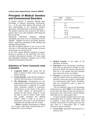
- 1. Lecture notes, prepared by Dr. Jamal A. RASHID Principles of Medical Genetics Patau Denver and Chromosomal Disorders classificatio classification At present, practice of medicine depends upon n knowledge of anatomy, physiology, biochemistry, A 1-3 etc…,. In the near future dealing with diseases will B 4-6 need understanding of the molecular anatomy, C 7-12 + X chromosome physiology, and biochemistry of the human genome. The greatest explosion had occurred in the field of D 13-15 genetics than in any other paediatric field during the E 16-18 previous decade. F 19-20 Genetically determined disorders, although G 21-22 + Y chromosome individually rare (some occur at a frequency of one in 200,000 – 300,000), the sum of all genetic disorders makes a significant contribution to the morbidity and mortality among children. The field of medical genetics is new. It was for the first time in 1956 when the normal number of human chromosomes was found to be 46. In the USA, among different categories of genetic diseases in neonates, it was found that 0.5% had a non-lethal chromosomal disorder, 0.4% had single gene Mendelian inheritance conditions and 1% had mutlifactorial (polygenic) disorders. • Medical Genetics: is the study of the inheritance of disease. Definitions of Terms Commonly Used • Inheritance: means the passage of hereditary in Genetics traits from one generation to another. It is the process by which, one acquires characteristics • Congenital Defect: only means that the from his or her parents and then transmits defect is present at birth, it does not suggest these traits to his or her own children. genetic aetiology. • The gene: it is the basic unit of heredity; it is • Genetic disorder: means that the condition is made up of DNA. Genes are ultramicroscopic caused by abnormal genes or chromosomes. structures. Each gene occupies a certain place • The chromosome: chromosomes are rod- on a chromosome called “locus”. Hereditary shaped bodies situated inside the cell nucleus. traits are controlled by pairs of genes on Each chromosome is composed of 2 strands chromosome pairs. called chromatids attached at centromere. The two alternative genes on the homlogous Chromosomes are the bearer of the genes. chromosome are called “allele”. If the alleles code The normal number of chromosomes is for the same trait, these are said to be present in a constant for each species, being 46 in man homozygous state, while if they code for different (23 pairs in each of somatic “body” cells). forms they are in a “heterozygous state”. This number constitutes the “Diploid If a gene clinically manifests itself even in the number” which is made of 22 pairs of heterozygous state, it is called a “Dominant” gene “autosomes” and a pair of sex chromosomes or character. A gene that cannot express itself designated XX in females and XY in males. clinically when present in heterozygous state, but Denver classified chromosomes in human only when in a homozygous state is called into 22 pairs, while Patau classified them into 7 “Recessive” gene or character. groups from A to G as follows: • Carrier State: individuals heterozygous for a recessive trait. 1
- 2. • Mutation: means a permanent heritable Gamete (ovum or sperm) Formation change in a gene, that causes it to have a Each somatic (body) cell in human contains 46 different effect from that it had previously, chromosomes, 23 have come from each parent. Cell this changed gene can be passed to the division in the somatic cells is mitosis (non-reduction following generations. Mutation may occur division) and each daughter cell contains 46 spontaneously or be induced by mutagens as chromosomes as the mother (original) cell. various medicines or chemicals, e.g., mustard While in the germ cells of both sexes, the cell division gas, ionizing radiation (X-ray or UVL). is of reduction (meiosis) variety, the resulting cell • Deletion: means loss of part of a (ovum or sperm) will contain only 23 chromosomes, chromosome, mostly through breakage. i.e., the number of chromosomes is halved. • Karyotype: It is the description of the The female gamete (ovum) has 22 autosomes and one chromosomes of an individual. Thus the X-chromosome, while the male gamete (sperm) can normal human male karyotype is 22 XY, have 2 types of chromosomal pattern: 22+X or 22+ Y. while the normal human female karyotype is Fertilization of the ovum by an X-bearing sperm will 22XX. result in a female offspring, while if fertilized by a Y- • Mitosis: the type of cell division which takes bearing sperm a male offspring will result. Thus each place in the somatic cells and in which each offspring (boy or girl) inherits half their chromosomes of the two daughter cells carries full from their father and the other half from their mother. compliment of 46 chromosomes as their That is why they have some characteristics of both. mother cells. • Meiosis: the reduction division which occurs Genetics and Disease in the germ cells during the process of Human diseases in general may have a purely genetic gametogenesis. This division in human cause without any role of the environment in their reduces the number of chromosomes to 23 in causation. On the other hand some conditions are the ova or sperm. caused by pure environmental factors without any role • Lyon Hypothesis: says that one of the two X of genetic predisposition as most infections and chromosomes in human female is mainly or accidents. completely inactivated early in embryonic life In-between these 2 extremes there are many childhood (before 16th day of gestation). This process diseases, causation of which requires both genetic and takes place randomly, resulting in 2 environmental factors (polygenic or multifactorial). populations of somatic cells, one population Another group of diseases, which is relatively more with the maternal and the other with the significant in young children than in adults, is the paternal X-chromosome inactivated. But once group of chromosomal disorders which are generally decision is made, all the descendants of that linked to the genetically determined diseases. cell will have the same X-chromosome inactivated. • Mosaic: a karyotype state in which two or Chromosomal Disorders more cell lines are present in the same Although rare in clinical practice a large proportion of individual. aborted fetuses are found to have chromosomal • Phenotype: is the physical appearance or disorders. The younger the GA of the aborted foetus make up of an individual. the higher the percentage of chromosomal • Alleles: are alternative forms of a gene at a abnormalities, thus the incidence decreases from 50% given locus. during the first 2 months to less than 5% by 7 months • Sex chromatid “Barr Body”: it is a darkly of gestation and 5% of stillborns have a chromosomal stained body seen near the nuclear margin in anomaly. a high percentage of the cells of normal It has been found that 65% of the fetuses with Down female. The number of Barr bodies seen in a syndrome and 95% of those with Turner syndrome are cell is equal to the number of X- spontaneously aborted. chromosomes minus one, i.e., in a normal Chromosomes contain a large number of genes. Loss female there is one Barr body in each somatic or gain of a whole chromosome due to abnormalities cell, and there is no Barr body in male cells. in cell division may cause so great disturbances in the genetic constitution of the foetus that it may result in 2
- 3. abortion, stillbirth, death soon after birth, or survival 5. Translocation: one chromosome or a segment with some malformations, mental retardation or of it may translocate itself and join another infertility. chromosome. Thus one chromosome will be Of all live born babies 0.5% has a chromosomal absent or shorter, while another chromosome anomaly. appears longer. If no loss or gain of the Chromosomal abnormalities are in general sporadic genetic material occurs in this process the and therefore the risk of recurrence in the offspring is translocation is called balanced and the person low. is phenotypically normal. The translocated chromosome may be transmitted to either gamete during meiosis, and when this gamete Mechanisms of Chromosomal (with translocated chromosome) mates a Anomalies normal gamete, the resulting zygote may Chromosomal disorders may be numerical or either have excess or deficiency of the genetic structural and generally arise by some of the material. Such an offspring will be abnormal following mechanisms: in both karyotype and phenotype. 1. Inversion: a break may occur along the 6. Deletion: break-off and loss of a fragment of length of the chromosome arm. The broken a chromosome. If a large portion of a pieces may rearrange themselves in a new chromosome is lost, it would be lethal or way. If there is neither loss nor gain of causes a grave disability. genetic material, there may be no significant clinical manifestations. 2. Isochromosome: Normally chromosomes Classification of Chromosomal divide longitudinally during mitosis. Rarely a Abnormalities transverse division may occur through the I. Numerical Abnormalities: centromere, thus instead of making 2 normal a. Excess of one or more autosome (autosomal chromosomes, two new types of trisomy): this may occur by non-disjunction chromosomes are formed, one having both or by translocation mechanism. Examples of the long arms and the other both the short autosomal trisomies include: arms, these are called isochromosomes. i. Trisomy 21 (Down syndrome) Features may occur as there will be some ii. Trisomy 18 (Edward syndrome) excess and some deficiency of genetic iii. Trisomy 13 (Patau syndrome) materials. b. Autosomal monosomy: means absence of one 3. Nondisjunction: during meiosis both of the autosomal chromosomes, this members of a pair of chromosomes may fail abnormality is incompatible with life. to separate and go jointly to either of the c. Numerical sex chromosome anomalies: daughter cells. Thus one of the daughter cells i. Extra-chromosomes: will have 22, and the other 24 chromosomes. 1. XXY (Klinefelter syndrome) Then after fertilization by a normal gamete 2. XYY (having 23 chromosomes) the resultant 3. XXX zygote will either have 47 or 45 4. XXXY chromosomes. 5. XXXXY 4. Mosaicism: this state happens if the process ii. Deficient chromosome: XO (Turner of non-disjunction occurs in the first mitotic syndrome) instead of the meiotic division, resulting in 2 d. Triploidy: means presence of 3 haploid sets of cells, one with 47 and other with 45 chromosomes in one cell, i.e., presence of 69 chromosomes. As each one of these cells will chromosomes. This condition is not reproduce similar cells by further normal compatible with life. mitosis, 2 cell lines will be observed, a line e. Tetraploidy means presence of 4 haploid sets with 45 and another with 47 chromosomes. of chromosomes in one cell, i.e., 92 There may be more than 2 cell lines, one with chromosomes, which is again incompatible normal and the others with abnormal with life. chromosomes complements. f. Mosaicism II. Structural Abnormalities 3
- 4. a. Translocation (balanced structural change) f. Protruding tongue from a relatively b. Deletion (unbalanced structural changes) small mouth, furrowed tongue, small teeth g. Prominent malformed ears Features of Some Chromosomal h. Small nose and flat nasal bridge Disorders i. Broad short neck Down Syndrome “Mongolism”, “Trisomy G”, 4. Hands and Feet: short broad hands, single palmer crease (Simian crease) in 85%, “Trisomy 21” clinodactyly (short incurved little finger due It is the first autosomal trisomy described in man by to hypoplasia of its middle phalanx)& a gap John Down in 1966. The name mongolism is between the first and second toe. sometimes used as the patients look like those of 5. Greater liability to have: Mongolian race. a. Associated CHD, most commonly the 21 and 22 trisomies can not clinically be AV canal differentiated. b. Congenital intestinal obstruction The incidence of Down syndrome is 1 in 700 in most especially duodenal stenosis, or parts of the world. Hirschsprung disease Maternal age at pregnancy strongly contributes to the c. Leukaemia incidence, thus: d. Frequent respiratory infection 6. Special non-aggressive forms of moderate to Maternal Age Incidence of Down severe mental retardation syndrome 20 1 in 2000 They are characteristically friendly individuals who 30 1 in 1000 show an unusual enjoyment of music. Babies with 35 1 in 350 Down syndrome look all alike. Mosaic cases have less 40 1 in 100 severe features. 95% result from non-disjunction, 3% 45 1 in 40 from translocation, and 2% from mosaicism. None- >45 1 in 35 mosaic cases can be clinically diagnosed at birth from the appearance and the striking hypotonia. This increase of incidence in elderly mothers is attributed to the exposure of the maternal oocyte to Management the harmful environmental factors for a longer period Parents should be told about the illness on the second of time, since Graffian follicles are present in the or third day after birth, or the husband is told so that foetal life and remain throughout the reproductive life he will then later on explain the condition to his wife. of the woman. On the other hand the sperm of man The fact that there is no curative treatment for Down has a short life span and has therefore lesser chances syndrome must be explained to the parents. Children for exposure to harmful influences. with Down syndrome have a shorter life span than the The age of the father has lesser effect on the average in that community, even in the absence of incidence of Down syndrome. associated severe congenital anomalies. Genetic counseling may be helpful if one of the Clinical Features parents is found to have translocation of chromosome 1. General: hypotonia during infancy and mental 21. retardation throughout life are constant features Edward Syndrome (Trisomy 18 or “E” 2. Short stature Trisomy) 3. Craniofacies: Much less common than Down syndrome; it occurs at a. Brachycephaly a frequency of 1: 6000 births, being more common in b. Upward and outward slanting females. 90% are due to non-disjunction. As in Down palpebral fissures syndrome, maternal age seems to be important. c. Prominent epicanthic folds Clinical characters include: d. Strabismus e. Speckled irides (Brushfield spots) - Prominent occiput - CHD 4
- 5. - Receding chin and triangular face A variant of cri-du-chat is caused by deletion of the - Short sternum and single umbilical artery short arm of chromosome number 4, differentiated - Flexion deformities of the fingers, which are from cri-du-chat by absence of the characteristic cry rigidly fixed across the palm. The middle and by common presence of midline fusion defects finger overriding the index. (scalp, nose, lips, palate, and in males the penis). - Characteristic “rocker-bottom” feet, the sole is convex and the heel is prominent, Turner Syndrome (Ovarian Dysgenesis) simulating a rocking chair Occurs at a frequency of 1 in 2500 births and their chromosomal pattern is XO, i.e., they have 45 The majority of affected babies die during the first 3 chromosomes and are chromatin negative females. months of life. Most of the XO fetuses are aborted during the first trimester. The cause is probably non-disjunction in Patau Syndrome (Trisomy 13-15 or D spermatogenesis than in oogenesis. Trisomy) At birth diagnosis is suspected from lymphoedema of - It occurs at a frequency of 1 in 10,000 births. the feet and hands, and lax neck skin. Other features Characterized by gross deformities of the later in life include: hand and face. - Short stature - There is microcephaly with a relatively large - Shield-like chest, widely spaced nipples “onion-nose’ receding jaw, cleft lip and - Neck webbing and low posterior hairline palate are common. - Cubitus valgus (increased carrying angle of - The eyes may be absent, small or of normal the arms) size but with coloboma or cataract. The finger - Short fourth metacarpals and metatarsals nails are narrow and hyperconvex. - Mild mental retardation - Capillary haemangioma - The nails are small, narrow, and deeply set - CHD is common - Associated congenital anomalies especially - Flexion deformities of the fingers coarctation of the aorta and horse-shoe - There may be associated agenesis of corpus kidneys. callosum - The ovaries are either absent or severely - The majority of affected babies die during hypoplastic early infancy - Sexual infantilism becomes evidence at the time of expected puberty, thus there will be: 75% of cases result from non-disjunction, 20% from o Primary amenorrhoea and infertility translocation, and 5% from mosaicism. The risk for o Underdeveloped breasts, mostly recurrence in other children is only high in the consisting of fat presence of parental translocation. o Appearance of pubic and axillary hair, since these result from adrenal Cri-du-chat Syndrome androgens (these are absent in An extremely rare disorder caused by deletion of the hypopituitarism, since there is also short arm of chromosome 5. Clinically cri-du-chat lack of adrenal androgens) syndrome is characterized by: o Infantile external genitalia - Severe mental, motor, and growth retardation o Severely hypoplastic uterus - Microcephaly with broad head o Raised urinary gonadotrophins (FSH) - Prominent epicanthic folds, ocular hypertelorism and antimongoloid slant Patients with Turner syndrome, who are heterozygous (Downward and outward sloping palpebral for XLR disorders, may show the disease. fissures) - Broad face, saddle nose and micrognathia Treatment - Low set, malformed and rotated ears with No hormonal treatment is indicated in childhood, but accessory auricles at the usual age of puberty substitution therapy with - Simian crease in 50% oestrogen (daily orally for 6 months or until - Characteristic cry similar to mewing cat or menstruation occurs, subsequently, cyclic oestrogen- kittle progesterone therapy is administered). 5
- 6. Fragile X Syndrome Klinefelter Syndrome “XXY Syndrome” This is a familial (XLR) form of chromosomal It occurs at a frequency of 1 in 500 male births. 80% disorder and is considered to be the second most have XXY, 10% mosaic, and 10% XXYY or XXXY common cause of mental retardation after Down karyotype, so they are chromatic +ve males. syndrome. One third of female carriers may show mild Clinical features affection. On the other hand between 20-50% of boys who have the fragile X are asymptomatic and transmit They look like normal boy’s early in life and the the chromosome to all of their daughters, who are also condition is seldom diagnosed before adolescence, asymptomatic. However in the subsequent generation because the external genitalia are normal. The only both male and female offspring of those daughters clues to the diagnosis before puberty are unusually begin to show the illness. long legs, behavioral problems, and when investigated The following are some of the reported features: they are found to be chromatic positive. After - Mental retardation pubertal age there will be: - Macrocephaly - A long boy with sparse body (including - Prognathia (prominent jaw) axillary and pubic) hair, long limbs and knock - Short stature knees - Large protruding ears - Behavioral disorders: social inadequacy and - Macro-orchidism, especially after puberty immature personality. The degree of - Rapid and repetetive speech intellectual deficit is directly proportional to - Generally they are pleasant and socially the number of the extra X chromosomes engaging, although some of them avoid eye - Microorchidism (small sized testes) contact and simulate autism occasionally with cryptorchidism - Decreased androgen level in the blood, They have a normal life span. No curative treatment is resulting in temporal recession of the hair available, although it has been found that folic acid - Although they are capable of erection, may improve the psychological state. Prenatal performance of intercourse, and ejaculation of diagnosis is possible. sperm fluid, they are sterile - Raised urinary excretion of FSH - Gynaecomastia, broad pelvis, horizontal limit Single Gene Inheritance of pubic hair, and feminine voice The Mendelian laws of inheritance are applied to all - Commonly they suffer chronic pulmonary living creatures including human beings. conditions as asthma, emphysema, and All of the 46 human chromosomes which are present bronchiectasis in every single cell of the body carry genes. The autosomes arrange in pairs according to their shape, Patients with Klinefelter syndrome have normal and the members contain the same gene loci and are survival and there is no increased risk to have other known as homologous chromosomes. affected children. Every autosomal gene locus occurs twice in every cell of the body. If both loci possess the same genetic Treatment information, the individual is considered homozygous Replacement of testosterone since 11-12 years of age. (i.e., identical), while if the gene loci carry different 50mg every 3 weeks, slowly increased until a information (e.g., one normal gene with another maintenance of 250 mg/3 weeks is reached. abnormal one) the individual is considered heterozygous. Therefore, every individual can be XYY Syndrome considered as one of the following: This occurs at a frequency of 1-2/1000 boys. They are 1. Homozygous for the normal gene reproductively functional and more or less normal in 2. Homozygous for the abnormal gene outlook, but they are more criminal in behavior and 3. Heterozygous, i.e., he carries both normal and this is involved in the crimes against property. They abnormal gene on the homologous generally do not commit any crimes against their chromosome siblings, and they do not respond to the corrective During gamete formation, every gamete receives one measures taken against their crimes. chromosome from each pair and consequently carrys half of the “diploid” number of chromosomes. The 6
- 7. distribution of the genes to the gametes and the mating process by the sperm follows the laws of probability. The four known forms of Mendelian single gene inheritance are: Autosomal dominant Autosomal recessive. X-Linked recessive. X-Linked dominant. Autosomal Dominant In which a single gene is quite sufficient to make the trait or the disease to express itself clinically. It is usually not possible to differentiate between hetero and homozygous cases clinically. The most common situation is when one parent is affected; there will be affection of 50% of their children, regardless of their sex. Homozygous cases of AD inherited disorders may rarely be so severely affected that they die early in life, or even prenatally. In many families, AD conditions arise as a result of new mutation, followed by its inheritance to the following generations. Another explanation, apart from new mutation, to the absence of the disease in the parents is that AD conditions may skip a generation to appear again in another generation. Expression of AD conditions may to various degrees be sex influenced or sex limited. The degree of expression of these conditions are extremely variable even in the members of the same family. AD dominant conditions are generally less severe than the AR ones and the pedigrees of the former are vertical while in AR they are horizontal. Some AD conditions are characterized by delayed age of onset. The phenomena of being phenotypically normal in spite of carrying the defective AD trait gene and passing it on, is known as “incomplete penetrance”. Children who results from two heterozygous parents Examples of AD genetic diseases in human: run a 75% risk of inheriting the anomaly within which 1. Achondroplasia figure 25% will be homozygous. 2. Dubin-Johnson syndrome 3. Congenital spherocytosis 4. Marfan syndrome 5. Neurofibromatosis 6. Tuberous sclerosis 7. A and B blood groups and Rh 7
- 8. Autosomal Recessive Up to date now there are 950 AR diseases. AR disorders manifest only in homozygous state, i.e., in the presence of 2 defective genes on the homlogous autosomal chromosomes. Most of the IEM are inherited by AR mode, and the commonest situation is when both parents are clinically normal but heterozygous for the abnormal gene (carriers), such mating will result in: - ¼ of their offspring are affected (homozygous for the gene) - ¼ are normal (do not carry any abnormal gene) - ½ are clinically normal like their parent (heterozygous carriers) Examples of AR disorders 1. Albinism For obvious reasons, AR disorders are more common 2. Phenylketoneurea in consanguineous marriages, as the chance for the 3. Galactosaemia relatives of a heterozygous carrier individual to be 4. Thalassaemia similarly heterozygous is much higher in non- 5. Cystic fibrosis relatives. 6. Crigler-Najjar syndrome If one parent is homozygous (affected) for an AR 7. Wilson disease disorder and the other parent is homozygous normal 8. Werdnig-Hoffmann syndrome (free of abnormal gene), then all of their children will 9. O blood group be heterozygous (carriers), and thus clinically normal. AR as AD disorders affect both sexes equally and the The heterozygous states of some AR former is generally more severe, appears earlier in life disorders can be detected as in: and its pedigree is horizontal. 1. Beta thalassaemia: high HbA2 Careful examination of the heterozygous states may 2. Calactossaemia: low galactose-1-PUT show some variation from the normal homozygote in 3. Glycogen storage disease: high glycogen some AR conditions. 4. Cystinosis; high cystine 5. PKU: high s. phenylalanine X-Linked Disorders The genotypic difference between man and woman is that the two sex chromosomes of male are X and Y, and those of female are two X chromosomes. Practically there is no such ting as Y chromosomal mode of inheritance, with few exceptions which are so far unimportant. Therefore, sex chromosome-linked modes are generally termed X-linked, of which X-linked recessive is the one with the greatest practical importance. Up till now about 170 XL-disorders are known. XLR XLR disorders nearly always are confined to male individuals as the mutant recessive gene on the only X chromosome in males is not suppressed by a normal 8
- 9. allele. While in females, these disorders are not manifest clinically as the mutant gene is suppressed by a normal allele and thus these heterozygous females act as carriers of mutant gene, half of their male children will inherit the mutant gene and express the condition clinically, while half of heir female children (who inherit the mutant gene) become carriers like their mothers. There is no father-to-sun transmission in both XL recessive and dominant forms of disorders, as the only X chromosome of the male (father) goes to the daughters and not the sun, who receives the Y from his father. All of the daughters of an affected father obligatorily are carriers. 3. Mother is homozygous affected (extremely X-linked disorders are similar to AD disorders in that rare) and father is normal they show variation in clinical expression. Not all affected males with XLR disorders have carrier mothers, the abnormal gene might have originated by new mutation. In fact in diseases in which the affected male does not survive long enough to reproduce, the chance of new mutations to maintain the disease is high. Demonstrations 1. Mother is carrier and father is normal (most common situation) In XLR disorders, absence of family history indicates: 1. New mutation in X chromosome 2. No male children in the previous known generations 3. No transmission to male by chance On rare occasions females are clinically affected by XLR disorders, these situations may be due to: 1. A homozygous female (received an X from her carrier mother and other X from her 2. Father affected and the mother is affected father) homozygous normal (free of abnormal gene) 2. Heterozygous state in Turner syndrome 3. Random inactivation of the normal X chromosome of a heterozygous female (Lyon hypothesis). The carrier state can be identified in some of the XLR disorders as: - Haemophilia A: ↓ Factor VIII - Haemophilia B: ↓ Factor IX - G6PDD: ↓ RBC G6PD - Duchenne Muscular Dystrophy: ↑ s. CPK 9
- 10. Clinical Examples of XLR disorders Sex-Influenced Genes 1. Haemophilia A and B There are 2 other modes of sex-related inheritance that 2. Color blindness have nothing to do with sex chromosomes, these are: 3. Duchenne muscular dystrophy 1. Sex-controlled inheritance: the same gene 4. Hunter syndrome gives an effect in the male different from that 5. Nephrogenic diabetes insipidus in the female, e.g., the tone of the voice. One 6. Ocular albinism gene determines this character in both sexes, but it gives a high pitch in female and a low XLD pitch in male 2. Sex-limited inheritance: this is an extreme This is the rarest form of singe gene Mendelian degree of sex-controlled inheritance, e.g., the inheritance. XLD mode varies from XLR mode in gene which determines a heavy beard in males that not only the heterozygous males, but also the has no effect in female, i.e., it expresses itself heterozygous females manifest the disease. So LD only in one sex disorders affect both sexes equally, although males are more severely affected than females. As in XLR, there is no father to son transmission, but all Genetic Counseling daughters of an affected father are heterozygous and It is the process by which patients or relatives at risk clinically affected, as well as half of the sisters of the of a disorder which may be hereditary are advised of: father. Heterozygous female (clinically affected) will 1. The consequences of the disorder pass the trait to 50% of her children regardless of their 2. The probability of developing the disorder sex. 3. The probability of transmitting the disorder Thus male patients affected by an XLD disorder must 4. The ways of prevention or at least minimizing have inherited the disorder from their mothers, while its incidence or effects female patients may have inherited the disease from either father or mother. In order to make genetic counseling informative, accurate diagnosis of the proband is essential. Clinical Examples of XLD Disorders 1. Vitamin D resistant rickets 2. Oro-facio-digital syndrome Y-Linked Mode of Inheritance: As Y chromosome is only one and in males, the gene on it has no corresponding locus on the X chromosome, the mode of Y-lined transmission is quite simple. As a female has no Y chromosome, she cannot exhibit the condition. If the male Y chromosome carries the abnormal gene the condition will be clinically expressed, so the question of dominant or recessive cannot arise. The gene simply follows the path of the Y chromosome, i.e., it is handed on from the affected father to all his sons. The gene which is present on Y chromosome is known as “holandric gene” and the best known example of Y-linked disorder in human is the growth of hair on the outer rim of the external ear (trichosis). Another example is the gene of histocompatibility “H-Y gene”. 10